


 Close
Close
 16th January 2024
Immune-Mediated Respiratory Diseases
There are a variety of rare immune-mediated respiratory diseases, including the ANCA (anti-neutrophil cytoplasmic antibody) associated vasculitides. This section will discuss granulomatosis with polyangiitis (GPA) and eosinophilic granulomatosis with polyangiitis (EGPA) but will not cover other conditions in this category as they do not tend to present with primarily respiratory symptoms.
Granulomatosis with polyangiitis (GPA)
Background
Epidemiology
This rare condition was previously termed ‘Wegener’s disease’ and has also been referred to as necrotising respiratory granulomatosis. It is the most common of the above ANCA associated vasculitides, and the vasculitis involved affects small and medium-sized vessels. Incidence of GPA varies between countries, but in the UK, approximately 700 people are diagnosed with GPA annually. The condition is autoimmune in nature, with onset most commonly occurring in those aged between 40 and 60 years. It is rare for the condition to occur in children. People of any ethnicity or sex can develop GPA, although there is evidence the condition is more common in those of Caucasian descent.
References
- Comarmond, C. and Cacoub, P. (2014) “Granulomatosis with polyangiitis (Wegener): Clinical aspects and treatment,” Autoimmunity Reviews, 13(11), pp. 1121–1125. Available at: https://doi.org/10.1016/j.autrev.2014.08.017.
- Falk, R., Merkel, P. and King Jr, T. (2022) Granulomatosis with polyangiitis and microscopic polyangiitis: Clinical manifestations and diagnosis, UpToDate. Edited by R. Glassock et al. Available at: https://www.uptodate.com/contents/granulomatosis-with-polyangiitis-and-microscopic-polyangiitis-clinical-manifestations-and-diagnosis?search=gpa&source=search_result&selectedTitle=1~150&usage_type=default&display_rank=1 (Accessed: January 15, 2023).
- Kubaisi, B., Abu Samra, K. and Foster, C.S. (2016) “Granulomatosis with polyangiitis (Wegener’s disease): An updated review of Ocular disease manifestations,” Intractable & Rare Diseases Research, 5(2), pp. 61–69. Available at: https://doi.org/10.5582/irdr.2016.01014.
- Molloy, E. (2022) BMJ Best Practice Granulomatosis with polyangiitis. Available at: https://bestpractice.bmj.com/topics/en-gb/327/pdf/327/Granulomatosis%20with%20polyangiitis.pdf (Accessed: January 15, 2023).
- Pearce, F.A. et al. (2016) “The incidence, prevalence and mortality of granulomatosis with polyangiitis in the UK clinical practice research datalink,” Rheumatology [Preprint]. Available at: https://doi.org/10.1093/rheumatology/kew413.
- Qasim, A. and Patel, J. (2022) Anca positive vasculitis – statpearls – NCBI bookshelf, ANCA Positive Vasculitis. StatPearls. Available at: https://www.ncbi.nlm.nih.gov/books/NBK554372/ (Accessed: February 12, 2023).
Pathophysiology
The exact pathophysiology of GPA is unclear, although it has been suggested that exposure to an environmental agent, e.g., silica, or another agent such as infection, initiates excessive cytokine production, leading to granuloma formation, which can occlude vessels and cause necrosis. Alongside this, neutrophils are activated by ANCA, which release cellular products, resulting in vessel damage and inflammation. B cells are also thought to be involved in these processes. No immune complexes are deposited, which helps differentiate this condition from others with similar clinical presentations, e.g., Systemic Lupus Erythematosus (SLE).
Additionally, genetic factors may play a part in disease manifestation via association with genes such as HLA-DP and HLA-DQ. Currently, the condition is believed to be immune-mediated for a number of reasons. Firstly, it is associated with other immune-mediated conditions, and secondly, patients demonstrate good response to immunosuppressive treatment.
References
- Comarmond, C. and Cacoub, P. (2014) “Granulomatosis with polyangiitis (Wegener): Clinical aspects and treatment,” Autoimmunity Reviews, 13(11), pp. 1121–1125. Available at: https://doi.org/10.1016/j.autrev.2014.08.017.
- Kubaisi, B., Abu Samra, K. and Foster, C.S. (2016) “Granulomatosis with polyangiitis (Wegener’s disease): An updated review of Ocular disease manifestations,” Intractable & Rare Diseases Research, 5(2), pp. 61–69. Available at: https://doi.org/10.5582/irdr.2016.01014.
- Molloy, E. (2022) BMJ Best Practice Granulomatosis with polyangiitis. Available at: https://bestpractice.bmj.com/topics/en-gb/327/pdf/327/Granulomatosis%20with%20polyangiitis.pdf (Accessed: January 15, 2023).
- National Institute for Health and Care Excellence (NICE) (2021) Final scope for the appraisal of avacopan for treating anti-neutrophil cytoplasmic autoantibody- associated vasculitis [ID1581]. National Institute for Health and Care Excellence (NICE). Available at: https://www.nice.org.uk/guidance/ta825 (Accessed: January 15, 2023).
- National Organization for Rare Disorders (NORD) (2020) Granulomatosis with polyangiitis – national organisation for rare disorders, Granulomatosis with Polyangiitis. National Organization for Rare Disorders (NORD). Available at: https://rarediseases.org/rare-diseases/granulomatosis-with-polyangiitis/ (Accessed: January 15, 2023).
- Vasculitis UK (2019) Granulomatosis with polyangiitis (GPA), Vasculitis UK. Available at: https://www.vasculitis.org.uk/about-vasculitis/gpa-granulomatosis-with-polyangiitis (Accessed: January 15, 2023).
Presentation
As this condition demonstrates multi-organ system involvement, people may present with a wide variety of clinical symptoms and signs. Normally, at least two organ systems are involved at the time of diagnosis, with 90% of patients with GPA experiencing symptoms and signs indicating involvement of the respiratory tract consistent with diagnoses such as LRTI or pneumonia. The table below summarises the different systems that can be involved with some of the associated signs and symptoms that may be present.
| Affected organ/system | Symptoms | Signs |
|---|---|---|
| Eyes | Double vision, pain, blurry vision (as part of a presentation of scleritis or episcleritis) | Erythema, corneal ulcerations, reduced visual acuity, retinal haemorrhage. |
| Nervous system | Headaches, seizures (rare), altered limb sensation, e.g., burning. | Focal weakness, peripheral sensorimotor neuropathy on examination. |
| Upper respiratory tract (ear, nose, throat) | Ear and/or sinus pain, hearing loss, hoarse voice. | Epistaxis, nasal discharge, crusting, perforated tympanic membrane, saddle nose deformity. |
| Lower respiratory tract | Shortness of breath, cough, chest pain, haemoptysis (due to alveolar haemorrhage). | Stridor, wheeze, focal dullness and crackles on examination, reduced air entry on auscultation. |
| Skin | Pain/discomfort/itching over lesion sites. | Lower limb purpura, subcutaneous nodules, oral ulcers. |
| Musculoskeletal | Joint and muscle pain and/or stiffness. | Joint swelling and muscle weakness, signs of DVT. |
| Other | Fatigue, night sweats. | Fever, weight loss, asymptomatic haematuria (as part of a presentation of glomerulonephritis). |
Table 1: Summary table of the most common organs/systems affected in GPA, with associated signs and symptoms that can be seen in GPA. Adapted from: Falk, R., Merkel, P. and King Jr, T. (2022) Granulomatosis with polyangiitis and microscopic polyangiitis: Clinical manifestations and diagnosis, UpToDate. Edited by R. Glassock et al. Available at: https://www.uptodate.com/contents/granulomatosis-with-polyangiitis-and-microscopic-polyangiitis-clinical-manifestations-and-diagnosis?search=gpa&source=search_result&selectedTitle=1~150&usage_type=default&display_rank=1 (Accessed: January 15, 2023) and Molloy, E. (2022) BMJ Best Practice Granulomatosis with polyangiitis. Available at: https://bestpractice.bmj.com/topics/en-gb/327/pdf/327/Granulomatosis%20with%20polyangiitis.pdf (Accessed: January 15, 2023).
References
- Comarmond, C. and Cacoub, P. (2014) “Granulomatosis with polyangiitis (Wegener): Clinical aspects and treatment,” Autoimmunity Reviews, 13(11), pp. 1121–1125. Available at: https://doi.org/10.1016/j.autrev.2014.08.017.
- Falk, R., Merkel, P. and King Jr, T. (2022) Granulomatosis with polyangiitis and microscopic polyangiitis: Clinical manifestations and diagnosis, UpToDate. Edited by R. Glassock et al. Available at: https://www.uptodate.com/contents/granulomatosis-with-polyangiitis-and-microscopic-polyangiitis-clinical-manifestations-and-diagnosis?search=gpa&source=search_result&selectedTitle=1~150&usage_type=default&display_rank=1 (Accessed: January 15, 2023).
- Garlapati P, Qurie A. (2022) Granulomatosis with Polyangiitis. Available at: https://pubmed.ncbi.nlm.nih.gov/32491759/ (Accessed: January 15, 2023).
- Kubaisi, B., Abu Samra, K. and Foster, C.S. (2016) “Granulomatosis with polyangiitis (Wegener’s disease): An updated review of Ocular disease manifestations,” Intractable & Rare Diseases Research, 5(2), pp. 61–69. Available at: https://doi.org/10.5582/irdr.2016.01014.
- Molloy, E. (2022) BMJ Best Practice Granulomatosis with polyangiitis. Available at: https://bestpractice.bmj.com/topics/en-gb/327/pdf/327/Granulomatosis%20with%20polyangiitis.pdf (Accessed: January 15, 2023).
Diagnosis
Although GPA is a rare condition, it should be considered in any patient with atypical or persistent pneumonia. Several classification systems exist but were not intended for diagnostic use; therefore, they have not been included. The main investigations which should be performed as part of confirming a diagnosis are summarised below.
| Investigation | Findings in keeping with GPA |
|---|---|
| Urinalysis | Microscopic haematuria, proteinuria, and red blood cell casts can be seen in renal involvement. |
| Bloods | Normocytic normochromic anaemia can occur. Creatinine, CRP and/or ESR may be elevated, but serum albumin may be low. LFTs tend to be normal in GPA.
Up to 75% of patients with GPA are c-ANCA/PR3-ANCA positive on immunoassay testing, and in conjunction with typical symptoms and signs, this is usually sufficient for diagnosis. A negative result does not exclude GPA as a diagnosis, and ANCA negative patients can become positive over time. |
| Imaging | Chest CT has higher sensitivity than plain film; cavitating nodules, opacification (particularly alveolar) and pleural effusion may be seen. Depending on the clinical presentation, other area-specific imaging may also be required. |
| Biopsy of the affected organ | This is recommended where possible to ensure confirmation of diagnosis prior to treatment initiation. It is also useful in cases where patients are ANCA negative, but GPA remains suspected. Common sample sites are skin, kidney or lung. In positive cases, skin biopsy shows vasculitis in small skin vessels, kidney biopsy shows glomerulonephritis and occasionally granulomas, and lung biopsy shows granulomatous inflammation. |
Table 2: Summary of investigations used in diagnosis of GPA, with explanation of findings. Adapted from: Falk, R., Merkel, P. and King Jr, T. (2022) Granulomatosis with polyangiitis and microscopic polyangiitis: Clinical manifestations and diagnosis, UpToDate. Edited by R. Glassock et al. Available at: https://www.uptodate.com/contents/granulomatosis-with-polyangiitis-and-microscopic-polyangiitis-clinical-manifestations-and-diagnosis?search=gpa&source=search_result&selectedTitle=1~150&usage_type=default&display_rank=1 (Accessed: January 15, 2023) and Molloy, E. (2022) BMJ Best Practice Granulomatosis with polyangiitis. Available at: https://bestpractice.bmj.com/topics/en-gb/327/pdf/327/Granulomatosis%20with%20polyangiitis.pdf (Accessed: January 15, 2023).
Depending on the symptoms present, other investigations may be needed e.g., lung function or bronchoscopy may be needed to further explore signs like shortness of breath and haemoptysis, to rule out other conditions such as PE.
Moreover, a positive ANCA result on immunoassay can also occur in other disorders, e.g., IBD, infective endocarditis and malignancy. A careful history and examination are needed when considering GPA as a diagnosis, and if there is clinical suspicion of alternative disease/conditions, these should be excluded with further investigations if needed.
References
- Comarmond, C. and Cacoub, P. (2014) “Granulomatosis with polyangiitis (Wegener): Clinical aspects and treatment,” Autoimmunity Reviews, 13(11), pp. 1121–1125. Available at: https://doi.org/10.1016/j.autrev.2014.08.017.
- Falk, R., Merkel, P. and King Jr, T. (2022) Granulomatosis with polyangiitis and microscopic polyangiitis: Clinical manifestations and diagnosis, UpToDate. Edited by R. Glassock et al. Available at: https://www.uptodate.com/contents/granulomatosis-with-polyangiitis-and-microscopic-polyangiitis-clinical-manifestations-and-diagnosis?search=gpa&source=search_result&selectedTitle=1~150&usage_type=default&display_rank=1 (Accessed: January 15, 2023).
- Molloy, E. (2022) BMJ Best Practice Granulomatosis with polyangiitis. Available at: https://bestpractice.bmj.com/topics/en-gb/327/pdf/327/Granulomatosis%20with%20polyangiitis.pdf (Accessed: January 15, 2023).
- Qurratulain, Q., Ahmed, A. and Jones, Q. (2021) “Lesson of the Month: Severe granulomatosis with polyangiitis (GPA): A Diagnostic Challenge during the COVID-19 pandemic,” Clinical Medicine, 21(1), pp. 79–80. Available at: https://doi.org/10.7861/clinmed.2020-0793.
Differential diagnoses
A number of other conditions can present similarly to GPA. Some of these are summarised in the diagram below.
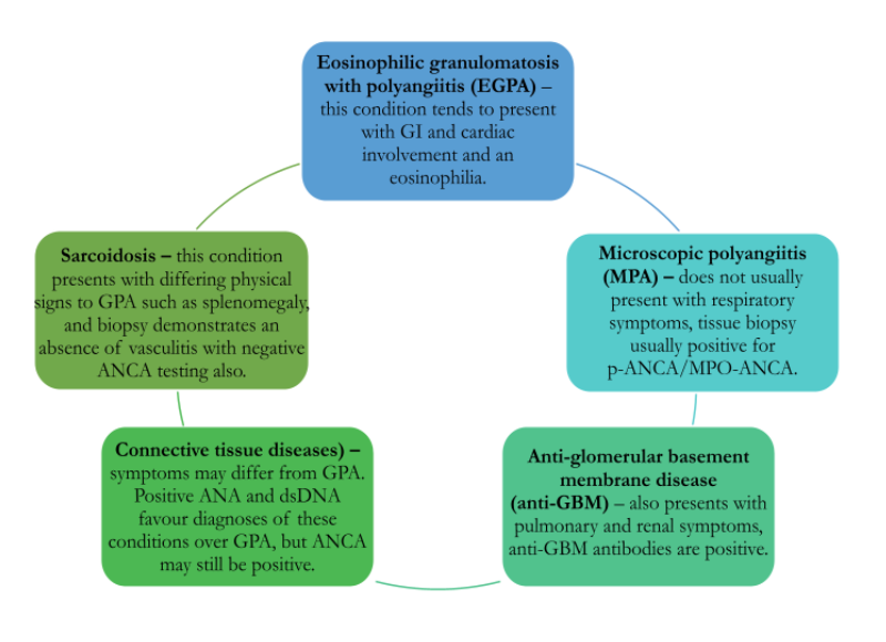
Adapted from: Falk, R., Merkel, P. and King Jr, T. (2022) Granulomatosis with polyangiitis and microscopic polyangiitis: Clinical manifestations and diagnosis, UpToDate. Edited by R. Glassock et al. Available at: https://www.uptodate.com/contents/granulomatosis-with-polyangiitis-and-microscopic-polyangiitis-clinical-manifestations-and-diagnosis?search=gpa&source=search_result&selectedTitle=1~150&usage_type=default&display_rank=1 (Accessed: January 15, 2023) and Molloy, E. (2022) BMJ Best Practice Granulomatosis with polyangiitis. Available at: https://bestpractice.bmj.com/topics/en-gb/327/pdf/327/Granulomatosis%20with%20polyangiitis.pdf (Accessed: January 15, 2023).
References
- Falk, R., Merkel, P. and King Jr, T. (2022) Granulomatosis with polyangiitis and microscopic polyangiitis: Clinical manifestations and diagnosis, UpToDate. Edited by R. Glassock et al. Available at: https://www.uptodate.com/contents/granulomatosis-with-polyangiitis-and-microscopic-polyangiitis-clinical-manifestations-and-diagnosis?search=gpa&source=search_result&selectedTitle=1~150&usage_type=default&display_rank=1 (Accessed: January 15, 2023).
- Molloy, E. (2022) BMJ Best Practice Granulomatosis with polyangiitis. Available at: https://bestpractice.bmj.com/topics/en-gb/327/pdf/327/Granulomatosis%20with%20polyangiitis.pdf (Accessed: January 15, 2023).
Management
Untreated GPA has a poor prognosis; therefore, it is vital a diagnosis is made quickly and treatment begun. The first stage of treatment focuses on achieving remission from the disease, and the second stage focuses on maintaining remission and preventing disease relapse.
It is recognised that during active disease, irreversible damage to various organs may have occurred and will remain regardless of treatment. Therefore, the goal of remission is instead to ensure resolution of symptoms and signs, accepting that some measured parameters may permanently remain abnormal or take significant time to improve. Any recurrence of clinical symptoms/signs would be deemed indicative of relapse, regardless of the organ affected.
Achieving remission
Several medications are required to induce remission for people with GPA, as monotherapy is usually ineffective. Steroids such as prednisolone plus further immunosuppressive medications are the mainstay of this stage of treatment. For severe disease, remission can take up to 6 months to achieve after treatment begins; if remission does not occur, an alternative medication regimen should be trialled.
The induction regime used depends on the status of disease: non-severe, severe or refractory to treatment. Non-severe disease is defined by no evidence of organ or life-threatening disease, e.g., lack of evidence of progressive neuropathy or active bleeding in any organ. Severe disease is defined as disease-causing an active threat to organs or life, e.g., pulmonary or GI haemorrhage, active glomerulonephritis, pericarditis, etc. Refractory disease is disease that has failed to effectively respond to an approved induction therapy regime. The induction regimes commonly used to achieve remission in GPA are detailed below.

Adapted from: Molloy, E. (2022) BMJ Best Practice Granulomatosis with polyangiitis. Available at: https://bestpractice.bmj.com/topics/en-gb/327/pdf/327/Granulomatosis%20with%20polyangiitis.pdf (Accessed: January 15, 2023) and Merkel, P., Kaplan, A. and Falk, R. (2022) Granulomatosis with polyangiitis and microscopic polyangiitis: Induction and maintenance therapy. Edited by G. Appel et al. Up To Date. Available at: https://www.uptodate.com/contents/granulomatosis-with-polyangiitis-and-microscopic-polyangiitis-induction-and-maintenance-therapy?search=gpa&source=search_result&selectedTitle=2~150&usage_type=default&display_rank=2 (Accessed: January 15, 2023).
Maintaining remission
Patients can be commenced on an appropriate maintenance therapy regime of immunotherapy agents, usually initially with steroids. The regime used will be decided on how successful and tolerated the medications used during the induction phase of treatment were.
Suitable medications to maintain remission are rituximab, methotrexate or azathioprine. IV cyclophosphamide is only used to induce remission, so patients on this medication will need to be switched to an alternative medication to maintain remission about 6 months after their last intravenous dose. If methotrexate was used to induce remission, it can be continued for maintenance as well. Mycophenolate is usually only commenced/continued for remission for those who cannot tolerate other immunosuppressive medications or those with resistant disease.
Duration of therapy needed can vary, and current evidence suggests maintenance treatment should be continued for at least 2 years after remission is achieved. If any disease relapse occurs, lifelong maintenance therapy is advised, even if only one has occurred. To treat a relapse, another remission induction regime will need to be commenced until the GPA is controlled again.
The steroids started during the induction period can be slowly weaned down if symptoms remain well controlled, but this must take place over a significant period of time. It is preferable for patients to remain on a low-dose maintenance steroid if needed to reduce risk of relapse if no contraindications to this are present and suitable monitoring can take place.
Monitoring recommendations
Full blood count, renal and liver function, plus inflammatory markers and urinalysis, should be tested to monitor for side effects from treatment and any evidence of recurring disease approximately every 3 months. Some of the potential conditions that can develop as a result of the above immunosuppressive medications and steroids are osteoporosis, bone marrow toxicity, renal failure and infection.
Other treatment considerations
All of the treatment agents discussed in this section have toxicity risks, especially cyclophosphamide, which can have a number of serious side effects, such as increased risk of bladder cancer. It is also known to contribute to infertility; therefore, for those wishing to preserve their fertility, rituximab should be offered instead in the induction phase of treatment. Any patient receiving rituximab or cyclophosphamide should also be commenced on pneumocystis jirovecii prophylactic medication to prevent pneumocystis jirovecii pneumonia (PCP). Patients on a daily steroid dose of 20 milligrams or more, alongside methotrexate, azathioprine or mycophenolate therapy, should also be considered for PCP prophylaxis.
Methotrexate should not be given to those with an eGFR <60mL/min and is contraindicated in pregnancy. Azathioprine could be a suitable alternative agent in these cases.
References
- Garlapati P, Qurie A. (2022) Granulomatosis with Polyangiitis. Available at: https://pubmed.ncbi.nlm.nih.gov/32491759/ (Accessed: January 15, 2023).
- Kubaisi, B., Abu Samra, K. and Foster, C.S. (2016) “Granulomatosis with polyangiitis (Wegener’s disease): An updated review of Ocular disease manifestations,” Intractable & Rare Diseases Research, 5(2), pp. 61–69. Available at: https://doi.org/10.5582/irdr.2016.01014.
- Merkel, P., Kaplan, A. and Falk, R. (2022) Granulomatosis with polyangiitis and microscopic polyangiitis: Induction and maintenance therapy. Edited by G. Appel et al. Up To Date. Available at: https://www.uptodate.com/contents/granulomatosis-with-polyangiitis-and-microscopic-polyangiitis-induction-and-maintenance-therapy?search=gpa&source=search_result&selectedTitle=2~150&usage_type=default&display_rank=2 (Accessed: January 15, 2023).
- Molloy, E. (2022) BMJ Best Practice Granulomatosis with polyangiitis. Available at: https://bestpractice.bmj.com/topics/en-gb/327/pdf/327/Granulomatosis%20with%20polyangiitis.pdf (Accessed: January 15, 2023).
Eosinophilic granulomatosis with polyangiitis (EGPA)
Background
Epidemiology
Eosinophilic granulomatosis with polyangiitis (EGPA) is another type of autoimmune ANCA associated small and medium vessel vasculitis. This condition was previously known as Churg-Strauss syndrome and is the least common of the ANCA associated vasculitides referenced in the previous section. It is also classed as one of several hypereosinophilic diseases.
It is a multi-system disease; however, the most commonly involved organ is the lung. The estimated incidence is 1-3 cases per 1 million people in the UK, but higher in the asthmatic population. Presentation typically occurs in asthmatic patients who are 40 to 60 years of age, with slightly higher incidence in patients of male sex compared to female sex. It rarely occurs in children or those older than 65 years of age. The 5-year survival rate with appropriate treatment can be up to 100% in some cases.
References
- Jacobs, K. and Kligerman, S. (2019) “Immune-mediated lung diseases,” Seminars in Ultrasound, CT and MRI, 40(3), pp. 213–228. Available at: https://doi.org/10.1053/j.sult.2018.11.011.
- Keogh, K. (2022) Eosinophilic granulomatosis with polyangiitis (Churg-Strauss syndrome), Eosinophilic granulomatosis with polyangiitis (Churg-Strauss syndrome) – Symptoms, diagnosis and treatment | BMJ Best Practice. Available at: https://bestpractice.bmj.com/topics/en-gb/942 (Accessed: January 15, 2023).
- King Jr, T. and Paul Dieffenbach (2022) Epidemiology, pathogenesis, and pathology of eosinophilic granulomatosis with polyangiitis (Churg-Strauss), UpToDate. Edited by K. Flaherty, R. Glassock, and B. Bochner. UpToDate. Available at: https://www.uptodate.com/contents/epidemiology-pathogenesis-and-pathology-of-eosinophilic-granulomatosis-with-polyangiitis-churg-strauss?search=egpa&source=search_result&selectedTitle=3~143&usage_type=default&display_rank=3 (Accessed: January 15, 2023).
Pathophysiology
As with the other autoimmune vasculitides, the exact aetiology and pathophysiology of EGPA are unknown. It has been proposed that exposure to environmental triggers, e.g., infection, results in raised serum immunoglobulin (IgE), TH1 and TH2 cytokines (interleukin 4, 5 and 13) levels. Th2 cytokines contribute to the maturation and activation process of eosinophils, which directly release toxic proteins. These proteins drive inflammation; therefore, raised levels of eosinophils due to raised IL-5 results in higher levels of toxic protein release and, hence, more inflammation. Levels of regulatory T cells, which protect against autoimmune disease development, have been found to be lower in those with EGPA.
As with GPA, ANCA is also thought to play a role in this disease, likely via development of vessel inflammation due to neutrophil activation. Genetic factors may also be involved; for example, the interleukin (IL)10 gene and HLA class of immune cells are both associated with EGPA.
It has been suggested that omalizumab and leukotriene receptor antagonists use can reveal undiagnosed cases of EGPA if they improve asthma symptoms to the point that oral steroid weaning can occur, and clinical manifestations of EGPA then become apparent.
References
- Keogh, K. (2022) Eosinophilic granulomatosis with polyangiitis (Churg-Strauss syndrome), Eosinophilic granulomatosis with polyangiitis (Churg-Strauss syndrome) – Symptoms, diagnosis and treatment | BMJ Best Practice. Available at: https://bestpractice.bmj.com/topics/en-gb/942 (Accessed: January 15, 2023).
- King Jr, T. and Paul Dieffenbach (2022) Epidemiology, pathogenesis, and pathology of eosinophilic granulomatosis with polyangiitis (Churg-Strauss), UpToDate. Edited by K. Flaherty, R. Glassock, and B. Bochner. UpToDate. Available at: https://www.uptodate.com/contents/epidemiology-pathogenesis-and-pathology-of-eosinophilic-granulomatosis-with-polyangiitis-churg-strauss?search=egpa&source=search_result&selectedTitle=3~143&usage_type=default&display_rank=3 (Accessed: January 15, 2023).
Presentation
It has been proposed that EGPA presentations can be split into three different phases, depending on clinical symptoms and signs at the time of diagnosis. The three phases are summarised below:
- Phase 1: Prodrome – tends to begin between the ages of 20 and 30 and precedes the final phase by about a decade. The features of this phase are asthma symptoms, e.g., wheeze, with sinusitis and allergic rhinitis.
- Phase 2: Eosinophilia – involves eosinophilic infiltration of the organs, especially the lungs.
- Phase 3: Organ-specific vasculitis – small to medium vessel vasculitis develops. Other symptoms like fatigue and weight loss can occur concurrently with this final phase, and asthma exacerbations often increase with greater severity.
Patients typically present with a history of asthma and associated symptoms e.g., cough and wheeze. They may also have a number of other associated signs and symptoms depending on their stage of disease, including:
- Purulent/bloody nasal discharge (due to sinusitis/allergic rhinitis)
- Haemoptysis
- Symptoms of PE or DVT – EGPA increases the risk of VTE.
- Orthopnoea, crackles and peripheral oedema may be present if heart failure secondary to vasculitis-induced cardiomyopathy has developed – cardiac involvement in EGPA has high mortality and poor prognosis.
- Abdominal pain and change in bowel habit
- Renal insufficiency and glomerulonephritis – occur in about a quarter of patients with EGPA, also indicates poor prognosis.
- Peripheral neuropathy and weakness
- New-onset motor deficits, e.g., foot drop
- Arthralgia and myalgia
- Fatigue
- Purpuric skin rash and nodules on extensor surfaces
- Lymphadenopathy
References
- Keogh, K. (2022) Eosinophilic granulomatosis with polyangiitis (Churg-Strauss syndrome), Eosinophilic granulomatosis with polyangiitis (Churg-Strauss syndrome) – Symptoms, diagnosis and treatment | BMJ Best Practice. Available at: https://bestpractice.bmj.com/topics/en-gb/942 (Accessed: January 15, 2023).
- King Jr, T. (2022) Clinical features and diagnosis of eosinophilic granulomatosis with polyangiitis (Churg-Strauss), UpToDate. Edited by K. Flaherty et al. UpToDate. Available at: https://www.uptodate.com/contents/clinical-features-and-diagnosis-of-eosinophilic-granulomatosis-with-polyangiitis-churg-strauss?search=egpa&source=search_result&selectedTitle=1~143&usage_type=default&display_rank=1#H17 (Accessed: January 15, 2023).
Diagnosis
The American College of Rheumatology (ACR) have proposed an updated 2022 classification criteria for EGPA; however, this was only intended to distinguish between vasculitides rather than for diagnostic use.
The table below summarises some investigations used to diagnose EGPA, with the expected associated findings.
| Investigation | Results/findings |
|---|---|
| Urinalysis | Microscopic haematuria or proteinuria may be present, indicative of glomerulonephritis. |
| Lung function | Demonstrates reversible airway obstruction, in line with an asthma presentation. |
| Blood tests | Eosinophil count is usually more than 10% of the total white cell count (WCC), ESR and CRP can help evaluate inflammation levels but are non-specific for EGPA, deranged U&Es may indicate glomerulonephritis, proBNP will be raised if concurrent heart failure is present, serum IgE is usually raised. p-ANCA/MPO-ANCA is positive in at least one-third of patients with EGPA on immunoassay. |
| Imaging | Chest X-ray may show consolidation and nodules (consistent with granulomas) – these findings can be transient and migratory. Any abnormalities should be further evaluated with a dedicated HRCT of the chest, which can show centrilobular nodules, ground glass opacification and bronchial wall thickening. Bronchoscopy is usually performed in those with opacities to rule out alveolar haemorrhage and malignancy. CT pulmonary angiography may be needed if pulmonary embolism is suspected.
Transthoracic echocardiography (TTE) should be performed to evaluate symptoms of heart failure – wall motion abnormalities are common in EGPA. |
| Biopsy | Skin lesions are usually easily sampled, but renal and/or lung biopsies can also be obtained if severe and progressive disease is apparent. Histology typically shows a mixture of granulomas, eosinophilic infiltrates and vasculitis. |
Table 3: Summary of investigations performed to investigate suspected EGPA. Adapted from: Keogh, K. (2022) Eosinophilic granulomatosis with polyangiitis (Churg-Strauss syndrome), Eosinophilic granulomatosis with polyangiitis (Churg-Strauss syndrome) – Symptoms, diagnosis and treatment | BMJ Best Practice. Available at: https://bestpractice.bmj.com/topics/en-gb/942 (Accessed: January 15, 2023) and King Jr, T. (2022) Clinical features and diagnosis of eosinophilic granulomatosis with polyangiitis (Churg-Strauss), UpToDate. Edited by K. Flaherty et al. UpToDate. Available at: https://www.uptodate.com/contents/clinical-features-and-diagnosis-of-eosinophilic-granulomatosis-with-polyangiitis-churg-strauss?search=egpa&source=search_result&selectedTitle=1~143&usage_type=default&display_rank=1#H17 (Accessed: January 15, 2023).
References
- Grayson, P.C. et al. (2022) “2022 American College of Rheumatology/European Alliance of Associations for Rheumatology classification criteria for eosinophilic granulomatosis with polyangiitis,” Annals of the Rheumatic Diseases, 81(3), pp. 309–314. Available at: https://doi.org/10.1136/annrheumdis-2021-221794.
- Jacobs, K. and Kligerman, S. (2019) “Immune-mediated lung diseases,” Seminars in Ultrasound, CT and MRI, 40(3), pp. 213–228. Available at: https://doi.org/10.1053/j.sult.2018.11.011.
- Keogh, K. (2022) Eosinophilic granulomatosis with polyangiitis (Churg-Strauss syndrome), Eosinophilic granulomatosis with polyangiitis (Churg-Strauss syndrome) – Symptoms, diagnosis and treatment | BMJ Best Practice. Available at: https://bestpractice.bmj.com/topics/en-gb/942 (Accessed: January 15, 2023).
- King Jr, T. (2022) Clinical features and diagnosis of eosinophilic granulomatosis with polyangiitis (Churg-Strauss), UpToDate. Edited by K. Flaherty et al. UpToDate. Available at: https://www.uptodate.com/contents/clinical-features-and-diagnosis-of-eosinophilic-granulomatosis-with-polyangiitis-churg-strauss?search=egpa&source=search_result&selectedTitle=1~143&usage_type=default&display_rank=1#H17 (Accessed: January 15, 2023).
Differential diagnoses
A number of conditions can present similarly to EGPA, including those that also present with eosinophilia. Some of these conditions and their key differences to EGPA are summarised below.
- Hypereosinophilic syndrome (HES) – in HES, asthma and vasculitis complications do not occur. Additionally, specific gene mutations can be tested for that can be present in HES but would not be present in EGPA.
- Allergic bronchopulmonary aspergillosis (ABPA) – ABPA does not present with vasculitis and tends to affect only the upper and lower airways rather than any other organ systems. Positive allergy and specific serum IgE testing for Aspergillosis fumigatus would support a diagnosis of ABPA over EGPA.
- Microscopic polyangiitis (MPA) – asthma and peripheral eosinophilia are absent, as are lung infiltrates and nodules. Lung function is usually normal.
- Granulomatosis with polyangiitis (GPA) – GPA does not present with asthma; lung function will likely be normal. Additionally, alveolar haemorrhage and glomerulonephritis are common in GPA but not in EGPA. Those with GPA are likely to have a positive c-ANCA/PR3-ANCA result, compared to those with EGPA, who tend to have a positive p-ANCA/MPO-ANCA result instead.
References
- Keogh, K. (2022) Eosinophilic granulomatosis with polyangiitis (Churg-Strauss syndrome), Eosinophilic granulomatosis with polyangiitis (Churg-Strauss syndrome) – Symptoms, diagnosis and treatment | BMJ Best Practice. Available at: https://bestpractice.bmj.com/topics/en-gb/942 (Accessed: January 15, 2023).
- King Jr, T. (2022) Clinical features and diagnosis of eosinophilic granulomatosis with polyangiitis (Churg-Strauss), UpToDate. Edited by K. Flaherty et al. UpToDate. Available at: https://www.uptodate.com/contents/clinical-features-and-diagnosis-of-eosinophilic-granulomatosis-with-polyangiitis-churg-strauss?search=egpa&source=search_result&selectedTitle=1~143&usage_type=default&display_rank=1#H17 (Accessed: January 15, 2023).
Management
As with GPA, steroids and immunosuppressive medications are the mainstay of treatment for EGPA. Similar to GPA management, the initial treatment offered also focuses on inducing remission and then maintaining it. The exact approach taken will depend on whether the patient’s disease is classed as severe (organ and/or life-threatening) or non-severe.
Remission induction
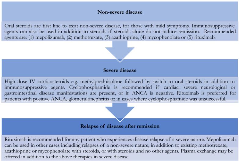
Adapted from: Keogh, K. (2022) Eosinophilic granulomatosis with polyangiitis (Churg-Strauss syndrome), Eosinophilic granulomatosis with polyangiitis (Churg-Strauss syndrome) – Symptoms, diagnosis and treatment | BMJ Best Practice. Available at: https://bestpractice.bmj.com/topics/en-gb/942 (Accessed: January 15, 2023).
Remission maintenance
For patients who required steroids alone to successfully induce remission, depending on their symptoms and clinical parameters, e.g., CRP, steroids can usually be gradually weaned down. This can take many months and will be guided by the clinical presentation when the dose is reviewed. Some patients may never be able to be fully weaned and require a lifelong low dose of steroids to keep symptoms under control. Some patients needing a maintenance dose may be offered immunosuppressive therapy too, to facilitate a lower maintenance steroid dose.
Any patient who needed an immunosuppressive agent alongside steroid therapy to successfully induce remission normally needs to continue this dual regime to maintain remission. The agent used to induce remission can normally be continued into maintenance unless it was rituximab. For this group of patients, it is recommended that for maintenance, they switch to methotrexate, azathioprine or mycophenolate instead – contraindications to any of these medications would need to be explored prior to deciding which would be recommended. Cyclophosphamide is not used for long-term maintenance due to toxicity issues; therefore, these patients should also switch to one of the three other agents mentioned above.
Immunosuppressive agents are typically continued for up to 18 months to maintain remission, although some patients need longer courses or indefinite immunosuppression if they have multiple relapses. Steroids can still be tapered as they would be if they were used as a monotherapy, guided by the patient’s symptoms and response.
Monitoring recommendations and other treatment considerations
These are the same as those for GPA, e.g., considering osteoporosis screening due to long-term steroid treatment exposure. As in GPA, methotrexate should not be given to those with an eGFR <60mL/min and is contraindicated in pregnancy.
References
- Berger, A. (2000) “Science commentary: Th1 and th2 responses: What are they? ” BMJ, 321(7258), pp. 424–424. Available at: https://doi.org/10.1136/bmj.321.7258.424.
- Chung, S.A. et al. (2021) “2021 American College of Rheumatology/Vasculitis Foundation guideline for the management of Antineutrophil cytoplasmic antibody–associated vasculitis,” Arthritis & Rheumatology, 73(8), pp. 1366–1383. Available at: https://doi.org/10.1002/art.41773.
- Gioffredi, A. et al. (2014) “Eosinophilic granulomatosis with polyangiitis: An overview,” Frontiers in Immunology, 5. Available at: https://doi.org/10.3389/fimmu.2014.00549.
- Keogh, K. (2022) Eosinophilic granulomatosis with polyangiitis (Churg-Strauss syndrome), Eosinophilic granulomatosis with polyangiitis (Churg-Strauss syndrome) – Symptoms, diagnosis and treatment | BMJ Best Practice. Available at: https://bestpractice.bmj.com/topics/en-gb/942 (Accessed: January 15, 2023).
- King Jr, T. (2022) Eosinophilic granulomatosis with polyangiitis (Churg-Strauss): Treatment and prognosis, UpToDate. Edited by K. Flaherty et al. UpToDate. Available at: https://www.uptodate.com/contents/eosinophilic-granulomatosis-with-polyangiitis-churg-strauss-treatment-and-prognosis?search=egpa&source=search_result&selectedTitle=2~143&usage_type=default&display_rank=2#H1409426116
Hypersensitivity pneumonitis (HP)
Background
Epidemiology
Hypersensitivity pneumonitis (or extrinsic allergic alveolitis) is an immune-mediated respiratory syndrome caused by allergen inhalation and subsequent reaction. This leads to alveolar and bronchiole inflammation via a non-IgE mediated process. The most common allergen is organic dust; however, there are hundreds of known causative agents, so identifying the allergen can be difficult in some cases. Pigeon fancier’s lung and Farmer’s lung are examples of hypersensitivity pneumonitis caused by pigeon exposure (avian protein antigen in faeces) and grain/straw mould (usually due to Aspergillus, Alternaria or Trichosporon fungi) respectively.
Prevalence and incidence of the condition overall are thought to be low; however, mild cases can masquerade as other conditions, such as viral illness and, therefore, remain undetected for a long period of time. Smoking may be a protective factor against developing acute HP; some evidence suggests that smokers exposed to causative allergens form fewer antibodies against antigens present and, therefore, do not manifest a clinical presentation of HP. However, in cases of established disease, smoking may influence a more severe presentation and cause recurrent episodes of exacerbation.

Adapted from: King Jr, T. (2021) Hypersensitivity pneumonitis (extrinsic allergic alveolitis): Epidemiology, causes, and pathogenesis, UpToDate. Edited by K. Flaherty and P. Dieffenbach. UpToDate. Available at: https://www.uptodate.com/contents/hypersensitivity-pneumonitis-extrinsic-allergic-alveolitis-epidemiology-causes-and-pathogenesis?search=hypersensitivity+pneumonitis&source=search_result&selectedTitle=1~150&usage_type=default&display_rank=1 (Accessed: January 15, 2023).
References
- Grammer, L. (2022) Hypersensitivity pneumonitis, Hypersensitivity pneumonitis – Symptoms, diagnosis and treatment | BMJ Best Practice. BMJ Best Practice. Available at: https://bestpractice.bmj.com/topics/en-gb/647 (Accessed: January 15, 2023).
- King Jr, T. (2021) Hypersensitivity pneumonitis (extrinsic allergic alveolitis): Epidemiology, causes, and pathogenesis, UpToDate. Edited by K. Flaherty and P. Dieffenbach. UpToDate. Available at: https://www.uptodate.com/contents/hypersensitivity-pneumonitis-extrinsic-allergic-alveolitis-epidemiology-causes-and-pathogenesis?search=hypersensitivity+pneumonitis&source=search_result&selectedTitle=1~150&usage_type=default&display_rank=1 (Accessed: January 15, 2023).
- Selman, M., Pardo, A. and King, T.E. (2012) “Hypersensitivity Pneumonitis Insights in Diagnosis and Pathobiology,” American Journal of Respiratory and Critical Care Medicine, 186(4), pp. 314–324. Available at: https://doi.org/10.1164/rccm.201203-0513ci.
Pathophysiology
Not every individual exposed to the same allergen will go on to develop HP – indeed, most of those exposed will develop no clinical signs or symptoms post-exposure. In susceptible individuals, it has been proposed that in acute cases, exposure to the antigens in an allergen causes a type 3 hypersensitivity reaction, leading to IgG antibody production for that allergen’s antigens. Then, subsequent exposure to the same allergen results in a type 4 hypersensitivity reaction with increased CD4+ cell activity and cytokine release, leading to granuloma formation and other chronic pathological changes.
A genetic component to the disease has been suggested in a number of reports; however, no definite genetic risk factor appears to have been identified with a consistent link to HP development yet.
References
- Grammer, L. (2022) Hypersensitivity pneumonitis, Hypersensitivity pneumonitis – Symptoms, diagnosis and treatment | BMJ Best Practice. BMJ Best Practice. Available at: https://bestpractice.bmj.com/topics/en-gb/647 (Accessed: January 15, 2023).
- Jacobs, K. and Kligerman, S. (2019) “Immune-mediated lung diseases,” Seminars in Ultrasound, CT and MRI, 40(3), pp. 213–228. Available at: https://doi.org/10.1053/j.sult.2018.11.011.
Selman, M., Pardo, A. and King, T.E. (2012) “Hypersensitivity Pneumonitis Insights in Diagnosis and Pathobiology,” American Journal of Respiratory and Critical Care Medicine, 186(4), pp. 314–324. Available at: https://doi.org/10.1164/rccm.201203-0513ci.
Presentation
The presentation of HP can be broadly classed as acute, subacute and chronic, and the clinical presentation of an individual with HP may differ depending on their disease classification status. The below diagram lists common symptoms experienced in HP.

Adapted from: Grammer, L. (2022) Hypersensitivity pneumonitis, Hypersensitivity pneumonitis – Symptoms, diagnosis and treatment | BMJ Best Practice. BMJ Best Practice. Available at: https://bestpractice.bmj.com/topics/en-gb/647 (Accessed: January 15, 2023).
The above symptoms can begin as soon as four hours after exposure to an allergen. They are usually accompanied by physical signs such as: diffuse crackles, mid-inspiratory noises and clubbing (advanced disease). Wheeze is not normally a prominent feature of HP, and if present, other diagnoses such as severe asthma, should be considered instead.
References
- Grammer, L. (2022) Hypersensitivity pneumonitis, Hypersensitivity pneumonitis – Symptoms, diagnosis and treatment | BMJ Best Practice. BMJ Best Practice. Available at: https://bestpractice.bmj.com/topics/en-gb/647 (Accessed: January 15, 2023).
- Selman, M., Pardo, A. and King, T.E. (2012) “Hypersensitivity Pneumonitis Insights in Diagnosis and Pathobiology,” American Journal of Respiratory and Critical Care Medicine, 186(4), pp. 314–324. Available at: https://doi.org/10.1164/rccm.201203-0513ci.
Diagnosis
Firstly, a detailed history should be taken, particularly in relation to identifying if any confirmed exposure to a causative agent has occurred at any point. In two-thirds of cases, the allergen is never found but known exposure to a known causative agent makes this diagnosis likely alongside relevant clinical signs and symptoms.
Testing for sensitivity to known allergens is not always recommended, as false positive and negative results are common. The diagram below details the typical panel of tests ordered when working up a patient with suspected HP, alongside an explanation of expected findings in someone with this diagnosis.
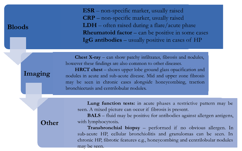
Adapted from: Grammer, L. (2022) Hypersensitivity pneumonitis, Hypersensitivity pneumonitis – Symptoms, diagnosis and treatment | BMJ Best Practice. BMJ Best Practice. Available at: https://bestpractice.bmj.com/topics/en-gb/647 (Accessed: January 15, 2023).
References
- Grammer, L. (2022) Hypersensitivity pneumonitis, Hypersensitivity pneumonitis – Symptoms, diagnosis and treatment | BMJ Best Practice. BMJ Best Practice. Available at: https://bestpractice.bmj.com/topics/en-gb/647 (Accessed: January 15, 2023).
- Hsieh, C. and Kamangar, N. (2022) Hypersensitivity pneumonitis, Background, Pathophysiology, Etiology. Medscape. Available at: https://emedicine.medscape.com/article/299174-overview (Accessed: January 15, 2023).
- King Jr, T. et al. (2022) Hypersensitivity pneumonitis (extrinsic allergic alveolitis): Clinical manifestations and diagnosis, UpToDate. UpToDate. Available at: https://www.uptodate.com/contents/hypersensitivity-pneumonitis-extrinsic-allergic-alveolitis-clinical-manifestations-and-diagnosis?search=hypersensitivity+pneumonitis&source=search_result&selectedTitle=2~150&usage_type=default&display_rank%5D (Accessed: January 15, 2023).
- Selman, M., Pardo, A. and King, T.E. (2012) “Hypersensitivity Pneumonitis Insights in Diagnosis and Pathobiology,” American Journal of Respiratory and Critical Care Medicine, 186(4), pp. 314–324. Available at: https://doi.org/10.1164/rccm.201203-0513ci.
Differential diagnoses
HP should be considered as part of the initial work up for any patient presenting with confirmed exposure to one of the identified causative allergens, alongside clinical findings. Other conditions to consider include:
- Sarcoidosis – this condition usually demonstrates symptoms and signs not limited to the respiratory system, e.g., erythema nodosum. A transbronchial biopsy may be needed in some cases to accurately diagnose HP.
- Idiopathic pulmonary fibrosis (IPF) – in IPF, HRCT chest would show more evidence of abnormalities in the lower zones of the chest rather than the upper zones, and centrilobular nodules would likely not be seen. However, the imaging can look very similar and respiratory opinion may be needed. Additionally, BALS in IPF would not show lymphocytosis.
- Pneumoconiosis – this usually only occurs in patients with an obvious exposure history for this condition, e.g., miners who worked with mineral dust. Chest imaging tends to show signs of asbestosis and silicosis, e.g., calcified hilar lymph nodes.
- Viral pneumonia – the symptoms of viral pneumonia tend to last for some time (at least a week) and would not repeatedly occur once resolved. In HP, symptoms may last only a couple of days but occur repeatedly in an episodic fashion every time the patient is exposed to the allergen. A respiratory virus panel can be helpful to deduce if any viral infection is present.
- Other more common respiratory diseases, e.g., asthma, COPD – these should also be considered if the history does not contain any clear evidence of an allergen and other suspicious factors are present, e.g., history of atopy for asthma, smoking history in COPD, etc., alongside imaging and other initial investigations.
References
- Grammer, L. (2022) Hypersensitivity pneumonitis, Hypersensitivity pneumonitis – Symptoms, diagnosis and treatment | BMJ Best Practice. BMJ Best Practice. Available at: https://bestpractice.bmj.com/topics/en-gb/647 (Accessed: January 15, 2023).
- Jacobs, K. and Kligerman, S. (2019) “Immune-mediated lung diseases,” Seminars in Ultrasound, CT and MRI, 40(3), pp. 213–228. Available at: https://doi.org/10.1053/j.sult.2018.11.011.
- King Jr, T. et al. (2022) Hypersensitivity pneumonitis (extrinsic allergic alveolitis): Clinical manifestations and diagnosis, UpToDate. UpToDate. Available at: https://www.uptodate.com/contents/hypersensitivity-pneumonitis-extrinsic-allergic-alveolitis-clinical-manifestations-and-diagnosis?search=hypersensitivity+pneumonitis&source=search_result&selectedTitle=2~150&usage_type=default&display_rank%5D (Accessed: January 15, 2023).
- Ungprasert, P., Ryu, J.H. and Matteson, E.L. (2019) “Clinical manifestations, diagnosis, and treatment of sarcoidosis,” Mayo Clinic Proceedings: Innovations, Quality & Outcomes, 3(3), pp. 358–375. Available at: https://doi.org/10.1016/j.mayocpiqo.2019.04.006.
Management
The mainstay of treatment for HP revolves around reducing/removing exposure to any identified causative allergens. Patients are advised to avoid either an allergen they are known to be sensitive to, or other potential allergens that may be present in their environment. This can be difficult, especially if the allergen is linked to their livelihood. Total avoidance can result in regression of disease and/or prevent progression to fibrosis, so the importance of avoiding causative allergens must be emphasised and encouraged.
Acute and sub-acute HP
For mild cases of acute HP, steroids are not recommended as the clinical presentation will often improve once the causative allergen is removed. If symptoms are recurrent and persistent, despite allergen avoidance, a short course of oral steroids can be offered if no contraindications are present. These can usually be tapered off unless fibrosis is present – patients in this group normally require long-term low-dose therapy.
Prednisolone is the 1st line oral steroid choice, and dosing is 0.5-1mg/kg/day on average. When symptoms improve, the dose can be tapered by about 5mg every other day over 4-6 weeks, depending on the ongoing clinical presentation.
Chronic HP
For those with chronic HP of a fibrotic nature, smoking avoidance/cessation, seasonal respiratory vaccinations and chest physiotherapy are recommended in addition to the above management strategy. For some, long-term oxygen therapy may eventually also be needed. Use of immunosuppressive agents such as azathioprine and mycophenolate mofetil (MMF) are not currently part of standard care, and further data is needed to support their use in this condition.
For those with very severe disease, lung transplant may be an option, depending on comorbidities, functional status and overall suitability for transplant. There have been several reports of the condition reoccurring in the transplanted organ, likely due to ongoing exposure to the allergen post-procedure.
References
- American Lung Association (2022) How Hypersensitivity Pneumonitis Is Treated, American Lung Association. Available at: https://www.lung.org/lung-health-diseases/lung-disease-lookup/hypersensitivity-pneumonitis/treating-and-managing (Accessed: January 15, 2023).
- Grammer, L. (2022) Hypersensitivity pneumonitis, Hypersensitivity pneumonitis – Symptoms, diagnosis and treatment | BMJ Best Practice. BMJ Best Practice. Available at: https://bestpractice.bmj.com/topics/en-gb/647 (Accessed: January 15, 2023).
- King Jr, T. (2022) Hypersensitivity pneumonitis (extrinsic allergic alveolitis): Treatment, prognosis and prevention, UpToDate. Edited by K. Flaherty and P. Dieffenbach. UpToDate. Available at: https://www.uptodate.com/contents/hypersensitivity-pneumonitis-extrinsic-allergic-alveolitis-treatment-prognosis-and-prevention?search=hypersensitivity%20pneumonitis&source=search_result&selectedTitle=3~150&usage_type=default&display_rank=3#H3199492110
Allergic bronchopulmonary aspergillosis (ABPA)
Background
Epidemiology
Allergic bronchopulmonary aspergillosis (ABPA) is a type of hypersensitivity immune response respiratory disorder specifically associated with the Aspergillus fungus. The main species responsible is usually Aspergillus fumigatus, but other species, such as Aspergillus flavus, have also been identified as causative agents of ABPA. Other associated hypersensitivity respiratory disorders include HP and IgE mediated asthma. ABPA normally only occurs in those with cystic fibrosis (CF) or asthma– in those with asthma, prevalence ranges from 1-5%, and in those with CF, prevalence is approximately 1-9%. The main affected age group tends to be teenagers (if they have CF) or young adults (with asthma). It can also affect children and older patients, but occurrence is less common within these age groups.
ABPA is an emerging condition in India, and there is high prevalence in the Southwest areas of America also. Under recognition of signs and symptoms of the disease means rates may be much higher worldwide than is currently known.
References
- Akuthota, P. (2022) Clinical manifestations and diagnosis of allergic bronchopulmonary aspergillosis, UpToDate. Edited by C. Kauffman, P. Barnes, and P. Dieffenbach. UpToDate. Available at: https://www.uptodate.com/contents/clinical-manifestations-and-diagnosis-of-allergic-bronchopulmonary-aspergillosis?search=abpa&source=search_result&selectedTitle=1~57&usage_type=default&display_rank=1 (Accessed: January 15, 2023).
- Jack, J. and Bajaj, T. (2022) Allergic Bronchopulmonary Aspergillosis, Allergic Bronchopulmonary Aspergillosis. StatPearls. Available at: https://www.ncbi.nlm.nih.gov/books/NBK542329/ (Accessed: January 15, 2023).
- Radojicic, C. (2020) Allergic bronchopulmonary aspergillosis, Allergic bronchopulmonary aspergillosis – Symptoms, diagnosis and treatment | BMJ Best Practice. BMJ Best Practice. Available at: https://bestpractice.bmj.com/topics/en-gb/836/pdf/836/Allergic%20bronchopulmonary%20aspergillosis.pdf (Accessed: January 15, 2023).
Pathophysiology
ABPA is an IgE and IgG immune-mediated hypersensitivity response to Aspergillus fumigatus fungus. It is not a systemic nor invasive fungal infection, and the fungi can be found in multiple environmental sources, including soil, grass and sewage. The spores are airborne and must be inhaled.
Exposure in individuals without atopy does not usually lead to antibody formation. However, in those with atopy, such as asthmatics, antibody formation after exposure to the fungal allergen is common. Conditions like asthma and CF tend to cause impaired mucus secretion and clearance, increasing the likelihood that spores will become trapped in these individuals’ airways and allow colonisation.
Once in the airway, the presence of fungal spores triggers an intense inflammatory response on top of existing inflammation caused by underlying asthma or CF. Th2 cells are recruited to release interleukin (IL)-5, which stimulates eosinophil and B cell recruitment. The presence of fungal enzymes causes IL-8 release, which stimulates neutrophil recruitment. These interleukins also stimulate synthesis of IgG and IgE antibodies, as do B cells. The inflammatory products released by the eosinophils and neutrophils recruited cause the bronchi and bronchioles to fill with eosinophilic plugs and hyphae, which leads to bronchiectasis and obstruction, and the signs and symptoms associated with the disease.
References
- Akuthota, P. (2022) Clinical manifestations and diagnosis of allergic bronchopulmonary aspergillosis, UpToDate. Edited by C. Kauffman, P. Barnes, and P. Dieffenbach. UpToDate. Available at: https://www.uptodate.com/contents/clinical-manifestations-and-diagnosis-of-allergic-bronchopulmonary-aspergillosis?search=abpa&source=search_result&selectedTitle=1~57&usage_type=default&display_rank=1 (Accessed: January 15, 2023).
- Jack, J. and Bajaj, T. (2022) Allergic Bronchopulmonary Aspergillosis, Allergic Bronchopulmonary Aspergillosis. StatPearls. Available at: https://www.ncbi.nlm.nih.gov/books/NBK542329/ (Accessed: January 15, 2023).
- Moss, R.B. (2005) “Pathophysiology and immunology of allergic bronchopulmonary aspergillosis,” Medical Mycology, 43(s1), pp. 203–206. Available at: https://doi.org/10.1080/13693780500052255.
- Radojicic, C. (2020) Allergic bronchopulmonary aspergillosis, Allergic bronchopulmonary aspergillosis – Symptoms, diagnosis and treatment | BMJ Best Practice. BMJ Best Practice. Available at: https://bestpractice.bmj.com/topics/en-gb/836/pdf/836/Allergic%20bronchopulmonary%20aspergillosis.pdf (Accessed: January 15, 2023).
- Tillie-Leblond, I. and Tonnel, A.-B. (2005) “Allergic bronchopulmonary aspergillosis,” Allergy, 60(8), pp. 1004–1013. Available at: https://doi.org/10.1111/j.1398-9995.2005.00887.x.
Presentation
The condition is clinically hard to distinguish from CF, and for these patients, the symptoms associated with ABPA may be no different to their usual symptoms other than potentially more pronounced. A careful history and examination will be needed alongside relevant investigations to determine if ABPA is the cause of their symptoms. In those with asthma, ABPA tends to only develop after asthma treatment is begun. Symptoms of the disease can include:
- Upper airway symptoms, e.g., in keeping with rhinitis or sinusitis
- Shortness of breath if fibrosis is present
- Wheeze – may not always be present
- Productive cough
- Haemoptysis
- Pleuritic chest pain
- Malaise
- Fever
In advanced disease, signs such as finger clubbing, cyanosis and peripheral oedema can occur. Crackles may also be heard if fibrosis is present on auscultation.
References
- Akuthota, P. (2022) Clinical manifestations and diagnosis of allergic bronchopulmonary aspergillosis, UpToDate. Edited by C. Kauffman, P. Barnes, and P. Dieffenbach. UpToDate. Available at: https://www.uptodate.com/contents/clinical-manifestations-and-diagnosis-of-allergic-bronchopulmonary-aspergillosis?search=abpa&source=search_result&selectedTitle=1~57&usage_type=default&display_rank=1 (Accessed: January 15, 2023).
- Jack, J. and Bajaj, T. (2022) Allergic Bronchopulmonary Aspergillosis, Allergic Bronchopulmonary Aspergillosis. StatPearls. Available at: https://www.ncbi.nlm.nih.gov/books/NBK542329/ (Accessed: January 15, 2023).
- Radojicic, C. (2020) Allergic bronchopulmonary aspergillosis, Allergic bronchopulmonary aspergillosis – Symptoms, diagnosis and treatment | BMJ Best Practice. BMJ Best Practice. Available at: https://bestpractice.bmj.com/topics/en-gb/836/pdf/836/Allergic%20bronchopulmonary%20aspergillosis.pdf (Accessed: January 15, 2023).
Diagnosis
ABPA should be suspected in any patient with CF or asthma presenting with several exacerbations, upper zone bronchiectasis, eosinophilia and raised IgE. It is rare to develop the condition without the underlying disease of CF or asthma. A number of investigations are needed to rule in or out a diagnosis of ABPA as follows:
- Bloods
- Eosinophilia – can be seen in blood tests and histology results. May be absent if steroids are taken regularly.
- Total serum IgE – also usually raised in ABPA
- Specific serum IgE and IgG to Aspergillus fumigatus may also be raised
- Imaging
- A chest X-ray will likely show non-specific features, and if ABPA is suspected, CT imaging should be performed.
- HRCT of the chest can show bronchiectasis (mainly in the central lobes), mucus plugging, centrilobular nodules and bronchial wall thickening and dilatation.
- Other investigations
- Sputum culture – may be positive for presence of Aspergillus fumigatus but can also show no growth.
- Skin test – to establish if an individual is sensitised to the fungus (type 1 hypersensitivity reaction occurs if so).
- HLA-DR molecules – this gene marker is associated with ABPA, but it is not a routine clinical test.
- Lung function tests – in ABPA, an obstructive pattern is usually seen, with reduced FEV1 and increased residual volume. However, fibrosis and/or bronchiectasis can cause a mixed or restrictive pattern to occur as well.
References
- Akuthota, P. (2022) Clinical manifestations and diagnosis of allergic bronchopulmonary aspergillosis, UpToDate. Edited by C. Kauffman, P. Barnes, and P. Dieffenbach. UpToDate. Available at: https://www.uptodate.com/contents/clinical-manifestations-and-diagnosis-of-allergic-bronchopulmonary-aspergillosis?search=abpa&source=search_result&selectedTitle=1~57&usage_type=default&display_rank=1 (Accessed: January 15, 2023).
- Jack, J. and Bajaj, T. (2022) Allergic Bronchopulmonary Aspergillosis, Allergic Bronchopulmonary Aspergillosis. StatPearls. Available at: https://www.ncbi.nlm.nih.gov/books/NBK542329/ (Accessed: January 15, 2023).
- Nawaz Khan, A. (2020) Pulmonary Aspergillosis Imaging, Practice Essentials, Radiograph, CT Scanning. Medscape. Available at: https://emedicine.medscape.com/article/353200-overview (Accessed: January 15, 2023).
- Radojicic, C. (2020) Allergic bronchopulmonary aspergillosis, Allergic bronchopulmonary aspergillosis – Symptoms, diagnosis and treatment | BMJ Best Practice. BMJ Best Practice. Available at: https://bestpractice.bmj.com/topics/en-gb/836/pdf/836/Allergic%20bronchopulmonary%20aspergillosis.pdf (Accessed: January 15, 2023).
Differentials
ABPA is a rare condition; however, the clinical presentation and imaging may be misleading and equally suggestive of other conditions also. The list below details some of the main differentials to consider when working a patient up for a possible diagnosis of ABPA.
- Eosinophilic pneumonia – this condition is more common in non-smoking women and not specific to the asthma or CF population. A chest X-ray may show bilateral infiltrates peripherally, and a biopsy would show alveolar eosinophils with minimal fibrosis.
- Eosinophilic granulomatosis with polyangiitis (EGPA) – EGPA is a type of vasculitis rather than a hypersensitivity condition; therefore, other organ systems are involved and contribute to the clinical presentation, whereas ABPA presents with purely respiratory symptoms.
- Severe asthma or CF exacerbation alone – haemoptysis would be unlikely, a skin prick test for Aspergillus fumigatus would be negative, serum IgE would not be elevated (unless the patient is on oral steroids), and specific IgE and IgG for Aspergillus fumigatus would not be elevated.
- Parasite infection – acute eosinophilia on blood testing could be indicative of parasitic infection and should be explored if symptoms and signs do not fit with ABPA. Infectious disease opinion may be needed.
References
- Akuthota, P. (2022) Clinical manifestations and diagnosis of allergic bronchopulmonary aspergillosis, UpToDate. Edited by C. Kauffman, P. Barnes, and P. Dieffenbach. UpToDate. Available at: https://www.uptodate.com/contents/clinical-manifestations-and-diagnosis-of-allergic-bronchopulmonary-aspergillosis?search=abpa&source=search_result&selectedTitle=1~57&usage_type=default&display_rank=1 (Accessed: January 15, 2023).
- Jack, J. and Bajaj, T. (2022) Allergic Bronchopulmonary Aspergillosis, Allergic Bronchopulmonary Aspergillosis. StatPearls. Available at: https://www.ncbi.nlm.nih.gov/books/NBK542329/ (Accessed: January 15, 2023).
- O’Connell, E.M. and Nutman, T.B. (2015) “Eosinophilia in infectious diseases,” Immunology and Allergy Clinics of North America, 35(3), pp. 493–522. Available at: https://doi.org/10.1016/j.iac.2015.05.003.
- Radojicic, C. (2020) Allergic bronchopulmonary aspergillosis, Allergic bronchopulmonary aspergillosis – Symptoms, diagnosis and treatment | BMJ Best Practice. BMJ Best Practice. Available at: https://bestpractice.bmj.com/topics/en-gb/836/pdf/836/Allergic%20bronchopulmonary%20aspergillosis.pdf (Accessed: January 15, 2023).
Management
Management will depend on what stage of disease an affected patient is at during diagnosis. Five distinct stages of disease have been proposed; table 4 below summarises each stage and the associated recommended management for it.
As part of overall management for ABPA, appropriate monitoring for medications such as steroids and azole antifungals must occur, as both can cause adrenal suppression, and azole antifungals can be hepatotoxic. Yearly spirometry is advised, and total IgE should be checked every 2 months after acute treatment for at least one year. Chest physiotherapy and oxygen therapy may need to be considered as part of optimising symptoms especially in the later stage of disease.
| Stage | Primary treatment | Other treatment considerations |
|---|---|---|
| Acute – patients with active features of disease e.g., cough, fever. |
|
|
| Remission – any asymptomatic patient, off steroids for more than 6 months and no infiltrates on imaging is considered to be in remission. |
|
|
| Relapse – patients who have increased serum IgE (more than 100% of their baseline) or worsening clinical symptoms are considered to be having an exacerbation. |
|
|
| Steroid dependent – only occurs in patients with asthma who cannot be weaned off steroids. Can occur alongside chronic lung changes and ongoing raised IgE despite treatment. Bronchiectasis will likely be present. |
|
|
| Fibrosis – patients in this stage are dependent on steroids to control symptoms and have chronic lung obstruction and restriction. Cavitating lesions and bullae may be seen on imaging, indicative of irreversible change and poor prognosis. |
|
|
Table 4: Summary of management algorithm for ABPA. Adapted from: Radojicic, C. (2020) Allergic bronchopulmonary aspergillosis, Allergic bronchopulmonary aspergillosis – Symptoms, diagnosis and treatment | BMJ Best Practice. BMJ Best Practice. Available at: https://bestpractice.bmj.com/topics/en-gb/836/pdf/836/Allergic%20bronchopulmonary%20aspergillosis.pdf (Accessed: January 15, 2023) and Akuthota, P. and Weller, P. (2022) Treatment of allergic bronchopulmonary aspergillosis, UpToDate. Edited by C. Kauffman et al. UpToDate. Available at: https://www.uptodate.com/contents/treatment-of-allergic-bronchopulmonary-aspergillosis?search=abpa&source=search_result&selectedTitle=2~57&usage_type=default&display_rank=2 (Accessed: January 15, 2023).
References
- Akuthota, P. and Weller, P. (2022) Treatment of allergic bronchopulmonary aspergillosis, UpToDate. Edited by C. Kauffman et al. UpToDate. Available at: https://www.uptodate.com/contents/treatment-of-allergic-bronchopulmonary-aspergillosis?search=abpa&source=search_result&selectedTitle=2~57&usage_type=default&display_rank=2 (Accessed: January 15, 2023).
- Jack, J. and Bajaj, T. (2022) Allergic Bronchopulmonary Aspergillosis, Allergic Bronchopulmonary Aspergillosis. StatPearls. Available at: https://www.ncbi.nlm.nih.gov/books/NBK542329/ (Accessed: January 15, 2023).
- Jacobs, K. and Kligerman, S. (2019) “Immune-mediated lung diseases,” Seminars in Ultrasound, CT and MRI, 40(3), pp. 213–228. Available at: https://doi.org/10.1053/j.sult.2018.11.011.
- Radojicic, C. (2020) Allergic bronchopulmonary aspergillosis, Allergic bronchopulmonary aspergillosis – Symptoms, diagnosis and treatment | BMJ Best Practice. BMJ Best Practice. Available at: https://bestpractice.bmj.com/topics/en-gb/836/pdf/836/Allergic%20bronchopulmonary%20aspergillosis.pdf (Accessed: January 15, 2023).
Complete Guide to Clinical Genetics ST3 Application and Recruitment (2027)
Written by Medibuddy. Last updated: October 2026. Clinical Genetics is

Complete Guide to Clinical Radiology ST3 Application and Recruitment (2027)
Recruitment paused: NHS England is not running Clinical Radiology ST3+ recru

Complete Guide to Paediatric Surgery ST3 Application and Recruitment in United Kingdom (2026)
Written by Medibuddy. Last updated: October 2026. If you’re planning


Take your subscriptions with you
Our mobile app allows you to access your interview and exam question banks wherever you are.

