


 Close
Close
 6th July 2023
Infections and Blisters
Bacterial Skin Infections
Cellulitis
An acute bacterial infection of the dermis and subcutaneous tissue. Commonly caused by Streptococcus pyogenes followed by Staphylococcus aureus. Atypical organisms are summarised below:
| Causative organism | Associations |
|---|---|
| Pseudomonas aeruginosa | Diabetic and immunocompromised patients, puncture wounds, hot tub exposure |
| Haemophilus influenzae | Children |
| Pasteurella multocida, Capnocytophaga | Cat scratches/bites, dog bites |
| Eikenella corrodens | Human bites (note: polymicrobial) |
| Aeromonas hydrophila | Injuries in freshwater |
| Vibrio vulnificus | Injuries in saltwater |
| Erysipelothrix rhusiopathiae | Butchers, fishermen, vets |
| Mycobacterium marinum | Aquarium keepers |
Table 1: Atypical organisms in cellulitis
Factors that predispose to cellulitis include diabetes mellitus, lymphoedema, immunodeficiency, and conditions that compromise the skin barrier (e.g. tinea pedis, eczema).
Presentation is usually on the lower limbs and unilateral. The skin is hot, tender and erythematous with indistinct borders. Bullae and petechiae may also form. Systemic features of infection may also be present.
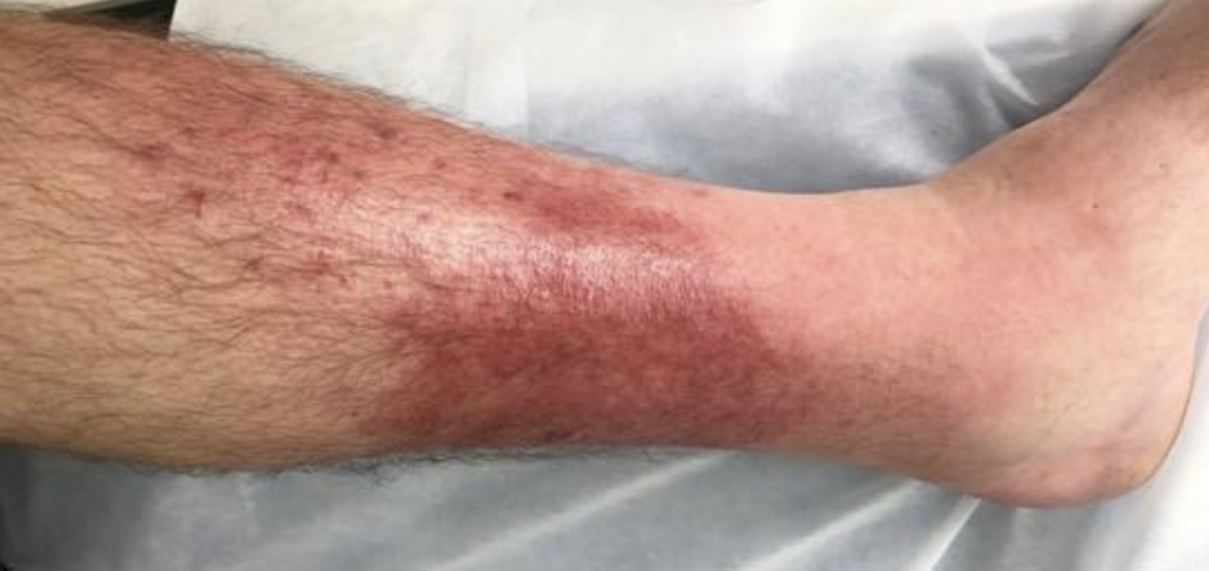
Left leg cellulitis
Image Source: Wikimedia Commons (2007). Cellulitis3, Licensed under a creative commons licence. Available for free at: https://commons.wikimedia.org/wiki/File:Cellulitis3.jpg (Accessed on: 14 March 2023).

Diagnosis is usually clinical but can be supported by blood tests (leukocytosis, elevated CRP) and wound MCS (if skin is broken). A blood culture is warranted if the patient is systemically unwell. General measures include marking the involved area, elevation, and simple analgesia. Patients with uncomplicated infection can be managed with oral antibiotics (e.g. flucloxacillin 500mg QDS). Systemically unwell patients require admission and intravenous antibiotics. Patients with at least 2 separate episodes of cellulitis in the previous 12 months can be trialled on prophylactic phenoxymethylpenicillin 250mg BD.
Erysipelas
A more superficial form of cellulitis involving the upper dermis and superficial lymphatics. Commonly caused by Group A beta-haemolytic streptococcus. Recent nasopharyngeal infections are a risk factor for facial erysipelas, however, it can also present on the lower limbs. Flu-like symptoms typically precede the appearance of skin changes. Erysipelas manifests as a well-demarcated erythematous patch which progresses to a tender, firm, fiery-red plaque. This may be accompanied by bullae or purpura. The workup and management of patients is the same as cellulitis.

Facial erysipelas
Image Source: Wikimedia Commons (2009). Facial erysipelas, Licensed under a creative commons licence. Available for free at: https://commons.wikimedia.org/wiki/File:Facial_erysipelas.jpg (Accessed on: 10 March 2022).

Necrotising Fasciitis
A life-threatening soft tissue and fascia infection that leads to tissue necrosis. 50% of cases occur in previously healthy individuals. Predisposing risk factors include skin injury, chronic conditions (e.g. diabetes, renal disease), IV drug abuse and immunosuppression. It is caused by a mixture of aerobic and anaerobic organisms. This can be categorised into 4 main types, shown in table 2:
| Type | Additional notes |
|---|---|
| I | Polymicrobial, caused by aerobic and anaerobic bacteria (Staphylococcus aureus, Haemophilus, Escherichia coli). Seen in immunocompromised patients and those with chronic disease. |
| II | Monomicrobial, commonly caused by Group A beta-haemolytic streptococci, followed by Staphylococcus aureus. Affect all age group and previously healthy individuals |
| III | Monomicrobial gram-negative bacteria. Usually from freshwater exposure, including marine organisms such as Vibrio vulnificus. Infection with Clostridium is also classified here and causes gangrenous necrotising fasciitis. |
| IV | Caused by fungal pathogens |
Table 2: Different categories of necrotising fasciitis
The primary feature of necrotising fasciitis is intense pain disproportionate to the clinical findings. The affected tissue is hot and swollen, with ill-defined erythema that rapidly becomes blackened and bullous. Subcutaneous crepitus develops from the accumulation of gas within the soft tissue.

Extensive necrotising fasciitis of the left leg
Image Source: Wikimedia Commons (2009). Necrotizing fasciitis left leg, Licensed under a creative commons licence. Available for free at: https://en.wikipedia.org/wiki/File:Necrotizing_fasciitis_left_leg.JPEG (Accessed on: 10 March 2022).

A high index of suspicion is required for diagnosis. This is supported by raised inflammatory markers on blood tests and the presence of gas in the subcutaneous tissue on imaging such as x-ray or CT. Wound MCS and blood cultures should also be taken. Diagnosis is confirmed during surgical exploration with the presence of friable superficial fascia and grey exudate. Survival is improved with immediate surgical debridement. Supportive measures include broad-spectrum intravenous antibiotics, resuscitation and management in an intensive care setting.
Staphylococcal Scalded Skin Syndrome (SSSS)
An acute blistering skin condition caused by staphylococcal toxin. Group II coagulase-positive staphylococci toxins damage desmoglein 1 and subsequently cause the upper epidermis to split and shed. Primarily seen in children under 5 years old, however, SSSS has been noted in immunodeficient and renal failure patients. 40% of adults are asymptomatic nasal carriers of S. aureus.
Areas typically affected include the groin, face, axilla and neck. SSSS develops within hours to days with widespread tender erythematous skin followed by large flaccid bullae. These rupture easily under gentle pressure (Nikolsky’s sign) leaving large denuded areas that resemble a scald. The patient is also systemically unwell.

SSSS in a neonate
Image Source: Wikimedia Commons (2016). OSC Microbio 21 02 SSSS, Licensed under a creative commons licence. Available for free at: https://commons.wikimedia.org/wiki/File:OSC_Microbio_21_02_SSSS.jpg (Accessed on: 10 March 2022).

Diagnosis is usually clinical. Swabs are taken from an area of possible primary infection (e.g. nasopharynx, surgical wound) and blood cultures obtained. Nasal swabs are performed on close contacts to identify asymptomatic carriers. Skin biopsy demonstrates intraepidermal cleavage at the granular layer. Supportive management is with emollients (e.g. 50% liquid paraffin & 50% white soft paraffin), fluid balance management and analgesia. A penicillinase-resistant, anti-staphylococcal antibiotic (e.g. flucloxacillin) is used to eradicate the infection. Recovery with skin healing is within 5 days.
Impetigo
A common and contagious superficial skin infection often caused by Staphylococcus aureus, followed by Streptococcus pyogenes. Methicillin-resistant Staphylococcus aureus (MRSA) has been isolated in an increasing number of cases. Impetigo mostly affects children but can occur at any age. Atopic eczema, scabies and any type of skin trauma predisposes to impetigo. Impetigo presents on the face, extremities and flexural surfaces. It is itchy and may present with mild fever and lymphadenopathy. The 3 main types are shown in table 3:
| Type | Presentation |
|---|---|
| Non-bullous impetigo | Clusters of vesicles or pustules that evolve rapidly and rupture into honey-coloured crusted plaques. Satellite lesions occur due to autoinoculation. Caused by Staphylococcus aureus, Streptococcus pyogenes or both. |
| Bullous impetigo | Staphylococcus aureus produces exfoliative toxins that cause bullae formation. Small vesicles develop into flaccid bullae. On rupture, these become covered with honey-coloured crust. |
| Ecthyma | Full-thickness ulcerative form of impetigo usually caused by Streptococcus pyogenes but can also be caused by Staphylococcus aureus. Starts as a non-bullous impetigo that develops into shallow punched-out ulcers with blackened centres. Mostly affects the legs and heals slowly. |
Table 3: Types of impetigo

Impetigo on the chin
Image Source: Wikimedia Commons (2020). Impetigo2020, Licensed under a creative commons licence. Available for free at: https://commons.wikimedia.org/wiki/File:Impetigo2020.jpg (Accessed on: 10 March 2022).

Impetigo is a clinical diagnosis. Bacterial swabs sent for MCS should be considered in persistent, recurrent and widespread cases. Patients should stay away from school/work until lesions dry out and scab over. Non-bullous impetigo is managed with a 5-day course of hydrogen peroxide 1% cream or topical fusidic acid 2% if not suitable. Bullous and more extensive impetigo require oral antibiotics for 5-7 days (e.g. flucloxacillin, clarithromycin).
Complications include cellulitis and SSSS. Rare complications like scarlet fever or glomerulonephritis can occur if the causative organism is group A beta-haemolytic streptococcus (Streptococcus pyogenes).
Lyme Disease
A tick-transmitted infection primarily caused by the spirochaete bacteria, Borrelia burgdorferi. Most cases have been reported in the USA and Europe. The most prevalent variant in Europe is B. garinii.
Infection is acquired in heavily wooded and leafy areas with peak cases occurring in summer. Ixodes ticks become infected by Borrelia from feeding on an infected animal (e.g. mouse, deer, sheep). Ticks then painlessly bite and feed on any part of the human body, usually a limb. Infection may be cleared by the host’s defences with no clinical manifestation, or the organism spreads via the skin, lymphatics and can disseminate in the blood to other organs.
There are 3 stages of Lyme disease, shown in table 4:
| Early Lyme disease (localised disease) | 3-36 days |
|
|
| Early disseminated Lyme disease | Days-weeks |
|
|
| Late-stage Lyme disease | Months-years |
|
|
Table 4: Stages of Lyme disease
A clinical diagnosis can be made in patients with erythema migrans as it often develops before positive serology tests. In patients suspected of having Lyme disease without erythema migrans, an enzyme-linked immunosorbent assay (ELISA) test for Lyme disease IgM and IgG should be sent. If this is positive, an immunoblot test for Lyme disease should be sent. If negative, this test should be repeated 4-6 weeks later. Management is with doxycycline or amoxicillin for 21 days and is highly effective in the early Lyme disease stage.

Erythema migrans
Image Source: Wikimedia Commons (2019). Erythema migrans – erythematous rash in Lyme disease – PHIL 9875, Licensed under a creative commons licence. Available for free at: https://commons.wikimedia.org/wiki/File:Erythema_migrans_-_erythematous_rash_in_Lyme_disease_-_PHIL_9875.jpg (Accessed on: 10 March 2022).

Viral Skin Infections
Herpes Simplex Virus (HSV)
A very common and contagious blistering viral infection. There are two types of HSV:
- HSV-1: usually causes oral and facial infection. Now also the most common cause of genital herpes in the UK.
- HSV-2: Most likely to cause recurrent anogenital disease. Previously the most common cause of anogenital herpes in the UK.
Both variants can affect any area of skin or mucosal membrane, and any age. Transmission is via direct or indirect contact with an individual actively shedding the virus (this may be asymptomatic). The incubation period is 2-12 days. After primary infection, the latent non-replicating HSV resides dormant in the dorsal root ganglion, where it can reactivate. Reactivation can be triggered by excessive UV exposure, illness, stress and immunosuppression.
HSV causes irregular vesicular eruptions on an erythematous base, commonly affecting the lips, mouth, fingers (herpetic whitlow) and anogenital area. Vesicles on mucous membranes quickly develop into painful erosions. Eruptions may be preceded by a few hours by a tingling or burning sensation, fever, malaise and local lymphadenopathy. Eruptions heal within 7-14 days.
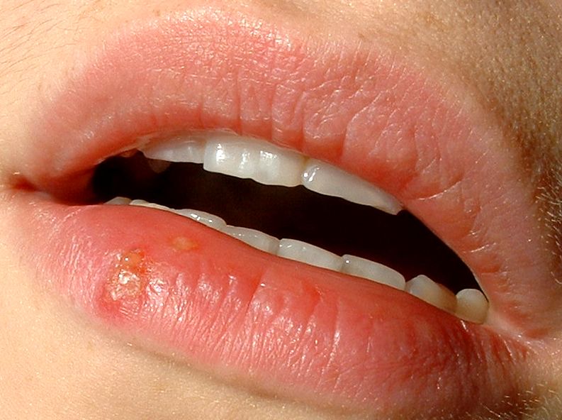
Herpes labialis
Image Source: Wikimedia Commons (2005). Herpes labialis, Licensed under a creative commons licence. Available for free at: https://commons.wikimedia.org/wiki/File:Herpes_labialis.jpg (Accessed on: 10 March 2022).

Diagnosis is usually clinical and can be confirmed on a viral PCR swab. Oral HSV infection can be considered for oral antivirals (e.g. aciclovir or valaciclovir). Patients with anogenital HSV infection should be referred to GUM and managed with 5/7 of oral antiviral within 5 days of the start of an episode or while new lesions are still forming. Suppressive antiviral treatment should be considered in patients having 6 or more attacks a year.
Eczema Herpeticum
A widespread and severe HSV infection of the skin that is mostly associated with atopic eczema. This is a dermatological emergency. Eczema herpeticum usually presents during the first episode of HSV infection but can result from recurrent infection. It has also been seen in non-eczematous conditions such as pemphigus vulgaris, burns and Darier disease.
Eczema herpeticum manifests as clusters of painful and itchy monomorphic vesicles. These rupture leaving small punched-out erosions and ulcerations overlying an erythematous base. Patients often present with extensive vesicles over the face, neck or trunk with associated fever, malaise and lymphadenopathy. Lesions can take up to 6 weeks to heal.
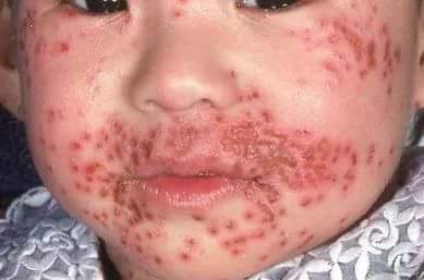
Eczema herpeticum
Image Source: Wikimedia Commons (2018). Eczema herpitcum, Licensed under a creative commons licence. Available for free at: https://commons.wikimedia.org/wiki/File:Eczema_herpitcum.jpg (Accessed on: 15 March 2023).

Diagnosis is usually clinical, however, a viral PCR swab can be taken for HSV. A bacterial swab should also be taken to exclude secondary bacterial infection. Prompt treatment is with oral aciclovir for 10-14 days or until lesions heal. IV aciclovir may be required if the patient is too unwell/deteriorating. An ophthalmologist should be consulted in suspected cases of eye involvement.
Herpes Zoster (Shingles)
A self-limiting blistering rash caused by the reactivation of the varicella zoster virus (VZV), also known as human herpesvirus type 3 (HHV3). Chickenpox is the acute, primary infection which produces widespread lesions at different stages of development. The virus remains dormant in the dorsal root ganglion in the spinal cord and, when reactivated, migrates down cutaneous sensory nerves to induce lesions.

Herpes zoster (shingles)
Image Source: Wikimedia Commons (2007). Herpes zoster chest, Licensed under a creative commons licence. Available for free at: https://commons.wikimedia.org/wiki/File:Herpes_zoster_chest.png (Accessed on: 10 March 2022).

Prior to the skin changes, there may be localised pain associated with fever and tender lymphadenopathy. Red papules then erupt into vesicles in a unilateral dermatomal distribution and are confined to the distribution of one or two sensory nerves. Commonly affects the face, neck and trunk. Involvement of the ophthalmic division of the trigeminal nerve is termed herpes zoster ophthalmicus, and involvement of the VII nerve is termed Ramsay Hunt syndrome. Lesions can take up to a month to resolve.
Herpes zoster is self-limiting and management can include analgesia and emollients. Oral antivirals can be commenced in more vulnerable patients presenting <72 hours from onset or severe cases. Complications include corneal scarring, Ramsay Hunt syndrome and post-herpetic neuralgia. The preventive shingles vaccine is offered in the UK to individuals over 70 years old.
Molluscum Contagiosum
This is caused by a pox virus and presents mostly in children and young adults. Transmitted via close and indirect contact (e.g. towels).
It presents as clusters of pearly umbilicated papules that occur on the face, trunk and extremities. Surrounding dermatitis is frequent. Larger and more widespread molluscum contagiosum occur in immunosuppressed patients.

Molluscum contagiosum
Image Source: Wikimedia Commons (2005). Molluscaklein, Licensed under a creative commons licence. Available for free at: https://commons.wikimedia.org/wiki/File:Molluscaklein.jpg (Accessed on: 10 March 2022).

Molluscum contagiosum is a clinical diagnosis. It is self-limiting and typically resolves without intervention within 18 months. Treatment with cryotherapy, imiquimod, benzoyl peroxide or curettage can also be used.
Viral Warts
This is caused by infection of the epidermal cells with human papillomavirus (HPV). It is transmitted through direct and indirect contact. The most common HPV types infecting the skin are 1, 2, 3, 4, 10, 27, 29 and 57. Anogenital warts are commonly caused by types 6 and 11.
Viral warts are highly contagious and most frequently present in children, immunosuppressed patients (e.g. long-term immunosuppressants, HIV) and in conditions where the skin barrier is defective. Cutaneous warts are papules or nodules with a papillomatous and hyperkeratotic surface. Plantar warts (verruca) are usually painful with dark punctate centres with hyperkeratotic surrounding skin. It is a clinical diagnosis and warts can resolve spontaneously. Other treatment options include podophyllin, imiquimod, cryotherapy or curettage and cautery.
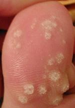
Viral warts
Image Source: Wikimedia Commons (2008). Dornwarzen, Licensed under a creative commons licence. Available for free at: https://commons.wikimedia.org/wiki/File:Dornwarzen.jpg (Accessed on: 15 March 2023).

Fungal Skin Infections
Tinea Infections (Dermatophytoses)
This refers to a skin infection caused by dermatophytes and is one of the most common causes of superficial fungal infections. Dermatophytes are fungi that require keratin for growth and therefore infect the stratum corneum, nails and hair.
Risk factors include diabetes, immunodeficiency, occlusive clothing and crowded conditions. It is more likely in children and the elderly. Dermatophyte infections present as itchy, annular lesions with raised borders. Transmission is via contact with an infected person, contaminated objects or infected animal. The feature of the fungal infection depends on the site:
| Type | Site affected | Most common organisms | Additional notes |
|---|---|---|---|
| Tinea capitis | Scalp | M. canis | Annular patches with scale, broken hair shaft. Kerion: inflammatory tinea capitis that causes a painful, boggy plaque with associated pustules and scarring alopecia. |
| Tinea manuum | Hands | T. rubrum | Diffuse scaling with associated itch. Frequently affecting the palmar surface. Can form annular rashes and blisters. |
| Tinea unguium (onychomycosis) | Nails | T. rubrum
T. interdigitale |
Often involves the nail of the big toe or the little toe. Causes a white/yellow discolouration, thickening and crumbling. |
| Tinea cruris | Groin, natal cleft, adjacent thigh | T. rubrum
T. interdigitale E. floccosum |
Male predominance. RF: obesity, wet or restrictive clothing, warm climates. Itchy, erythematous with raised edges. Can be hyperpigmented. |
| Tinea pedis (athlete’s foot) | Feet | T. rubrum
T. interdigitale E. floccosum |
Most common tinea infection. Usually between the 4th and 5th toes. Male predominance. Skin is itchy, moist, fissured and erythematous. It can also be associated with hyperkeratosis and blisters. |
| Tinea incognita | – | – | Tinea infection that has been inappropriately treated with corticosteroids. This modifies its appearance – less scaly, margins are less raised and there is extension of the original infection. |
Table 5: Summary of tinea infection sites and common organisms

Tinea skin infection
Image Source: Wikimedia Commons (2012). Yeartinfection, Licensed under a creative commons licence. Available for free at: https://commons.wikimedia.org/wiki/File:Yeartinfection.JPG (Accessed on: 10 March 2022).

Diagnosis can be established with skin scrapings, hair or nail clippings sent for microscopy and culture. First line management is with topical antifungals (e.g. terbinafine, clotrimazole, miconazole). Oral antifungals should be used in tinea capitis and severe or extensive disease. Oral terbinafine can cause hepatotoxicity so LFTs must be monitored.
Superficial Candida Albicans Infection
Candida albicans is a yeast that commonly infects the skin and is a commensal organism in the GI tract. Predisposing factors include extremes of age, opposing skin folds, obesity, diabetes, immunosuppression, use of broad-spectrum antibiotics and humid environments. Diagnosis is clinical but swab samples sent for microscopy can demonstrate budding yeast cells and pseudohyphae. Superficial infection may present in a number of sites:
| Site | Presentation | Management |
|---|---|---|
| Oral | White/red patches or plaque. May be associated with pain on swallowing, glossitis and angular stomatitis. RF: dentures, broad-spectrum antibiotics and use of steroid inhalers. | Consider if the patient is immunosuppressed.
1st line: miconazole gel, if not suitable – nystatin for 7/7 2nd line: PO fluconazole 7/7 |
| Intertrigo | Submammary, axillary, interdigital and inguinal folds. Manifests as itchy red plaques with satellite lesions. | 1st line: topical imidazole (clotrimazole, miconazole, ketoconazole).
2nd line/widespread disease/immunocompromised: PO fluconazole 14/7 |
| Nails | Presents as chronic paronychia. Affects individuals that work in wet environments (e.g. fishermen, gardeners). Red, swollen and tender nail fold with associated nail discolouration and ridging. | 1st line: topical antifungals
2nd line: PO fluconazole |
| Vulvovaginal /penile | Itchy white adherent plaques, cottage cheese-like discharge, inflamed mucus membranes. | 1st line: topical antifungals. Addition of PV clotrimazole in vulvovaginal disease.
2nd line: PO fluconazole |
Table 6: Summary of candida albicans infection and its management

Oral candida albicans infection
Image Source: Wikimedia Commons (2008). Oral thrush Aphthae Candida albicans. PHIL 1217 lores, Licensed under a creative commons licence. Available for free at: https://commons.wikimedia.org/wiki/File:Oral_thrush_Aphthae_Candida_albicans._PHIL_1217_lores.jpg (Accessed on: 10 March 2022).

Pityriasis (Tinea) Versicolor
Pityriasis versicolor commonly affects teenagers and young adults and is caused by a yeast, Malassezia furfur. It presents on the trunk, neck and arms and is characterised by multiple round pink, brown or hypopigmented macules and patches with a fine scale. It may be asymptomatic or mildly itchy.

Pityriasis versicolor
Image Source: Wikimedia Commons (2012). Tinea versicolor1, Licensed under a creative commons licence. Available for free at: https://commons.wikimedia.org/wiki/File:Tinea_versicolor1.jpg (Accessed on: 10 March 2022).

Diagnosis is clinical but microscopy from skin scrapings using potassium hydroxide will demonstrate hyphae and budding cells (“spaghetti and meatballs” appearance). Wood lamp examination will demonstrate a yellow-green fluorescence. Treatment is usually needed for remission. Initial management is with an antifungal shampoo (ketoconazole 2% shampoo OD for 5/7) or cream (e.g. clotrimazole). If extensive or topical treatment is ineffective, oral antifungal such as fluconazole or itraconazole can be used. Changes in skin pigmentation can take months to resolve and relapse is common.
Other Skin Infections
Scabies
A skin infestation caused by the mite, Sarcoptes scabiei. The fertilised female mite burrows into the stratum corneum laying up to 3 eggs daily. Eggs develop into adult mites within 2 weeks. Symptoms may take 4-8 weeks to develop.
Scabies can affect any age and presents mostly in the winter months. It is spread via direct contact and is more frequent in crowded conditions, institutions and in immunosuppressed patients.
Scabies classically present with intense itching that is worse at night and can persist for several weeks despite treatment. Sites of infestation include the fingers, wrist, ankles, nipples and genitalia. Irregular burrows around 1cm long may be evident, with a mite visible at one end. A hypersensitivity reaction produces a rash that can consist of erythematous papules, vesicles or nodules.

Scabies
Image Source: Wikimedia Commons (2005). Acarodermatitis Hand, Licensed under a creative commons licence. Available for free at: https://commons.wikimedia.org/wiki/File:Acarodermatitis_Hand.jpg (Accessed on: 10 March 2022).

Crusted scabies is due to an impaired host immune response and is more frequent in elderly and immunodeficient patients. It is highly contagious and represents hyper infestation with thousands/millions of mites. It is characterised by a scaly rash with minimal/absent itch.
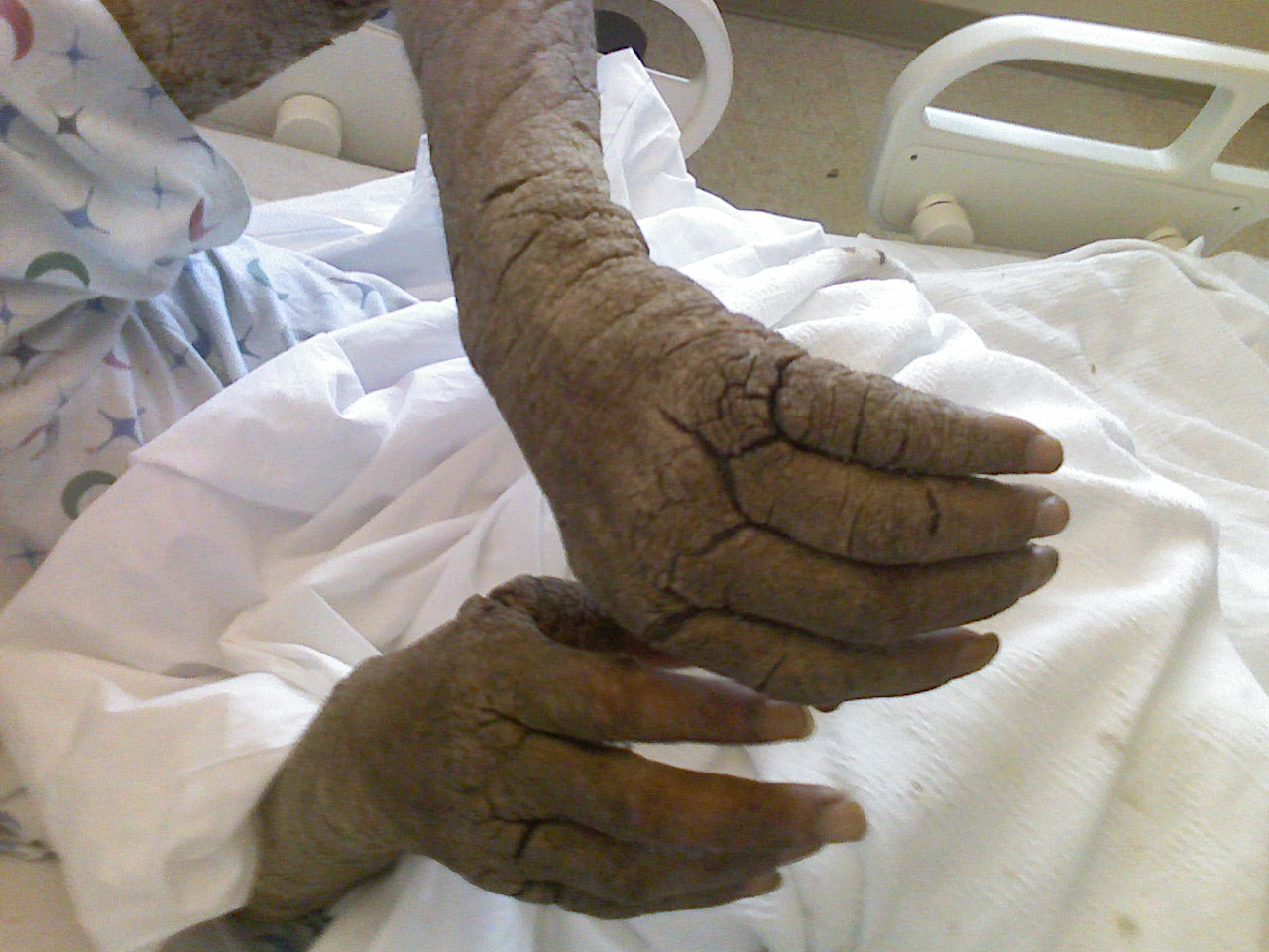
Crusted scabies in a patient with AIDS
Image Source: Wikimedia Commons (2008). Norwegian Scabies in Homeless AIDS Patient, Licensed under a creative commons licence. Available for free at: https://commons.wikimedia.org/wiki/File:Norwegian_Scabies_in_Homeless_AIDS_Patient.jpg (Accessed on: 10 March 2022).

Scabies is diagnosed clinically. Uncertain cases can be confirmed by dermatoscopy and skin scraping microscopy, which may demonstrate burrows, mites, ova and mite faeces. If crusted scabies is suspected, underlying immunodeficiency should be investigated. Management involves treating the individual, household members, close contacts and sexual contacts with permethrin 5% cream. Alternative: malathion 0.5% liquid. Crusted scabies requires oral ivermectin. Clothing/bedding/towels should be washed at least 60 degrees and dried in a hot dryer. Pruritus can be managed with crotamiton 10% cream, antihistamines and sometimes mild-topical corticosteroids.
Erythema Multiforme (EM)
EM is a self-limiting hypersensitivity reaction caused predominantly by infection, for which HSV causes the majority of cases. Other causes include certain medications or conditions (table 7). It is now considered a distinct entity from Stevens-Johnson syndrome and Toxic Epidermal Necrolysis.
| Infections | Medications | Conditions |
|---|---|---|
| Cytomegalovirus | Antibiotics | Inflammatory bowel disease |
| Epstein-Barr virus | Anticonvulsants | Malignancy |
| SARS-CoV-2 | NSAIDS | Hepatitis |
| Candidiasis | Vaccines |
Table 7: Triggers for erythema multiforme
Mycoplasma pneumoniae is often listed as a cause of EM, however this has recently been classified as its own distinct entity, mycoplasma pneumoniae-induced rash and mucositis (MIRM).
EM acutely manifests on the distal extremities as symmetrical erythematous papules that transform into target-like lesions. These have a dusky centre, a pale and oedematous intermediate ring and an erythematous outer ring. Lesions may be painful or itchy. EM minor has cutaneous manifestations only, whereas EM major has the addition of mucosal involvement. Diagnosis is clinical but investigations to review the cause may be required. Treatment is supportive. Precipitating factors should be managed and offending medication avoided. Ocular involvement requires ophthalmology input. Recurrent cases may benefit from antiviral therapy.

Cutaneous erythema multiforme
Image Source: Wikimedia Commons (2011). Erythema Multiforme EM 01 ajustement niveaux auto, Licensed under a creative commons licence. Available for free at: https://commons.wikimedia.org/wiki/File:Erythema_Multiforme_EM_01_ajustement_niveaux_auto.jpg (Accessed on: 10 March 2022).


Mucosal erythema multiforme
Image Source: Wikimedia Commons (2021). Erythema multiforme tongue, Licensed under a creative commons licence. Available for free at: https://commons.wikimedia.org/wiki/File:Erythema_multiforme_tongue.jpg (Accessed on: 10 March 2022).

Autoimmune Blistering Disorders
Pemphigus Vulgaris
A potentially fatal autoimmune blistering disorder, typically presenting between the ages of 30 and 60 years. The condition occurs due to intraepithelial blisters caused by IgG antibodies against adhesion proteins desmoglein 3 and/or desmoglein 1. Painful mucosal erosions which can precede skin involvement by months. However, note that mucosal involvement may be the only manifestation of this disorder in some patients. Skin involvement is characterised by flaccid, easily ruptured blisters that quickly evolve into itchy and painful erosions and crusting.

Pemphigus vulgaris
Image Source: Wikimedia Commons (2018). Pemphgoid vulgaris, Licensed under a creative commons licence. Available for free at: https://commons.wikimedia.org/wiki/File:Pemphgoid_vulgaris.jpg (Accessed on: 10 March 2022).

Skin biopsy is sent for histology and direct immunofluorescence. Diagnosis is confirmed on viewing intraepidermal deposition of IgG antibodies and suprabasal epidermal acantholysis. Serum antibody levels can also be taken.
Management involves high dose corticosteroids for disease control followed by a step-down regime. Other immunosuppressive agents such as azathioprine and cyclophosphamide are used as adjuvant or second-line.
| Variants of pemphigus | Additional notes |
|---|---|
| Pemphigus foliaceus | Superficial, scaly and relatively benign form of pemphigus. There is no mucosal involvement. |
| Drug-induced pemphigus | Pemphigus precipitated by a medication. Causative agents include penicillamine, ACE inhibitors and piroxicam. |
| Paraneoplastic pemphigus | Pemphigus is associated with underlying malignancy. |
Table 8: Variants of pemphigus
Bullous Pemphigoid
The most common autoimmune blistering disorder, typically affecting the elderly. IgG autoantibodies attack the basement membrane (specifically BP180 and BP230 proteins), resulting in subepidermal blistering. Bullous pemphigoid has been associated with:
- Neurological disorders: stroke, Parkinson’s disease, dementia, epilepsy and multiple sclerosis.
- Medications: antibiotics, penicillamine, ACE inhibitors, Beta-blockers, DPP-4 inhibitors (e.g. sitagliptin), diuretics, immune checkpoint inhibitors
- Physical triggers: trauma and burns
The onset of blisters is preceded by a non-bullous phase lasting weeks or months. This presents as intense itching which may be accompanied by erythema and/or urticated plaques. The bullous phase consists of localised or widespread tense bullae on erythematous or normal-looking skin. The trunk and flexor aspect of the limbs are typically affected. Mucosal involvement is uncommon.

Bullous pemphigoid
Image Source: Wikimedia Commons (2018). Drugs induce pemphigoud bullous, Licensed under a creative commons licence. Available for free at: https://commons.wikimedia.org/wiki/File:Drugs_induce_pemphigoud_bullous.jpg (Accessed on: 10 March 2022).

Investigation involves checking serum antibodies and skin biopsies for histology and direct immunofluorescence. Histology and immunofluorescence will demonstrate subepidermal blistering and IgG deposition at the basement membrane, respectively. Super-potent topical steroids can treat localised disease, whereas widespread disease will require oral prednisolone +/- immunosuppressants (e.g. Azathioprine). Doxycycline 200 mg daily is an alternative to immunosuppressive therapy, due to its anti-inflammatory properties, and it has fewer side effects compared to oral prednisolone.
Dermatitis Herpetiformis
A rare chronic autoimmune blistering skin condition that is associated with coeliac disease (a gluten-sensitive enteropathy) in 90% of patients. More common in men and typically presents between 15-40 years old. Loss of immune tolerance to gluten peptides triggers the production of IgA antibodies and an autoimmune process targeting both the skin and intestinal submucosa.
Classically presents as intensely itchy clusters of vesicles on erythematous or normal-looking skin. Vesicles are often broken through scratching leaving erosions. Distribution is symmetrical and commonly affects the extensors, including the elbows, knees, buttocks and scalp. Symptoms of gastrointestinal disturbance and malabsorption may also be present.

Dermatitis herpetiformis on the abdomen
Image Source: Wikimedia Commons (2010). Dermatitis-herpetiformis, Licensed under a creative commons licence. Available for free at: https://commons.wikimedia.org/wiki/File:Dermatitis-herpetiformis.jpg (Accessed on: 10 March 2022).

Skin biopsy demonstrates IgA deposits at the dermal papillae and subepidermal blisters on direct immunofluorescence. Coeliac disease should also be tested for. A lifelong gluten-free diet is strongly recommended in these patients. Dapsone is the first choice in cases that cannot be managed with dietary changes alone and will control eruptions and itch. However, regular blood monitoring is required due to the risk of haemolytic anaemia and agranulocytosis.
Other Blistering Disorders
Differential Diagnosis for a Blistering Rash
Vesicles and bullae are non-specific signs that arise from multiple causes, including physical, infectious, medication-related and immunobullous conditions.
Physical/chemical
- Friction blisters: commonly seen on palms, soles and heels
- Insect bites: may produce a localised blister
- Polymorphic light eruption: a common photosensitivity reaction that occurs hours to days following UV exposure. Presents on sun-exposed skin as itchy macules, papules, plaques and/or vesicles.
- Sunburn: presents with oedema, erythema and blistering in severe cases
- Burns: blistering seen in partial thickness burns
Infections
These conditions are explained in detail in the Infections section
- Bullous cellulitis/erysipelas: severe cases, associated with Group A beta-haemolytic streptococcus.
- Bullous impetigo: due to staphylococcal exfoliative toxins.
- Staphylococcal scalded skin syndrome (SSSS): an acute blistering rash caused by staphylococcal toxins. Primarily seen in children under 5 years old and noted in immunodeficient and renal failure patients.
- Herpes simplex virus (HSV) infection: a contagious vesicular condition affecting the skin and mucosal caused by HSV-1 and HSV-2.
- Eczema herpeticum: a dermatological emergency secondary to HSV infection presenting with widespread painful monomorphic blisters. Most frequently associated with atopic eczema.
- Herpes zoster infection: a self-limiting painful vesicular rash caused by the reactivation of the varicella zoster virus.
Drug eruptions
These conditions are explained in detail in the Allergy and sensitivity topic.
- Stevens-Johnson syndrome/Toxic Epidermal Necrolysis (SJS/TEN): an acute and life-threatening skin reaction that causes sheet-like skin and mucosal loss due to medications and infections. Culprit medications include allopurinol, anticonvulsants, antibiotics, allopurinol and NSAIDs.
- Fixed drug eruption: a cutaneous adverse drug reaction that recurs at the same site each time the individual is exposed to the drug. It is a delayed type IV hypersensitivity reaction and can present as a bullous, erythematous or pigmented rash. Culprit medications include antibiotics, analgesics, antihypertensives and anticonvulsants.
Immunobullous
- Pemphigus vulgaris: a potentially fatal autoimmune blistering disorder typically presenting <60 years. Intraepithelial blisters caused by IgG antibodies against desmoglein 3 and/or desmoglein 1.
- Pemphigus foliaceus: superficial, scaly and relatively benign form of pemphigus with no mucosal involvement.
- Drug-induced pemphigus: pemphigus precipitated by a medication. Causative agents include penicillamine, ACE inhibitors and piroxicam.
- Paraneoplastic pemphigus: pemphigus associated with underlying malignancy.
- Bullous pemphigoid: common autoimmune blistering typically present in the elderly. Subepidermal blistering caused by IgG antibodies against BP180 and/or BP230. Can be triggered by neurological disorders, medications and physical trauma (e.g. burns).
- Pemphigoid gestationis: Intensely itchy urticaria-like rash which develops into tense sub-epidermal blisters. Typically presents in the second and third trimester of pregnancy.
- Dermatitis herpetiformis: intensely itchy clusters of vesicles caused by gluten peptides triggering the production of IgA autoantibodies targeting the skin.
Other
- Eczema pompholyx: also called dyshidrotic eczema. This typically presents with clusters of vesicles on the palm and soles, palmoplantar hyperhidrosis and a history of atopic eczema.
- Porphyria cutanea tarda: the most common type of porphyria due to a defect in the liver enzyme, uroporphyrinogen decarboxylase. Excess porphyrin results in photosensitivity. This condition presents with painful blistering and crusting on sun-exposed skin, hyperpigmentation and red/tea-coloured urine.
Complete Guide to Clinical Genetics ST3 Application and Recruitment (2027)
Written by Medibuddy. Last updated: October 2026. Clinical Genetics is

Complete Guide to Clinical Radiology ST3 Application and Recruitment (2027)
Recruitment paused: NHS England is not running Clinical Radiology ST3+ recru

Complete Guide to Paediatric Surgery ST3 Application and Recruitment in United Kingdom (2026)
Written by Medibuddy. Last updated: October 2026. If you’re planning


Take your subscriptions with you
Our mobile app allows you to access your interview and exam question banks wherever you are.

