


 Close
Close
 6th July 2023
Bacterial Skin Infections
Cellulitis
An acute bacterial infection of the dermis and subcutaneous tissue. Commonly caused by Streptococcus pyogenes followed by Staphylococcus aureus. Atypical organisms are summarised below:
| Causative organism | Associations |
|---|---|
| Pseudomonas aeruginosa | Diabetic and immunocompromised patients, puncture wounds, hot tub exposure |
| Haemophilus influenzae | Children |
| Pasteurella multocida, Capnocytophaga | Cat scratches/bites, dog bites |
| Eikenella corrodens | Human bites (note: polymicrobial) |
| Aeromonas hydrophila | Injuries in freshwater |
| Vibrio vulnificus | Injuries in saltwater |
| Erysipelothrix rhusiopathiae | Butchers, fishermen, vets |
| Mycobacterium marinum | Aquarium keepers |
Table 1: Atypical organisms in cellulitis
Factors that predispose to cellulitis include diabetes mellitus, lymphoedema, immunodeficiency, and conditions that compromise the skin barrier (e.g. tinea pedis, eczema).
Presentation is usually on the lower limbs and unilateral. The skin is hot, tender and erythematous with indistinct borders. Bullae and petechiae may also form. Systemic features of infection may also be present.
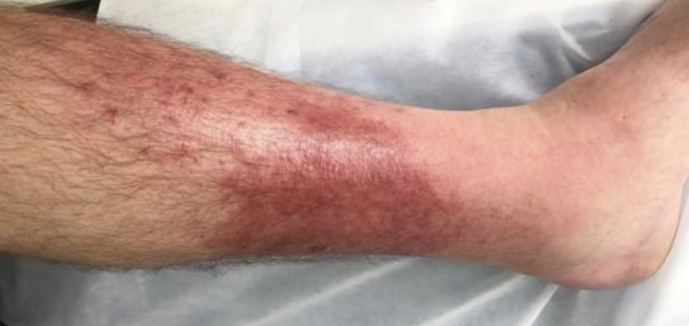
Left leg cellulitis
Image Source: Wikimedia Commons (2007). Cellulitis3, Licensed under a creative commons licence. Available for free at: https://commons.wikimedia.org/wiki/File:Cellulitis3.jpg (Accessed on: 14 March 2023).

Diagnosis is usually clinical but can be supported by blood tests (leukocytosis, elevated CRP) and wound MCS (if skin is broken). A blood culture is warranted if the patient is systemically unwell. General measures include marking the involved area, elevation, and simple analgesia. Patients with uncomplicated infection can be managed with oral antibiotics (e.g. flucloxacillin 500mg QDS). Systemically unwell patients require admission and intravenous antibiotics. Patients with at least 2 separate episodes of cellulitis in the previous 12 months can be trialled on prophylactic phenoxymethylpenicillin 250mg BD.
Erysipelas
A more superficial form of cellulitis involving the upper dermis and superficial lymphatics. Commonly caused by Group A beta-haemolytic streptococcus. Recent nasopharyngeal infections are a risk factor for facial erysipelas, however, it can also present on the lower limbs. Flu-like symptoms typically precede the appearance of skin changes. Erysipelas manifests as a well-demarcated erythematous patch which progresses to a tender, firm, fiery-red plaque. This may be accompanied by bullae or purpura. The workup and management of patients is the same as cellulitis.

Facial erysipelas
Image Source: Wikimedia Commons (2009). Facial erysipelas, Licensed under a creative commons licence. Available for free at: https://commons.wikimedia.org/wiki/File:Facial_erysipelas.jpg (Accessed on: 10 March 2022).

Necrotising Fasciitis
A life-threatening soft tissue and fascia infection that leads to tissue necrosis. 50% of cases occur in previously healthy individuals. Predisposing risk factors include skin injury, chronic conditions (e.g. diabetes, renal disease), IV drug abuse and immunosuppression. It is caused by a mixture of aerobic and anaerobic organisms. This can be categorised into 4 main types, shown in table 2:
| Type | Additional notes |
|---|---|
| I | Polymicrobial, caused by aerobic and anaerobic bacteria (Staphylococcus aureus, Haemophilus, Escherichia coli). Seen in immunocompromised patients and those with chronic disease. |
| II | Monomicrobial, commonly caused by Group A beta-haemolytic streptococci, followed by Staphylococcus aureus. Affect all age group and previously healthy individuals |
| III | Monomicrobial gram-negative bacteria. Usually from freshwater exposure, including marine organisms such as Vibrio vulnificus. Infection with Clostridium is also classified here and causes gangrenous necrotising fasciitis. |
| IV | Caused by fungal pathogens |
Table 2: Different categories of necrotising fasciitis
The primary feature of necrotising fasciitis is intense pain disproportionate to the clinical findings. The affected tissue is hot and swollen, with ill-defined erythema that rapidly becomes blackened and bullous. Subcutaneous crepitus develops from the accumulation of gas within the soft tissue.

Extensive necrotising fasciitis of the left leg
Image Source: Wikimedia Commons (2009). Necrotizing fasciitis left leg, Licensed under a creative commons licence. Available for free at: https://en.wikipedia.org/wiki/File:Necrotizing_fasciitis_left_leg.JPEG (Accessed on: 10 March 2022).

A high index of suspicion is required for diagnosis. This is supported by raised inflammatory markers on blood tests and the presence of gas in the subcutaneous tissue on imaging such as x-ray or CT. Wound MCS and blood cultures should also be taken. Diagnosis is confirmed during surgical exploration with the presence of friable superficial fascia and grey exudate. Survival is improved with immediate surgical debridement. Supportive measures include broad-spectrum intravenous antibiotics, resuscitation and management in an intensive care setting.
Staphylococcal Scalded Skin Syndrome (SSSS)
An acute blistering skin condition caused by staphylococcal toxin. Group II coagulase-positive staphylococci toxins damage desmoglein 1 and subsequently cause the upper epidermis to split and shed. Primarily seen in children under 5 years old, however, SSSS has been noted in immunodeficient and renal failure patients. 40% of adults are asymptomatic nasal carriers of S. aureus.
Areas typically affected include the groin, face, axilla and neck. SSSS develops within hours to days with widespread tender erythematous skin followed by large flaccid bullae. These rupture easily under gentle pressure (Nikolsky’s sign) leaving large denuded areas that resemble a scald. The patient is also systemically unwell.

SSSS in a neonate
Image Source: Wikimedia Commons (2016). OSC Microbio 21 02 SSSS, Licensed under a creative commons licence. Available for free at: https://commons.wikimedia.org/wiki/File:OSC_Microbio_21_02_SSSS.jpg (Accessed on: 10 March 2022).

Diagnosis is usually clinical. Swabs are taken from an area of possible primary infection (e.g. nasopharynx, surgical wound) and blood cultures obtained. Nasal swabs are performed on close contacts to identify asymptomatic carriers. Skin biopsy demonstrates intraepidermal cleavage at the granular layer. Supportive management is with emollients (e.g. 50% liquid paraffin & 50% white soft paraffin), fluid balance management and analgesia. A penicillinase-resistant, anti-staphylococcal antibiotic (e.g. flucloxacillin) is used to eradicate the infection. Recovery with skin healing is within 5 days.
Impetigo
A common and contagious superficial skin infection often caused by Staphylococcus aureus, followed by Streptococcus pyogenes. Methicillin-resistant Staphylococcus aureus (MRSA) has been isolated in an increasing number of cases. Impetigo mostly affects children but can occur at any age. Atopic eczema, scabies and any type of skin trauma predisposes to impetigo. Impetigo presents on the face, extremities and flexural surfaces. It is itchy and may present with mild fever and lymphadenopathy. The 3 main types are shown in table 3:
| Type | Presentation |
|---|---|
| Non-bullous impetigo | Clusters of vesicles or pustules that evolve rapidly and rupture into honey-coloured crusted plaques. Satellite lesions occur due to autoinoculation. Caused by Staphylococcus aureus, Streptococcus pyogenes or both. |
| Bullous impetigo | Staphylococcus aureus produces exfoliative toxins that cause bullae formation. Small vesicles develop into flaccid bullae. On rupture, these become covered with honey-coloured crust. |
| Ecthyma | Full-thickness ulcerative form of impetigo usually caused by Streptococcus pyogenes but can also be caused by Staphylococcus aureus. Starts as a non-bullous impetigo that develops into shallow punched-out ulcers with blackened centres. Mostly affects the legs and heals slowly. |
Table 3: Types of impetigo

Impetigo on the chin
Image Source: Wikimedia Commons (2020). Impetigo2020, Licensed under a creative commons licence. Available for free at: https://commons.wikimedia.org/wiki/File:Impetigo2020.jpg (Accessed on: 10 March 2022).

Impetigo is a clinical diagnosis. Bacterial swabs sent for MCS should be considered in persistent, recurrent and widespread cases. Patients should stay away from school/work until lesions dry out and scab over. Non-bullous impetigo is managed with a 5-day course of hydrogen peroxide 1% cream or topical fusidic acid 2% if not suitable. Bullous and more extensive impetigo require oral antibiotics for 5-7 days (e.g. flucloxacillin, clarithromycin).
Complications include cellulitis and SSSS. Rare complications like scarlet fever or glomerulonephritis can occur if the causative organism is group A beta-haemolytic streptococcus (Streptococcus pyogenes).
Lyme Disease
A tick-transmitted infection primarily caused by the spirochaete bacteria, Borrelia burgdorferi. Most cases have been reported in the USA and Europe. The most prevalent variant in Europe is B. garinii.
Infection is acquired in heavily wooded and leafy areas with peak cases occurring in summer. Ixodes ticks become infected by Borrelia from feeding on an infected animal (e.g. mouse, deer, sheep). Ticks then painlessly bite and feed on any part of the human body, usually a limb. Infection may be cleared by the host’s defences with no clinical manifestation, or the organism spreads via the skin, lymphatics and can disseminate in the blood to other organs.
There are 3 stages of Lyme disease, shown in table 4:
| Early Lyme disease (localised disease) | 3-36 days |
|
|
| Early disseminated Lyme disease | Days-weeks |
|
|
| Late-stage Lyme disease | Months-years |
|
|
Table 4: Stages of Lyme disease
A clinical diagnosis can be made in patients with erythema migrans as it often develops before positive serology tests. In patients suspected of having Lyme disease without erythema migrans, an enzyme-linked immunosorbent assay (ELISA) test for Lyme disease IgM and IgG should be sent. If this is positive, an immunoblot test for Lyme disease should be sent. If negative, this test should be repeated 4-6 weeks later. Management is with doxycycline or amoxicillin for 21 days and is highly effective in the early Lyme disease stage.

Erythema migrans
Image Source: Wikimedia Commons (2019). Erythema migrans – erythematous rash in Lyme disease – PHIL 9875, Licensed under a creative commons licence. Available for free at: https://commons.wikimedia.org/wiki/File:Erythema_migrans_-_erythematous_rash_in_Lyme_disease_-_PHIL_9875.jpg (Accessed on: 10 March 2022).

Complete Guide to Clinical Genetics ST3 Application and Recruitment (2027)
Written by Medibuddy. Last updated: October 2026. Clinical Genetics is

Complete Guide to Clinical Radiology ST3 Application and Recruitment (2027)
Recruitment paused: NHS England is not running Clinical Radiology ST3+ recru

Complete Guide to Paediatric Surgery ST3 Application and Recruitment in United Kingdom (2026)
Written by Medibuddy. Last updated: October 2026. If you’re planning


Take your subscriptions with you
Our mobile app allows you to access your interview and exam question banks wherever you are.

