


 Close
Close
 2nd July 2023
Inflammatory Disorders
Dermatitis/Eczema
The terms ‘eczema’ and ‘dermatitis’ are used synonymously. It describes an irritation of the skin because of abnormal epidermal barrier function. Eczema is typically characterised by itching and often visible excoriation marks. There are several classifications of eczema, the main ones of which will be discussed.
Atopic Eczema
The most common type of eczema which typically presents in childhood. It usually occurs from prolonged hypersensitivity to common environmental allergens, such as pollen or house dust mite. Individuals who suffer from atopy also have a predisposition to produce excess IgE causing the atopic response. Other atopic conditions include hay fever, asthma, or food allergies. Eczema is a clinical diagnosis based on history and examination. The main diagnostic criteria for atopic eczema include at least three of the following:
- Onset before 2 years of age
- History of itch in the skin creases (or cheeks if <4)
- History of asthma/hay fever (or in first degree relative if <4)
- Dry skin (xeroderma)
- Visible flexural dermatitis (cheeks, forehead, outer limbs if <4)
The distribution of the rash often varies with age. In babies and infants, it often presents on the face and trunk compared to the flexures in children. In adults, the rash can be widespread, affecting the face, trunk, and limbs (not restricted to flexures). An image of an eczematous rash can be seen below:

Image Source: Wikimedia Commons (2015). Dermatite o eczema atopico avambraccio 2015, Licensed under a creative commons licence. Available for free at: https://commons.wikimedia.org/wiki/File:Dermatite_o_eczema_atopico_avambraccio_2015.jpg (Accessed on: 14 March 2023).

Pompholyx Eczema
The most common variant of vesicular hand dermatitis which presents as multiple vesicles on the hands. It can similarly affect the feet.
It is most common in young adults, females, and those with a history of atopic eczema. The causes are usually multifactorial but can be related to sweating in hot, humid conditions, in response to stress, contact with certain irritants or allergens, as well as adverse reactions to drugs.

Pompholyx on the finger
Image Source: Wikimedia Commons (2011). Finger pompholyx view 2, Licensed under a creative commons licence. Available for free at: https://commons.wikimedia.org/wiki/File:Finger_pompholyx_view_2.tif (Accessed on: 14 March 2023).
Allergic Contact Eczema
Allergic contact eczema, or contact dermatitis, is an example of a delayed type IV hypersensitivity reaction. It occurs following contact with specific allergens. Repeated exposure to the allergen usually results in recurrence of the eczematous rash at the site of the allergen and can often spread beyond this. Typical sites include the earlobes from nickel jewellery or the wrists from rubber gloves.
Seborrhoeic Dermatitis
Chronic form of eczema that affects the sebaceous glands primarily on the scalp, face, and trunk. In adults, it results from the proliferation of the skin commensal yeast Malassezia. It typically presents with scale and erythema on the scalp and face, with oily skin affecting the latter. Treatment can be with various agents, which include keratolytics such as salicylic acid to remove scale, topical antifungal agents such as ketoconazole shampoo, and mild topical steroids to reduce inflammation.
Investigations and Management
This is dependent on the presenting features and likely type of eczema. Atopic eczema, as well as pompholyx, are primarily clinical diagnoses. However, patch tests can be performed if contact allergic dermatitis is suspected.
Eczema is managed with emollients and topical corticosteroids. Steroid creams are often used in more acute episodes of irritation as a short course of 5-15 days. The potency of corticosteroids prescribed will depend on the severity and site of the exacerbation. If topical therapies fail, phototherapy, usually with narrowband UVB, can have good results.
Complications
Eczema Herpeticum (EH)
An important and rare risk of atopic eczema. It results from a secondary infection of eczema with HSV. The presenting rash appears as small, punched-out lesions on a background of recently worsening eczema history from the patient. EH is a dermatological emergency and should be managed with urgent admission to hospital and intravenous antivirals (acyclovir). In any suspicious case, swabs should be taken from the skin for microscopy and culture.
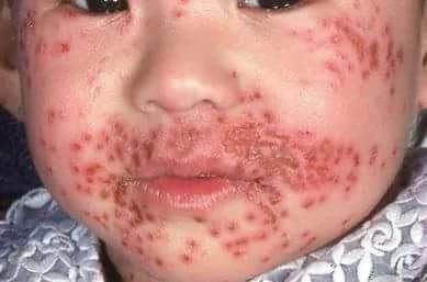
Eczema herpeticum showing the typical lesions of a secondary HSV infection in eczema.
Image Source: Wikimedia Commons (2018). Eczema herpitcum, Licensed under a creative commons licence. Available for free at: https://commons.wikimedia.org/wiki/File:Eczema_herpitcum.jpg (Accessed on: 14 March 2023).

Erythroderma
Erythroderma is a severe condition that presents with diffuse erythema and scaling. To be classified as erythroderma it must involve all or most of the skin surface area – over 90% as per the most common definitions. There are various causes, but two of the most common are exacerbation of pre-existing dermatitis or psoriasis, and hypersensitivity to drugs. It can develop acutely over a few hours, or it can evolve over days. It presents as skin that is red and warm to touch, often with severe pain or itching. Over time scaling of the skin can occur and crusted lesions can develop, and this is often more common in those who have underlying psoriasis.
Management involves hospitalisation requiring close monitoring of haemodynamic status as well as body temperature and treatment of any secondary cutaneous superinfections. Oral antihistamines can reduce itching, whereas inflammation and pruritus can be managed with topical corticosteroids.
Cutaneous Vasculitis and Connective Tissue Disorders
Cutaneous Vasculitis
A group of disorders resulting in inflamed blood vessels in the skin. There are many different causes and a wide variety of clinical presentations. It is thought that the inflammatory response in the blood vessel wall could be a result of:
- Direct injury from bacteria or viruses
- Indirect injury through activation of antibodies
- Indirect injury through activation of complement pathways
There are many different causes of cutaneous vasculitis, including infection, autoimmune conditions and vasculitides, medications, and malignancy. In some cases, it can be associated with systemic vasculitis. However, in many cases, there is no underlying cause, and the disease will be self-limiting.
It can be classified according to which type of blood vessel it affects, such as capillaries, small, medium, or large vessels; however, large vessel vasculitis very rarely results in cutaneous features. The main presenting feature of small vessel vasculitis is pigmented and often palpable purpura, whereas nodules and a rash known as livedo reticularis are more associated with medium vessel vasculitis.
Diagnosis is based on the clinical appearance but can be supported with a skin biopsy and blood tests to exclude other possible underlying causes. Treatment is through identification and removal of the underlying cause, including treatment of systemic vasculitis if it is a manifestation of a systemic disorder. Initial management of the rash when no underlying cause is identified is cold avoidance.
Connective Tissue Disorders (CTDs)
CTDs are a heterogenous group of conditions that occur because of problems with collagen, and some have a vascular component as well. There are many CTDs, often similar in presentation, and can largely be split into inherited (such as Marfan’s syndrome and Ehlers-Danlos syndrome) and acquired CTDs (there are many, for which three are focused on below).
Cutaneous Lupus Erythematosus (CLE)
CLE is an uncommon group of skin disorders and occurs from autoimmune processes. It can be associated with systemic lupus erythematosus (SLE). There are many different types of CLE. Some of the key forms have been described.
| Acute CLE | Presents as erythematous patches associated with a flare of acute SLE. These acute rashes include specific changes such as malar ‘butterfly’ rash presenting as redness and swelling over the cheeks and sparing the nasolabial folds and generalised acute CLE – a diffuse or papular erythema of the face, upper limbs, and trunk. |
| Subacute CLE | Less commonly associated with SLE, with only approximately 50% having a mild form. The typical presentation is widespread, non-scarring round or psoriasis-like plaques. It usually presents in areas of sun exposure and can be triggered or aggravated by sun exposure. |
| Chronic CLE | Discoid lupus is the most common form of chronic CLE, accounting for approximately 80% of cases and is much less commonly associated with SLE (1-2% of patients). Skin lesions are commonly on the scalp, ears, cheeks, nose, and lips. They are usually scaly plaques that heal slowly and can leave post-inflammatory pigmentation and scarring. |
| Chilblain lupus is a form of chronic CLE and causes cold-induced plaques on fingers and toes | |
A table summarising the key features in different forms of CLE.
Systemic Sclerosis
An autoimmune condition characterised by skin and internal organ fibrosis and vascular disease. There are two main subtypes according to the distribution of skin involvement: diffuse cutaneous systemic sclerosis (skin involvement across the whole body and early organ fibrosis) and limited cutaneous systemic sclerosis (skin involvement is limited to the face, hands, and feet).
The main skin manifestations are sclerodactyly, thickening and tightness of the skin of the fingers and toes, Raynaud’s phenomenon, telangiectasia on the face, chest and palms, and calcinosis on digits and extensor surfaces of arms. Patients will often exhibit microstomia, which can be defined as reduced interlabial distance of <4.5cm. In severe cases, patients can develop digital ulcers, which can result in dry gangrene and eventual loss of the tips of the fingertips.
Dermatomyositis
A rare multisystem disorder that can affect the skin as well as muscles and blood vessels. The exact underlying aetiology is not well understood. Triggers include medications, malignancy, viral infections, and certain trades, such as silica exposure. Additionally, most patients have disease-associated antibodies suggesting an autoimmune component, with ANA being the most common and anti-Mi-2 being the most specific. Skin manifestations can often have similarities with those seen in other CTDs, such as photosensitivity, calcinosis, and Raynaud’s phenomenon. More characteristic skin manifestations include Gottron’s papules and a heliotrope rash. The heliotrope rash presents on the face and upper eyelids as a bilateral lilac coloured discolouration. Gottron’s papules are characteristic purple papules and plaques that can be seen over the small joints of the hands.
Blood and Lymphatic Vessel Disorders
Venous ulcers
Venous ulceration can occur from venous insufficiency and hypertension in the lower limbs. Chronic venous hypertension alters capillary and lymphatic microcirculation, which can subsequently result in capillary leak, fibrin deposition, erythrocyte and leukocyte sequestration, thrombocytosis, and inflammation. In severe and more chronic cases, this can lead to tissue hypoxia and subsequent ulcer formation.
Identification and Symptoms
Diagnosis of chronic venous insufficiency is through the presence of symptoms, examination and venous doppler ultrasound of lower limbs. The main symptoms include leg pain, fatigue, and heaviness. Skin changes include venous eczema and discolouration, dilated superficial veins, and ultimately ulceration in severe cases. Venous ulcers tend to affect the medial aspect of the lower limb and have a poorly defined edge.

Venous insufficiency affecting the lower limbs with characteristic skin manifestations, including venous eczema, lipodermatosclerosis, and orange-brown macular pigmentation from haemosiderin deposition.
Image Source: Wikimedia Commons (2018). Chronicvenousinsufficiency, Licensed under a creative commons licence. Available for free at: https://commons.wikimedia.org/wiki/File:Chronicvenousinsufficiency.jpg (Accessed on: 14 March 2023).

Treatment
Treatment can be divided into various categories and aims. These include improvement of symptoms, reduction of overall leg oedema, and healing of ulcers.
General measures recommended to patients include leg elevation, regular exercise, avoidance of prolonged standing and appropriate skin care. If ulceration occurs, treatments include compression therapy, ulcer debridement and possible antibiotic therapy depending on appearance and symptoms of ulcers.
Lymphoedema
Defined as an ‘abnormal accumulation of interstitial fluid and fibroadipose tissues resulting from injury, infection, or congenital abnormalities of the lymphatic system’. When the lymphatic load exceeds the overall transport capacity of the lymphatic system, this results in the build-up of fluid in the interstitial tissues.
It is classified as either primary or secondary.
Primary causes are those without a precipitating cause and are congenital or inherited. It tends to present in children but can also present later in adulthood. This is referred to as Milroy disease.
Secondary lymphoedema occurs due to other conditions or treatments. The most common cause is cancer and cancer treatment, including radiotherapy and surgical excision of lymph nodes. Other causes include infection, inflammatory disorders, obesity, and chronic causes of lymphatic overload (venous insufficiency, trauma, and burns).
Identification and Diagnosis
The diagnosis can be made from a careful history and examination. Physical examination should include vascular, skin, and soft tissue examination with focus on the lymph nodes. Most cases are unilateral, often from precipitating events such as lymph node excision from cancer or radiotherapy. The circumference of the limbs should also be measured. Additional imaging is usually not required. However, in cases where imaging is required, CT and MR have been used but with variable results.
Management
The first line treatment is decongestive lymphatic therapy. This utilises multiple techniques to help control symptoms. The four main components are compression bandages, skin care, exercises to improve lymph drainage and specialised massage techniques to stimulate the flow of fluid in the lymphatic system.
Acne
Acne Vulgaris
Introduction
It typically presents in adolescent years and tends to be seen on the face, but it can also affect the neck, trunk, and proximal upper extremities (where sebaceous glands are abundant). It is an inflammatory disorder of the pilosebaceous unit – the hair follicle and sebaceous gland. Pathogenesis involves:
- Plugging of the follicle secondary to epidermal proliferation
- Excess sebum production
- Colonisation with anaerobic Propionibacterium acnes
- Inflammation
Contributing factors include androgen-mediated stimulation of sebaceous glands, cellular immune responses, dysbiosis of the microbiome of the pilosebaceous follicle and in some cases, it has been reported to be related to genetics, medication and possibly diet.
Investigation and Management
It is primarily a clinical diagnosis and is characterised by non-inflammatory comedones (open or closed) and inflammatory papules, pustules and/or nodules. It can be classified as mild, moderate, or severe, which informs management. There are a variety of topical treatments for mild lesions, such as with retinoids (adapalene), benzoyl peroxide and antibiotics (clindamycin).
If symptoms do not resolve with topical treatments, various systemic treatments can be considered:
- Oral antibiotics
- First line: tetracyclines such as doxycycline and lymecycline (contraindicated in pregnancy – can consider macrolide antibiotics however they are not recommended due to high levels of P. acnes resistance).
- Minocycline is not recommended as there is increased risk of adverse effects
- Anti-androgenic contraceptives in females, e.g. the combined oral contraceptive pill
- Retinoid therapy – isotretinoin
Isotretinoin, a systemic retinoid, is highly effective in severe cases. It is indicated when there is scarring and when other treatment options have failed. It can only be prescribed by specialist dermatology teams. As isotretinoin is teratogenic, women of childbearing age are required to use two methods of contraception and have monthly pregnancy tests. Regular blood monitoring of liver function tests and triglycerides are also required as it can cause derangement. Side effects include dry skin (most common) and low mood.
Complications
Several complications can occur, such as:
- Post inflammatory hyperpigmentation presenting as dark spots – photoprotection is essential
- Post inflammatory erythema presenting as residual, persistent, and pink to red discolouration of the skin
- Scarring which can be managed with minor surgical procedures, laser therapy and chemical peels as well as other interventions
Acne Rosacea
Introduction
A chronic inflammatory skin condition that typically affects the central face and eyes. It presents with centrofacial erythema, inflammatory papules and pustules, flushing, telangiectasia and phymatous skin changes, e.g. rhinophyma.
It occurs in approximately 5% of adults worldwide and tends to affect both genders equally. It typically presents after 30 years of age and is more prevalent with age.
The pathogenesis is multifactorial and is thought to include a combination of genetic susceptibility, altered microbiome of the skin and gut, and neurocutaneous mechanisms – alcohol, UV light, exercise, temperature changes and spicy food.
Investigation and Management
Rosacea is primarily a clinical diagnosis. Differential diagnoses include acne vulgaris, skin damage, seborrhoeic dermatitis, dermatomyositis, drug reactions and eczema, to name a few.
The main treatment is through lifestyle change – patients are encouraged to record a symptom diary to identify their triggers and subsequently avoid them. Patients should moisturise frequently and use daily SPF. Exfoliants, topical steroids and alcohol-based topical products should be avoided.
Medical treatment includes topical metronidazole in mild to moderate cases. In severe cases brimonidine is used (to reduce erythema). Systemic treatment options include a 2-month course of tetracyclines (or erythromycin) and should be used if symptoms have not resolved for a year with topical therapies. In cases of telangiectasia, laser treatments can be trialled. Complications include blepharitis, keratitis and conjunctivitis. Oral antibiotics are indicated in cases of ocular involvement.
Papulosquamous disorders
A group of disorders whose aetiology is not fully understood. They are named as such based on the descriptive features of the clinical lesions, which are characterised by scaly papules and plaques. Major conditions in this group include psoriasis, lichen sclerosus, lichen planus, pityriasis rosea and seborrhoeic dermatitis. Seborrhoeic dermatitis is covered in the topic ‘dermatitis/eczema’.
Psoriasis
There are various types of psoriasis. Psoriasis is a group of chronic inflammatory skin conditions characterised by clearly defined, red and scaly plaques. It is thought to be a multifactorial disorder that is an immune-mediated inflammatory disease. Plaque psoriasis is the most common type of psoriasis across all ethnic groups and populations.
The main types of psoriasis are summarised below:
| Type | Key Features | Image |
|---|---|---|
| Chronic plaque psoriasis |
|
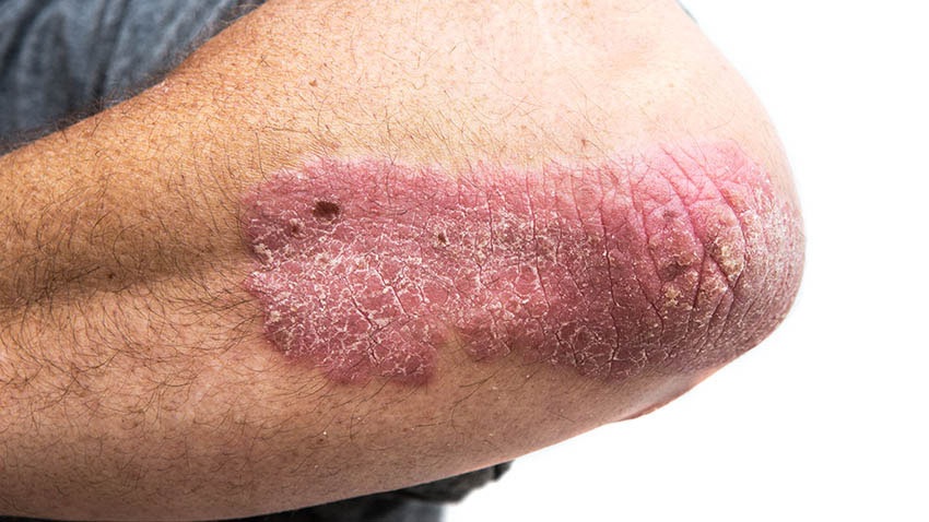
Image Source: Wikimedia Commons (2019). Psoriasis on elbow, Licensed under a creative commons licence. Available for free at: https://commons.wikimedia.org/wiki/File:Psoriasis_on_elbow.jpg (Accessed on: 14 March 2023). |
| Guttate psoriasis |
|

Image Source: Wikimedia Commons (2010). Guttate psoriasis, Licensed under a creative commons licence. Available for free at: https://commons.wikimedia.org/wiki/File:Guttate_psoriasis.jpg (Accessed on: 14 March 2023). |
| Flexural psoriasis |
|

Image Source: Wikimedia Commons (2013). Schuppenflechte am Knie, Licensed under a creative commons licence. Available for free at: https://commons.wikimedia.org/wiki/File:Schuppenflechte_am_Knie.JPG (Accessed on: 14 March 2023). |
| Scalp psoriasis |
|

Image Source: Wikimedia Commons (2021). Stelara before and after pictures-1, Licensed under a creative commons licence. Available for free at: https://commons.wikimedia.org/wiki/File:Stelara_before_and_after_pictures-1.png (Accessed on: 14 March 2023). |
| Nail psoriasis |
|

Image Source: Wikimedia Commons (2009). Psonail, Licensed under a creative commons licence. Available for free at: https://commons.wikimedia.org/wiki/File:Psonail.jpg (Accessed on: 14 March 2023) |
| Erythrodermic psoriasis |
|
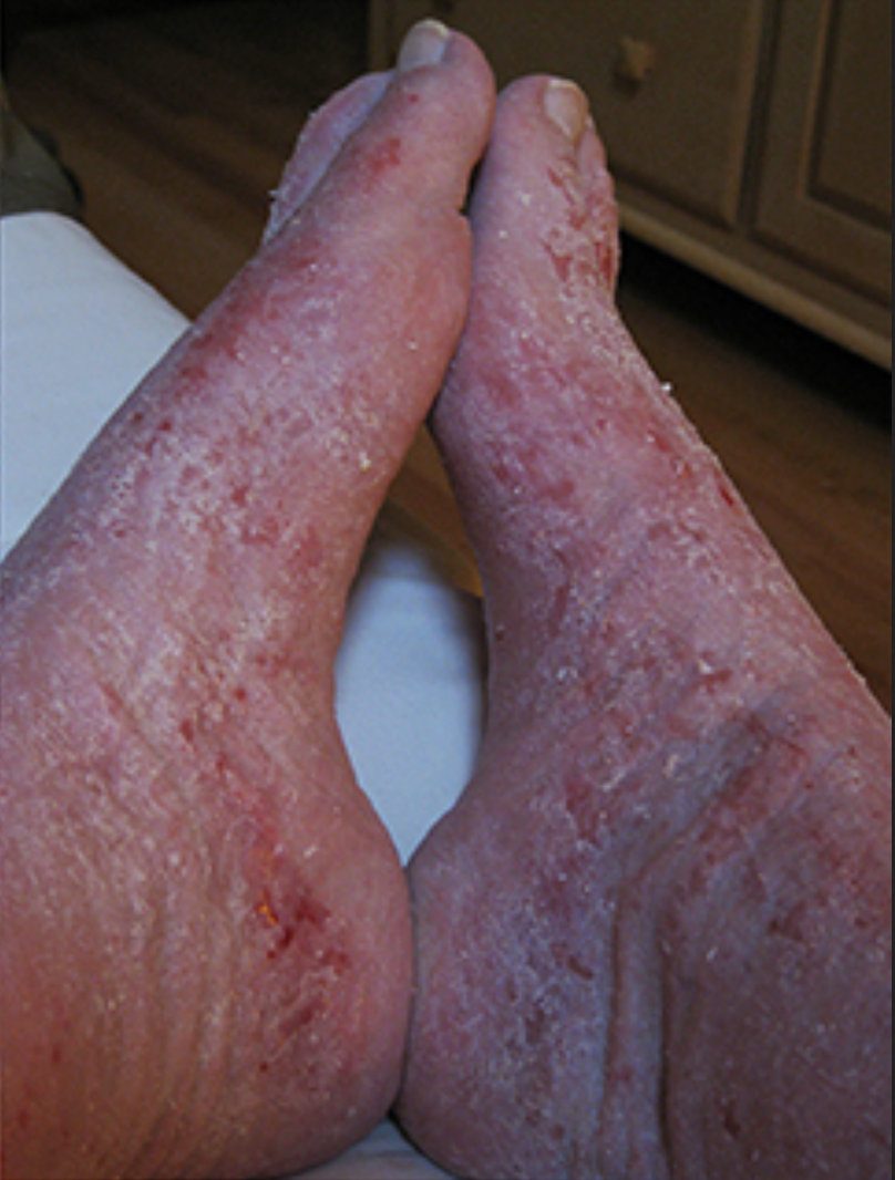
Image Source: Wikimedia Commons (2017). Red (burning) Skin Syndrome – Feet Collage, Licensed under a creative commons licence. Available for free at: https://commons.wikimedia.org/wiki/File:Red_(burning)_Skin_Syndrome_-_Feet_Collage.jpg (Accessed on: 14 March 2023) |


_Skin_Syndrome_-_Feet_Collage.jpg)
Types of psoriasis and common features
Psoriasis is primarily a clinical diagnosis; however, diagnosis can be supported through skin biopsy. Additionally, it can present as psoriatic arthritis without skin involvement in up to 15% of cases. The main features of psoriatic arthritis include inflammation at the distal interphalangeal joints as well as nail changes. It can also affect other joints and vary in its presentation of either symmetrical or asymmetrical oligo/polyarthritis. Severity of psoriasis can be assessed through history, examination, and assessment tools, such as the Psoriasis Area and Severity Index (PASI) scoring tool.
Treatment depends on severity. Mild disease can be managed with topical agents such as emollients, coal tar preparations, salicylic acid, vitamin D analogues (calcipotriol), topical steroids or combination ointments/gels. Treatment can be escalated to phototherapy, systemic therapy with immunomodulatory medications such as DMARDs or in more severe cases biologics or targeted therapies such as anti-TNFa treatments. These are reserved for severe psoriasis that is resistant to conventional treatments.
Certain medications are known to exacerbate psoriasis. These include beta blockers, lithium, antimalarials, NSAIDs, ACE inhibitors and infliximab.
Lichen Sclerosus
A chronic inflammatory skin disorder that most commonly affects the genital and perianal areas and is ten times more common in women than in men. It tends to occur in women over 50. The causes are likely to be a combination of factors, including genetics, hormonal, irritant, traumatic and infectious elements. Some cases are thought to be autoimmune with 60-80% of patients having extracellular matrix protein-1 (ECM01) antibodies.
Clinical features
Non-hair-bearing parts of the vulva are affected and it usually presents as itching and soreness. Sometimes bruising and ulceration occurs from scratching and minimal friction. In some cases, urination can cause stinging and patients can experience uncomfortable sexual intercourse. It can spread to the surrounding skin of labia majora and inguinal folds, as well as the anal and perianal skin area but never involves vaginal mucosa. Patients are often more prone to infections such as thrush (candida albicans), herpes simplex and staphylococcus aureus. It has been associated with an increased risk of vulval, penile or anal squamous cell carcinoma in up to 5% of patients.
Investigation and management
It is primarily a clinical diagnosis, however, skin biopsy can help aid diagnosis.
The main treatment is initially with a strong topical steroid ointment such as clobetasol propionate 0.05%. Once symptoms are more controlled, steroid ointment can be reduced in strength.
Lichen Planus
A chronic inflammatory skin condition that affects the skin and mucosal surfaces. It can cause many different clinical presentations, but all the conditions share similar features on histopathological testing. The different types include:
- Cutaneous lichen planus
- Mucosal lichen planus
- Lichen planus of nails
- Lichenoid drug eruption
Lichen planus is common and is thought to affect around 1 in 100 people worldwide, primarily in adults over the age of 40. The most common type is oral lichen planus, which is more common in women.
It is an autoimmune disorder mediated by T-cell attacking protein within the skin and mucosal keratinocytes. There are various contributing factors, including genetics, stress, skin injury, localised skin infection, systemic viral infection, contact allergy and various drugs.
Clinical features
Lesions on the skin and mucosal surfaces are often described as papules and polygonal plaques that appear shiny, flat-topped, and firm. They often have fine white lines that form a lace-like pattern across the lesions, called Wickham striae (see image). Symptoms can range from no symptoms to intense itching. Oral lichen planus tends to involve the cheeks and sides of the tongue. The most common drugs that can cause lichenoid reactions are gold, hydroxychloroquine, and captopril.

Oral Lichen planus on mucosal surfaces
Image Source: Wikimedia Commons (2019). Lichen planus, Licensed under a creative commons licence. Available for free at: https://commons.wikimedia.org/wiki/File:Lichen_planus.jpg (Accessed on: 14 March 2023).

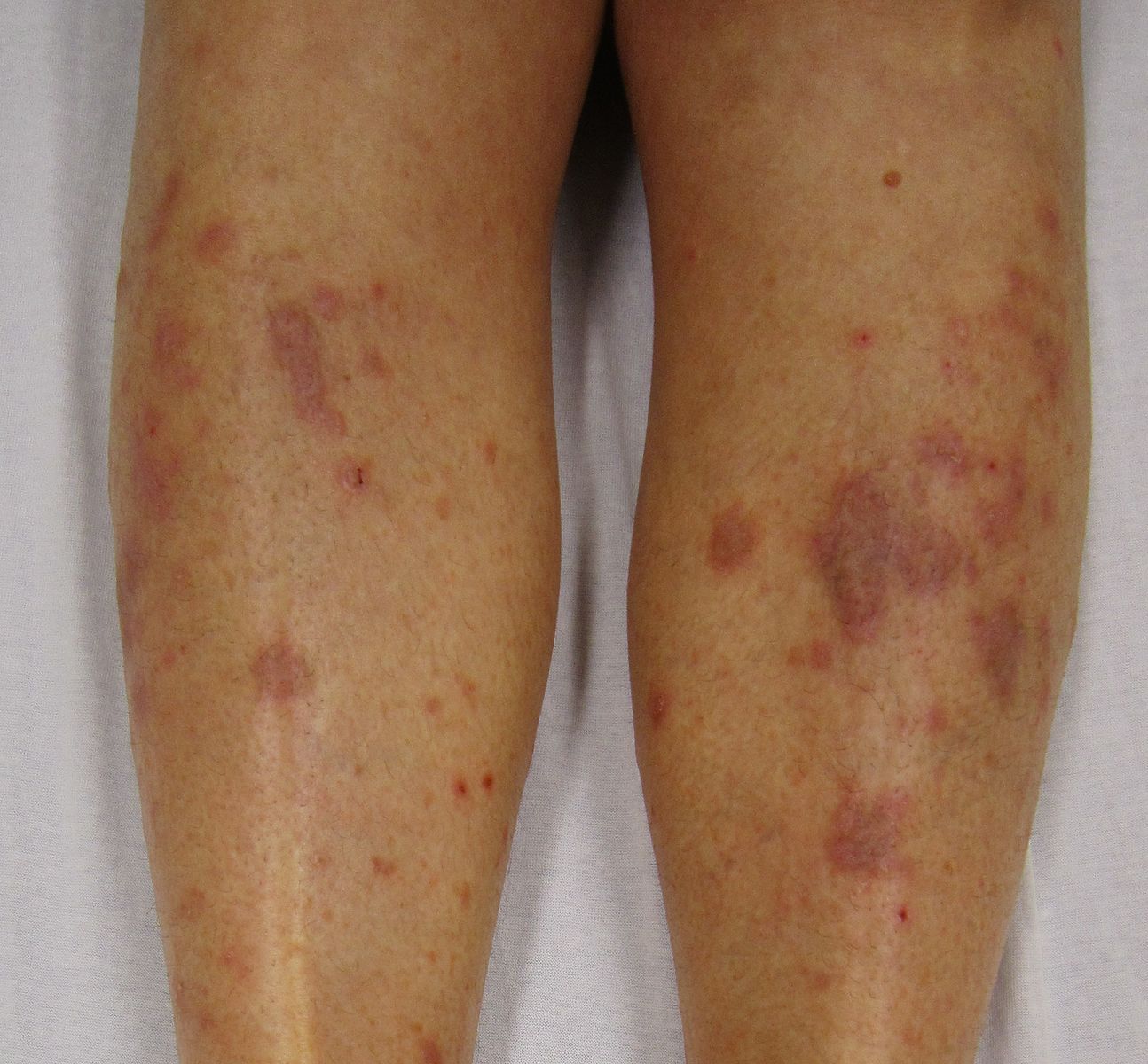
Lichen planus seen on legs – violaceous polygonal plaques
Image Source: Wikimedia Commons (2010). Lichen Planus (2), Licensed under a creative commons licence. Available for free at: https://commons.wikimedia.org/wiki/File:Lichen_Planus_(2).JPG (Accessed on: 14 March 2023).
.JPG)
Investigations and management
Primarily a clinical diagnosis which can be confirmed with a skin biopsy to look for epidermal lichenoid tissue reactions. Treatment is with potent topical agents such as steroids, topical calcineurin inhibitors (such as tacrolimus), topical retinoids and intralesional steroid injections.
A course of oral prednisolone or other immunosuppressive agents may be considered in more severe cases. Most lesions resolve within 2-3 years, but mucosal/oral lichen planus tends to be more persistent and more likely to recur.
Pityriasis Rosea
Aetiology and Symptoms
Pityriasis rosea is a common condition in teenagers and young adults, however, it can occur at any age. It is a self-limiting rash that tends to resolve in 6-10 weeks. The presenting feature is often a circular or oval ‘herald patch’ which is typically seen on the chest, abdomen or back, before a generalised rash occurs. The patch is raised, salmon-pink or red and 2-5cm in diameter. There is often a peripheral scale trailing just inside the edge of the lesion.
The exact cause is not well understood, but it is suggested that it can occur following viral (HHV-6/7) and bacterial infections, vaccinations, and drug-induced reactions (there are many associated drugs such as ACEi, NSAIDs, imatinib, metronidazole and isotretinoin, to name a few).
Most patients have no systemic symptoms when the rash occurs. However, mild-severe itching can occur in up to 25% of patients. It is a clinical diagnosis that can be supported with histological tests.
Treatments
It is a self-limiting condition. Management focuses on reducing pruritus in the 25% of patients who have severe itching. This can be done with emollients, non-scented soap substitutes and cautious sun exposure. Other treatments that may help reduce the duration of symptoms include topical steroid creams, oral steroids in severe cases and antivirals such as aciclovir. UVB therapy has been shown to be most effective at decreasing the duration of the rash that patients experience.
Complete Guide to Clinical Genetics ST3 Application and Recruitment (2027)
Written by Medibuddy. Last updated: October 2026. Clinical Genetics is

Complete Guide to Clinical Radiology ST3 Application and Recruitment (2027)
Recruitment paused: NHS England is not running Clinical Radiology ST3+ recru

Complete Guide to Paediatric Surgery ST3 Application and Recruitment in United Kingdom (2026)
Written by Medibuddy. Last updated: October 2026. If you’re planning


Take your subscriptions with you
Our mobile app allows you to access your interview and exam question banks wherever you are.

