


 Close
Close
 2nd July 2023
Papulosquamous disorders
A group of disorders whose aetiology is not fully understood. They are named as such based on the descriptive features of the clinical lesions, which are characterised by scaly papules and plaques. Major conditions in this group include psoriasis, lichen sclerosus, lichen planus, pityriasis rosea and seborrhoeic dermatitis. Seborrhoeic dermatitis is covered in the topic ‘dermatitis/eczema’.
Psoriasis
There are various types of psoriasis. Psoriasis is a group of chronic inflammatory skin conditions characterised by clearly defined, red and scaly plaques. It is thought to be a multifactorial disorder that is an immune-mediated inflammatory disease. Plaque psoriasis is the most common type of psoriasis across all ethnic groups and populations.
The main types of psoriasis are summarised below:
| Type | Key Features | Image |
|---|---|---|
| Chronic plaque psoriasis |
|
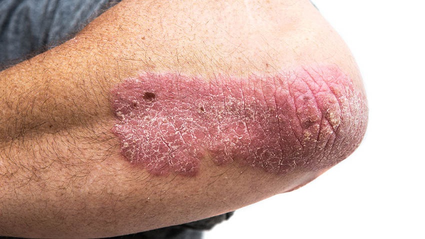
Image Source: Wikimedia Commons (2019). Psoriasis on elbow, Licensed under a creative commons licence. Available for free at: https://commons.wikimedia.org/wiki/File:Psoriasis_on_elbow.jpg (Accessed on: 14 March 2023). |
| Guttate psoriasis |
|

Image Source: Wikimedia Commons (2010). Guttate psoriasis, Licensed under a creative commons licence. Available for free at: https://commons.wikimedia.org/wiki/File:Guttate_psoriasis.jpg (Accessed on: 14 March 2023). |
| Flexural psoriasis |
|

Image Source: Wikimedia Commons (2013). Schuppenflechte am Knie, Licensed under a creative commons licence. Available for free at: https://commons.wikimedia.org/wiki/File:Schuppenflechte_am_Knie.JPG (Accessed on: 14 March 2023). |
| Scalp psoriasis |
|

Image Source: Wikimedia Commons (2021). Stelara before and after pictures-1, Licensed under a creative commons licence. Available for free at: https://commons.wikimedia.org/wiki/File:Stelara_before_and_after_pictures-1.png (Accessed on: 14 March 2023). |
| Nail psoriasis |
|

Image Source: Wikimedia Commons (2009). Psonail, Licensed under a creative commons licence. Available for free at: https://commons.wikimedia.org/wiki/File:Psonail.jpg (Accessed on: 14 March 2023) |
| Erythrodermic psoriasis |
|
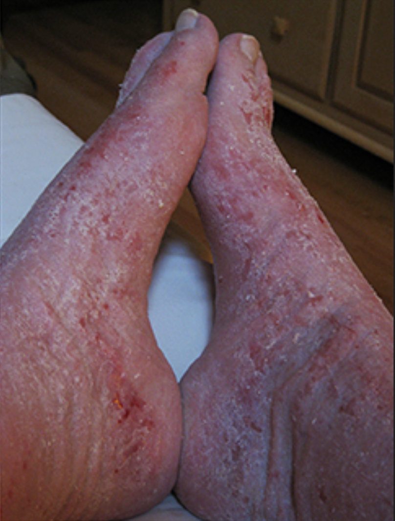
Image Source: Wikimedia Commons (2017). Red (burning) Skin Syndrome – Feet Collage, Licensed under a creative commons licence. Available for free at: https://commons.wikimedia.org/wiki/File:Red_(burning)_Skin_Syndrome_-_Feet_Collage.jpg (Accessed on: 14 March 2023) |


_Skin_Syndrome_-_Feet_Collage.jpg)
Types of psoriasis and common features
Psoriasis is primarily a clinical diagnosis; however, diagnosis can be supported through skin biopsy. Additionally, it can present as psoriatic arthritis without skin involvement in up to 15% of cases. The main features of psoriatic arthritis include inflammation at the distal interphalangeal joints as well as nail changes. It can also affect other joints and vary in its presentation of either symmetrical or asymmetrical oligo/polyarthritis. Severity of psoriasis can be assessed through history, examination, and assessment tools, such as the Psoriasis Area and Severity Index (PASI) scoring tool.
Treatment depends on severity. Mild disease can be managed with topical agents such as emollients, coal tar preparations, salicylic acid, vitamin D analogues (calcipotriol), topical steroids or combination ointments/gels. Treatment can be escalated to phototherapy, systemic therapy with immunomodulatory medications such as DMARDs or in more severe cases biologics or targeted therapies such as anti-TNFa treatments. These are reserved for severe psoriasis that is resistant to conventional treatments.
Certain medications are known to exacerbate psoriasis. These include beta blockers, lithium, antimalarials, NSAIDs, ACE inhibitors and infliximab.
Lichen Sclerosus
A chronic inflammatory skin disorder that most commonly affects the genital and perianal areas and is ten times more common in women than in men. It tends to occur in women over 50. The causes are likely to be a combination of factors, including genetics, hormonal, irritant, traumatic and infectious elements. Some cases are thought to be autoimmune with 60-80% of patients having extracellular matrix protein-1 (ECM01) antibodies.
Clinical features
Non-hair-bearing parts of the vulva are affected and it usually presents as itching and soreness. Sometimes bruising and ulceration occurs from scratching and minimal friction. In some cases, urination can cause stinging and patients can experience uncomfortable sexual intercourse. It can spread to the surrounding skin of labia majora and inguinal folds, as well as the anal and perianal skin area but never involves vaginal mucosa. Patients are often more prone to infections such as thrush (candida albicans), herpes simplex and staphylococcus aureus. It has been associated with an increased risk of vulval, penile or anal squamous cell carcinoma in up to 5% of patients.
Investigation and management
It is primarily a clinical diagnosis, however, skin biopsy can help aid diagnosis.
The main treatment is initially with a strong topical steroid ointment such as clobetasol propionate 0.05%. Once symptoms are more controlled, steroid ointment can be reduced in strength.
Lichen Planus
A chronic inflammatory skin condition that affects the skin and mucosal surfaces. It can cause many different clinical presentations, but all the conditions share similar features on histopathological testing. The different types include:
- Cutaneous lichen planus
- Mucosal lichen planus
- Lichen planus of nails
- Lichenoid drug eruption
Lichen planus is common and is thought to affect around 1 in 100 people worldwide, primarily in adults over the age of 40. The most common type is oral lichen planus, which is more common in women.
It is an autoimmune disorder mediated by T-cell attacking protein within the skin and mucosal keratinocytes. There are various contributing factors, including genetics, stress, skin injury, localised skin infection, systemic viral infection, contact allergy and various drugs.
Clinical features
Lesions on the skin and mucosal surfaces are often described as papules and polygonal plaques that appear shiny, flat-topped, and firm. They often have fine white lines that form a lace-like pattern across the lesions, called Wickham striae (see image). Symptoms can range from no symptoms to intense itching. Oral lichen planus tends to involve the cheeks and sides of the tongue. The most common drugs that can cause lichenoid reactions are gold, hydroxychloroquine, and captopril.

Oral Lichen planus on mucosal surfaces
Image Source: Wikimedia Commons (2019). Lichen planus, Licensed under a creative commons licence. Available for free at: https://commons.wikimedia.org/wiki/File:Lichen_planus.jpg (Accessed on: 14 March 2023).

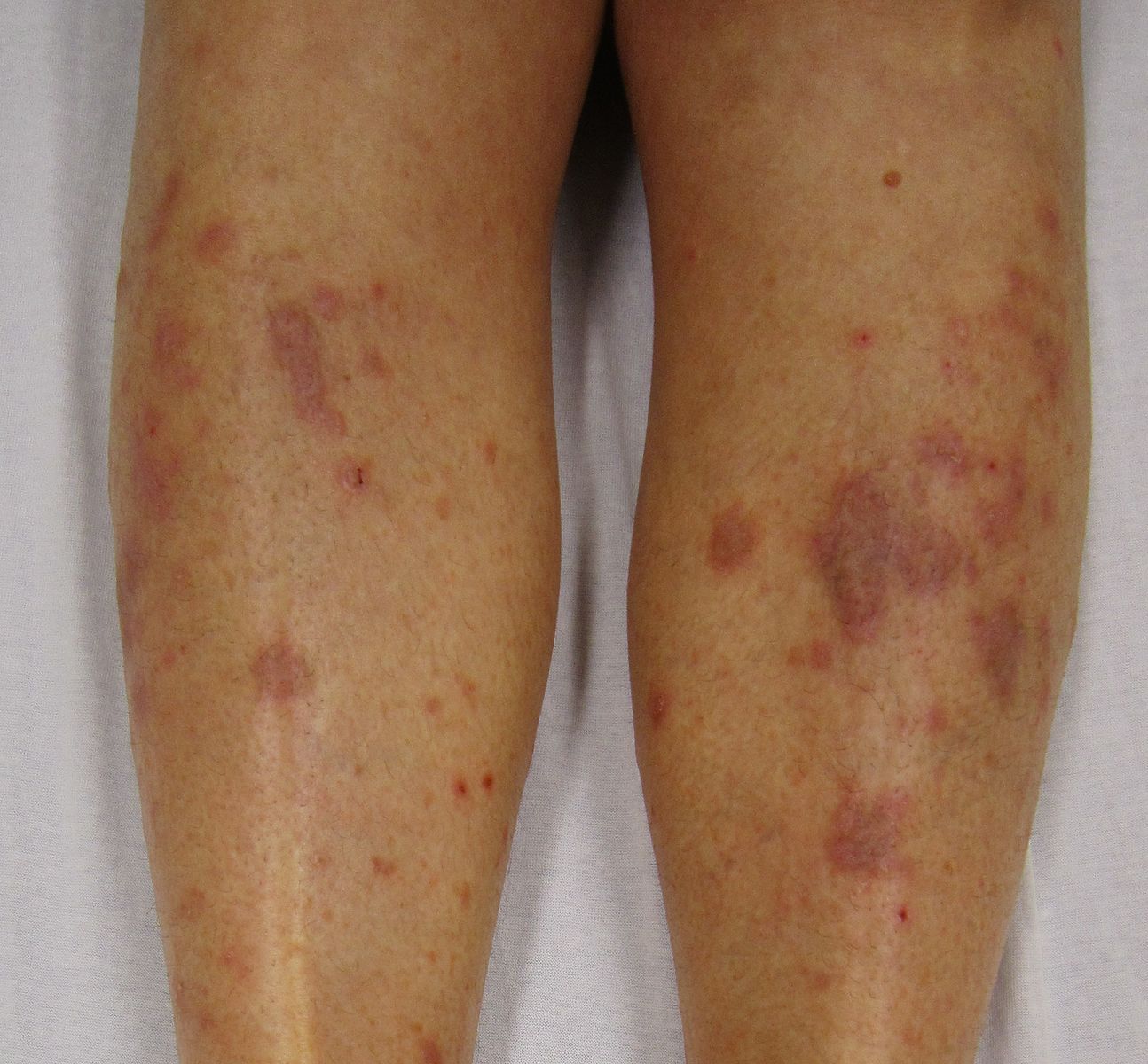
Lichen planus seen on legs – violaceous polygonal plaques
Image Source: Wikimedia Commons (2010). Lichen Planus (2), Licensed under a creative commons licence. Available for free at: https://commons.wikimedia.org/wiki/File:Lichen_Planus_(2).JPG (Accessed on: 14 March 2023).
.JPG)
Investigations and management
Primarily a clinical diagnosis which can be confirmed with a skin biopsy to look for epidermal lichenoid tissue reactions. Treatment is with potent topical agents such as steroids, topical calcineurin inhibitors (such as tacrolimus), topical retinoids and intralesional steroid injections.
A course of oral prednisolone or other immunosuppressive agents may be considered in more severe cases. Most lesions resolve within 2-3 years, but mucosal/oral lichen planus tends to be more persistent and more likely to recur.
Pityriasis Rosea
Aetiology and Symptoms
Pityriasis rosea is a common condition in teenagers and young adults, however, it can occur at any age. It is a self-limiting rash that tends to resolve in 6-10 weeks. The presenting feature is often a circular or oval ‘herald patch’ which is typically seen on the chest, abdomen or back, before a generalised rash occurs. The patch is raised, salmon-pink or red and 2-5cm in diameter. There is often a peripheral scale trailing just inside the edge of the lesion.
The exact cause is not well understood, but it is suggested that it can occur following viral (HHV-6/7) and bacterial infections, vaccinations, and drug-induced reactions (there are many associated drugs such as ACEi, NSAIDs, imatinib, metronidazole and isotretinoin, to name a few).
Most patients have no systemic symptoms when the rash occurs. However, mild-severe itching can occur in up to 25% of patients. It is a clinical diagnosis that can be supported with histological tests.
Treatments
It is a self-limiting condition. Management focuses on reducing pruritus in the 25% of patients who have severe itching. This can be done with emollients, non-scented soap substitutes and cautious sun exposure. Other treatments that may help reduce the duration of symptoms include topical steroid creams, oral steroids in severe cases and antivirals such as aciclovir. UVB therapy has been shown to be most effective at decreasing the duration of the rash that patients experience.
Complete Guide to Clinical Genetics ST3 Application and Recruitment (2027)
Written by Medibuddy. Last updated: October 2026. Clinical Genetics is

Complete Guide to Clinical Radiology ST3 Application and Recruitment (2027)
Recruitment paused: NHS England is not running Clinical Radiology ST3+ recru

Complete Guide to Paediatric Surgery ST3 Application and Recruitment in United Kingdom (2026)
Written by Medibuddy. Last updated: October 2026. If you’re planning


Take your subscriptions with you
Our mobile app allows you to access your interview and exam question banks wherever you are.

