


 Close
Close
 25th December 2023
Optic nerve disease
Optic neuropathy is damage to the optic nerve. The causes may be divided into:
Acquired
- Demyelinating/idiopathic optic neuritis
- Inflammatory/autoimmune
- Ischaemic optic neuropathy
Congenital
- Leber’s Hereditary Optic Neuropathy (LHON)
Optic neuritis (ON)
Inflammation of the optic nerve may be classified as typical or atypical. Typical ON often refers to either idiopathic or multiple sclerosis-associated disease. This most commonly occurs in young Caucasian women in temperate zones. Vision loss is progressive over hours-to-days, reaching a nadir by 2 weeks. The degree of vision loss is variable, ranging from moderate (6/18) to severe (<6/60). There is associated retrobulbar discomfort with eye movements and disturbance in colour vision. The optic disc may be mildly swollen or normal (retrobulbar ON). Uhthoff’ phenomenon describes a transient worsening of neurological symptoms associated with a rise in body temperature (exercise; bathing) and is associated with both multiple sclerosis (MS) and aquaporin-4 (AQP-4) optic neuritis.
Features of typical ON:
- Ages 20-50 years
- Unilateral
- Progression over hours-to-days
- Retrobulbar discomfort
- Colour vision disturbance
- Relative afferent pupillary defect
- Improvement begins within 2 weeks of onset
Investigations are centred on investigating for multiple sclerosis-associated ON:
- Magnetic resonance imaging (MRI) of the brain and orbits with contrast to look for demyelinating disease.
- In the active phase, the affected optic nerve will enhance.
- Evidence of infratentorial, periventricular and juxtacortical white matter lesions may suggest multiple sclerosis.
Management:
The optic neuritis treatment trial (ONTT) demonstrated that a combination of intravenous methylprednisolone (IVMP) followed by oral Prednisolone may accelerate visual recovery but did not affect final VA for a first episode of optic neuritis. Low-dose oral corticosteroids (1mg/kg) alone were associated with increased frequency of recurrence. For recurrent, idiopathic ON, initiation of treatment within 2 days of symptom onset may provide a better recovery of VA, although further multi-centre studies are warranted to confirm this. Oral corticosteroids in a bioequivalent dose may be as effective as IVMP. The decision to treat typical ON with corticosteroids is often taken on a case-by-case basis.
Prognosis:
In idiopathic or multiple sclerosis-associated optic neuritis, over 90% recover to a visual acuity of 6/12 or better without treatment. However, there may be permanent abnormalities of colour perception, contrast sensitivity or visual field defects. Following a first episode of optic neuritis, the following characteristics have a lower risk of developing multiple sclerosis:
- No demyelinating lesions on MRI
- Optic disc swelling
- Male gender
Atypical ON:
This describes clinical features not consistent with typical optic neuritis. A secondary cause for optic nerve inflammation should be sought. Traditionally, it was acceptable to observe the natural evolution of an ON event over 4-6 weeks. Increased recognition of AQP-4 and myelin oligodendrocyte glycoprotein (MOG) disease has broadened the spectrum of optic neuritis-related autoimmune disorders. AQP-4 and MOG-associated optic neuritis carry significant visual morbidity; prompt initiation of high-dose corticosteroid therapy may prevent a poor outcome. Early clinical suspicion is important because confirmatory antibody testing (aquaporin-4 and MOG antibodies) and neuro-radiological imaging may take weeks. Features which are suggestive of non-MS, autoimmune ON include:
- Bilateral disease
- Moderate-to-severe optic disc swelling
- Non-Caucasian ethnicity
- Age not in keeping with typical optic neuritis
Other investigations for atypical optic neuritis include FBC, ESR, CRP, vitamin B12, folate, HBA1c, LFTs, ANCA, ANA, ACE syphilis, genetic testing for Leber’s hereditary optic neuropathy, syphilis serology and lumbar puncture.
| Idiopathic or MS-related optic neuritis | AQP-4 optic neuritis | MOG-optic neuritis | |
|---|---|---|---|
| History | |||
| Age | 18-40 | 40+ | Any age |
| Ethnicity | Caucasian | Asian; Afro-Caribbean | Southeast Asia; Indian subcontinent |
| Retrobulbar pain | Yes | May be painless | Yes |
| Associated symptoms | May be a previous history or relapse-remitting symptoms | Nausea; vomiting; hiccups (area postrema syndrome) | |
| Examination | |||
| Visual acuity | Usually >6/60 | <6/60 | <6/60 |
| Laterality | Unilateral | Uni-or bilateral ON | Uni- or bilateral ON |
| Optic disc appearance | Normal Mild-to-moderate swelling |
Normal Mild-to-moderate swelling |
Moderate-to-severe disc swelling (disc or peripapillary haemorrhages; subretinal fluid) |
| Radiological findings | |||
| MRI Orbit | Distal optic nerve enhancement | Proximal optic nerve enhancement Chiasmitis |
Distal and proximal optic nerve enhancement Chiasm is spared Often bilateral May have perineural enhancement |
| MRI Head | Lesions in the corpus callosum, preventricular white matter, brainstem and cerebellum | Lesions around the periaqueduct and hypothalamus | Lesions of the cortical grey/juxtacortical white matter |
Source: Cooper SA, Leddy SG, Skipper NT, et al. Optic neuritis with potential for poor outcome. Practical Neurology 2022;22:190-200.
Ischaemic optic neuropathy
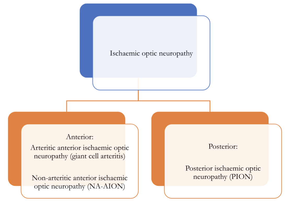
Ischaemic optic neuropathy is a significant cause of acute visual loss in those aged 50 years and over, affecting 2-10/100 000 people per year. It may be divided into anterior and posterior forms.
Giant Cell Arteritis (GCA)
GCA is a chronic granulomatous inflammation of medium-large-sized arteries originating from the arch of the aorta. Ocular involvement occurs in nearly 50%. Vision loss can be attributed to vasculitis affecting the posterior ciliary arteries, central retinal and/or the cilioretinal artery, and the ophthalmic artery respectively. It is important to understand the distinction between arteritic anterior ischaemic optic neuropathy and GCA: the latter is a systemic vasculitis, and ocular involvement is defined by the vision loss described above. Non-GCA causes of arteritic ischaemic optic neuropathy include ANCA-associated vasculitides.
Histopathology:
Early in the disease, the histopathological changes are confined to the region of the internal or external elastic lamina or adventitia. As the disease progresses, the wall of the artery of the artery becomes thickened by significant lymphocytic infiltrate. This reaction is directed against the internal elastic lamina. The vessel lumen may become obliterated by a marked fibroblastic proliferation in the intima. Multinucleated giant cells are present in only 50% of biopsies and are not a requirement for the diagnosis. Inflammation is often segmental, leading to skip lesions. Fibrinoid necrosis is not typical and indicates alternate diagnoses such as ANCA-associated vasculitis.
Clinical presentation:
Approximately 1/3ʳᵈ of patients with GCA will experience vision loss. This is instantaneous and profound (VA<6/60). Patients may experience a prior episode of amaurosis fugax. A brisk RAPD is evident. Fundus changes depend on the vascular territory involved:
Anterior ischaemic optic neuropathy (70-90%)
- Vasculitis of the short posterior ciliary arteries supplying the optic nerve head
- The optic disc displays a characteristic pallid swelling
- Swelling and pallor of the posterior pole: in approximately 1/3ʳᵈ of the population, the posterior ciliary arteries provide a cilioretinal artery branch which perfuses the posterior pole
Central retinal artery occlusion (15%)
- Inner retinal ischaemia produces a swollen, pale macula areas +/ a cherry-red spot
Posterior ischaemic optic neuropathy (5%)
- Vasculitis of the pial vessels and ophthalmic artery
- The optic nerve head appears normal during the early stages of disease
- Optic disc pallor manifests 6-8 weeks after the initial insult
Other key symptoms in GCA include constitutional symptoms (weight loss, fever, fatigue, anorexia, malaise), headache, scalp tenderness, and jaw claudication. The temporal arteries are often thick, nodular, pulseless and tender to palpation. GCA may also occur in a minority of patients with polymyalgia rheumatica, characterised by pain and stiffness of the shoulder and, less commonly, hip girdles. Approximately 20% of patients with vision loss will have no systemic symptoms.
Investigations:
Supportive laboratory investigations include marked elevation in acute inflammatory markers (ESR; CRP); normocytic, normochromic anaemia and mild thrombocytosis. ANCA and MP3 may be useful in excluding GPA and PAN. Patients with suspected GCA should have a prompt, confirmatory diagnostic test. This could be either:
- Temporal artery biopsy. This should be performed within 2 weeks to maximise diagnostic yield. A sample of at least 2cm is recommended to minimise a false-negative result due to skip lesions.
- Ultrasound. A hyperechoic ‘halo’ may be seen in the affected temporal artery. Ultrasound is user-dependent, with a sensitivity of ∼ 70% and specificity ∼87%. However, this drops markedly within one week of initiating corticosteroids.
Management:
Vision loss in giant cell arteritis is irreversible. Corticosteroids are the mainstay of treatment and should not be delayed pending a confirmatory test. A dose of 1mg/kg/day is continued until symptom resolution. There is usually a marked response within a week. Inflammatory markers should be monitored frequently; once normalised, corticosteroids may be cautiously tapered down. Most patients require at least a year of treatment. The role of intravenous corticosteroids (methylprednisolone 1g daily for three days) is uncertain. With established unilateral visual loss, many centres recommend intravenous treatment to protect the contralateral eye. However, there are no established studies demonstrating superiority of IVMP in preventing contralateral vision loss.
Tocilizumab (Subcutaneous injection 162 mg once weekly) is licensed for relapsed or remitting disease or cases intolerant of corticosteroids. IL-6 levels are significantly elevated in patients with GCA, and IL-6 is thought to play a central role in the pathogenesis of GCA by activation and recruitment of T-cells. There is no role for tocilizumab during the acute phase of disease.

Image Source: Wikimedia Commons (2017). Arteria temporalis superficialis, ramus frontalis, Licensed under a creative commons licence. Available for free at: https://commons.wikimedia.org/wiki/File:Arteria_temporalis_superficialis,_ramus_frontalis.jpg (Accessed on: 15 May 2022).

Image Source: Wikimedia Commons (2014). Giant cell arteritis — low mag, Licensed under a creative commons licence. Available for free at: https://commons.wikimedia.org/wiki/File:Giant_cell_arteritis_–_low_mag.jpg (Accessed on: 15 May 2022).
Non- Arteritic Ischaemic Optic Neuropathy (NA-AION)
NA-AION is attributed to relative hypoperfusion of the short posterior ciliary arteries supplying the optic nerve head. This results in oedema, ischaemia and infarction of the optic nerve fibres. Changes are often sectorial, affecting either the superior or inferior half of the optic nerve head.
Risk Factors:
Small optic nerve cup (‘disc at risk’) is the strongest risk factor for developing NA-ION. Cup-to-disc ratio is typically 0.2 or less. Optic nerve head oedema induced by ischemic injury is thought to cause axonal compression within a crowded scleral tunnel. Other notable associations include:
- optic disc drusen
- systemic hypertension
- diabetes mellitus
- obstructive sleep apnoea
- use of phosphodiesterase-5 inhibitors
Clinical presentation:
Vision loss is often painless and instantaneous. The following examination findings are present:
- VA: may be moderately-to-severely reduced (6/18 -(<6/60)
- Visual field: altitudinal visual field defect
- Colour vision: may or may not be affected
- Pupil examination: RAPD
- Optic nerve head: swollen; often sectorial and hyperaemic
Investigations:
All patients should have inflammatory markers (FBC, CRP, ESR) to exclude giant cell arteritis. Additionally, blood pressure should be documented.
Management:
Patients should have a full review of their medication history, with specific attention to the use of PDE-5 inhibitors and anti-hypertensive medications, especially the use of these medications at night. There is no accepted treatment for NA-AION. Corticosteroids are associated with an increased risk of second eye involvement. Aspirin does not aid in visual recovery nor reduce the risk to the contralateral eye. The Ischemic Optic Neuropathy Decompression Trial (IONDT) demonstrated optic nerve fenestration surgery is ineffective and potentially harmful.
Prognosis:
The Ischemic Optic Neuropathy Decompression Trial demonstrated a 40% improvement in visual acuity. The risk of second eye involvement is about 15% over 5 years. Poor baseline visual acuity and diabetes were risk factors for second eye involvement.
Posterior ischaemic optic neuropathy
Posterior ischaemic optic neuropathy affects the plial plexus, serving the retrolaminar optic nerve. The major risk factors are peri-operative hypotension and GCA. There may be unilateral or bilateral involvement. Visual acuity is often dramatically reduced. Initial examination reveals a RAPD but otherwise normal-appearing optic nerve head and fundus. Over 4-6 weeks, profound optic atrophy is revealed. All peri-operative patients complaining of visual loss should have a careful pupil examination. FBC, CRP and ESR should be performed in all patients >50 years of age. There are no treatment options.
Leber’s hereditary optic neuropathy
Leber’s hereditary optic neuropathy is a rare optic neuropathy of maternal inheritance arising from point mutations in mitochondrial DNA. Vision loss is attributed to free radical oxidate damage of the retinal ganglion cell layer. Patients may have concomitant cardiac abnormalities. There is often a family history of visual loss. The presentation is often a young male <50 years old with subacute bilateral, sequential vision loss (<6/60). Pupillary reactions are normal. Fundus examination may reveal peripapillary telangiectasia, arteriolar narrowing, optic disc oedema, or atrophy. Mitochondrial DNA analysis of a serum sample is required to confirm the diagnosis. An atypical optic neuropathy screen (see above) is recommended to exclude compressive and inflammatory optic neuropathies. An ECG should be performed for cardiac conduction defects associated with mitochondrial cytopathies. Patients should be advised to avoid smoking and avoid excess alcohol. Idebenone, a co-enzyme Q10 derivative, may be offered to maintain good residual vision. However, no clear benefit in final visual acuity has been demonstrated in randomised controlled trials.
References
- Age-Related Eye Disease Study Research Group. A randomised, placebo-controlled, clinical trial of high-dose supplementation with vitamins C and E and beta carotene for age-related cataract and vision loss: AREDS report no. 9. Arch Ophthalmol. 2001 Oct;119(10):1439-52.
- Aldrich EM, Lee AW, Chen CS, et al. Local intraarterial fibrinolysis administered in aliquots for the treatment of central retinal artery occlusion: the Johns Hopkins Hospital experience. Stroke 2008;39:1746–50.
- Beck RW, Cleary PA, Anderson MM, Keltner JL, Shults WT, Kaufman DI, Buckley EG, Corbett JJ, Kupersmith MJ, Miller NR. A randomised, controlled trial of corticosteroids in the treatment of acute optic neuritis. The Optic Neuritis Study Group. N Engl J Med. 1992 Feb 27;326(9):581-8.
- Beck RW, Cleary PA, Backlund JC. The course of visual recovery after optic neuritis. Experience of the Optic Neuritis Treatment Trial. Ophthalmology. 1994 Nov;101(11):1771-8.
- Beck RW, Cleary PA. Optic neuritis treatment trial. One-year follow-up results. Arch Ophthalmol. 1993 Jun;111(6):773-5.
- CATT Research Group. Martin DF, Maguire MG, Ying GS, Grunwald JE, Fine SL, Jaffe GJ. Ranibizumab and bevacizumab for neovascular age-related macular degeneration. N Engl J Med. 2011 19 May;364(20):1897-908.
- Cooper SA, Leddy SG, Skipper NT, et al. Optic neuritis with potential for poor outcome. Practical Neurology 2022;22:190-200.
- Hayreh SS, Podhajsky PA, Zimmerman B. Ocular manifestations of giant cell arteritis. Am J Ophthalmol 1998;125:509–20.
- Kertes PJ, Galic IJ, Greve M, Williams G, Baker J, Lahaie M, Sheidow T. Efficacy of a Treat-and-Extend Regimen With Ranibizumab in Patients With Neovascular Age-Related Macular Disease: A Randomised Clinical Trial. JAMA Ophthalmol. 2020 01 March;138(3):244-250.
- Klopstock T, Wai-Man Y, Dimitriadis K, et al. A randomised placebo-controlled trial of Idebenone in Leber’s Hereditary Optic Neuropathy. Brain 2011;134(Pt 9):2677-86.
- Nakamura M, Nakazawa T, Doi H, Hariya T, Omodaka K, Misu T, Takahashi T, Fujihara K, Nishida K. Early high-dose intravenous methylprednisolone is effective in preserving retinal nerve fiber layer thickness in patients with neuromyelitis optica. Graefes Arch Clin Exp Ophthalmol. 2010 Dec;248(12):1777-85.
- Park K, Tanaka K, Tanaka M. Uhthoff’s phenomenon in multiple sclerosis and neuromyelitis optica. Eur Neurol. 2014;72(3-4):153-6. doi: 10.1159/000361045. Epub 2014 28 August. PMID: 25195501.
- Petzold A, Braithwaite T, van Oosten BW, et al. Case for a new corticosteroid treatment trial in optic neuritis: review of updated evidence. J Neurol Neurosurg Psychiatry 2020;91:9–14.
- Schmidt-Erfurth U, Eldem B, Guymer R, Korobelnik JF, Schlingemann RO, Axer-Siegel R, Wiedemann P, Simader C, Gekkieva M, Weichselberger A., EXCITE Study Group. Efficacy and safety of monthly versus quarterly ranibizumab treatment in neovascular age-related macular degeneration: the EXCITE study. Ophthalmology. 2011 May;118(5):831-9.
- Schmidt-Erfurth U, Kaiser PK, Korobelnik JF, Brown DM, Chong V, Nguyen QD, Ho AC, Ogura Y, Simader C, Jaffe GJ, Slakter JS, Yancopoulos GD, Stahl N, Vitti R, Berliner AJ, Soo Y, Anderesi M, Sowade O, Zeitz O, Norenberg C, Sandbrink R, Heier JS. Intravitreal aflibercept injection for neovascular age-related macular degeneration: ninety-six-week results of the VIEW studies. Ophthalmology. 2014 Jan;121(1):193-201.
- Sivaprasad, S., Amoaku, W., Hykin, P. et al. The Royal College of Ophthalmologists Guidelines on retinal vein occlusions: executive summary. Eye 29, 1633–1638 (2015).
- Snodderly DM. Evidence for protection against age-related macular degeneration by carotenoids and antioxidant vitamins. Am J Clin Nutr. 1995 Dec;62(6 Suppl):1448S-1461S.
- Stiebel-Kalish H, Hellmann MA, Mimouni M, et al. Does time equal vision in the acute treatment of a cohort of AQP4 and MOG optic neuritis? Neurol Neuroimmunol Neuroinflamm Jul 2019, 6 (4) e572.
- Varma DD, Cugati S, Lee AW, et al. A review of central retinal artery occlusion: clinical presentation and management. Eye 2013;27:688–97.
Complete Guide to Clinical Genetics ST3 Application and Recruitment (2027)
Written by Medibuddy. Last updated: October 2026. Clinical Genetics is

Complete Guide to Clinical Radiology ST3 Application and Recruitment (2027)
Recruitment paused: NHS England is not running Clinical Radiology ST3+ recru

Complete Guide to Paediatric Surgery ST3 Application and Recruitment in United Kingdom (2026)
Written by Medibuddy. Last updated: October 2026. If you’re planning


Take your subscriptions with you
Our mobile app allows you to access your interview and exam question banks wherever you are.

