


 Close
Close
 17th January 2024
Pulmonary Embolism
Background
Epidemiology
Pulmonary embolism (PE) is the third most common cardiovascular disease, with an incidence of approximately 60-70 per 100,000 people (1). The vast majority occur provoked, with common risk factors including deep venous thrombosis (DVT) elsewhere, underlying active malignancy, prolonged immobility, recent surgery, lower limb trauma, inherited and acquired thrombophilias, use of the combined oral contraceptive pill (COCP), pregnancy and obesity.
Pathophysiology
Pulmonary embolism (PE) is a form of venous thromboembolism (VTE) affecting the pulmonary arterial system and can be either an acute or chronic problem associated with the risk factors detailed above. It most commonly occurs when emboli from elsewhere in the deep venous system travel to the pulmonary vasculature and become lodged in a pulmonary artery. This results in a ventilation-perfusion mismatch, as healthy lung tissue is insufficiently perfused with oxygenated blood and can cause progression to pulmonary infarction. Less commonly, non-thrombotic sources of emboli can arise, including air emboli, fat emboli, septic emboli and, in pregnant women, amniotic fluid emboli. In severe cases, PE can be associated with hypotension, syncope and sudden death, as pulmonary arterial pressure increases to the point where it causes right ventricular failure and compromises cardiac output.
Presentation
Clinical features of pulmonary embolism most commonly include tachypnoea, pleuritic chest pain, tachycardia, cough and haemoptysis. Features of the underlying cause are often present, in particular, unilateral lower limb pain and swelling in the case of DVT. Symptoms indicating severe illness include pre-syncope and syncope. Symptoms and signs on examination are important to elicit if present, as they form the basis of several scoring systems, such as the Wells’ score and PERC rule (see diagnosis section).
Common examination signs in PE (2):
- Tachycardia >100bpm
- Tachypnoea
- Hypoxia
- Hypotension
- Elevated jugular venous pressure (JVP)
- Loud P2
- Pleural rub
Diagnosis
PE should be part of the differential diagnosis in any adult presenting with chest pain, although it cannot be diagnosed on the basis of clinical history and examination alone. The diagnostic pathway followed depends on pre-test clinical probability; several clinical scoring tools have been developed to aid in this diagnostic process.
Clinical scoring tools
Wells’ score for PE
A risk stratification tool that determines recommended further investigations when used in the presence of clinical suspicion of PE. A score of >4 indicates that PE is likely and the clinician should proceed directly to CT pulmonary angiogram (CTPA), whilst a score of 4 or less suggests PE is less likely and that a d-dimer blood test should first be performed. If the d-dimer result is then negative, PE can be ruled out, whilst a positive d-dimer result would indicate further investigation +/- treatment is required.
Table 1 – Wells’ score for PE.
| Criteria | Points if present |
|---|---|
| Clinical signs and symptoms of DVT | 3 |
| PE is the number one diagnosis OR equally likely | 3 |
| Heart rate >100 | 1.5 |
| Immobilisation for at least 3 days OR surgery in the previous 4 weeks | 1.5 |
| Previous, objectively diagnosed PE or DVT | 1.5 |
| Haemoptysis | 1 |
| Malignancy within 6 months of treatment OR palliative | 1 |
Pulmonary Embolism rule-out criteria (PERC score)
The PERC score is strictly a rule-out criteria in patients who have a low pre-test probability of PE. When used in the correct patient group, a PERC score of 0 indicates the clinician can safely rule out a PE without the need for further investigations. Scoring even a single point means the PERC rule cannot be used to rule out PE, but importantly, it cannot be used to make a positive diagnosis in itself.
Table 2 – PERC rules.
| Age ≥ 50 | 1 |
| Heart rate ≥ 100 bpm | 1 |
| Oxygen saturations on room air <95% | 1 |
| Unilateral leg swelling | 1 |
| Haemoptysis | 1 |
| Recent surgery or trauma requiring general anaesthesia ≤ 4 weeks ago | 1 |
| Prior PE or DVT | 1 |
| Hormone use | 1 |
Table 1 Source: Wells’ Criteria for DVT – MDCalc [Internet]. Available from: https://www.mdcalc.com/wells-criteria-dvt
Table 2 Source: PERC Rule for Pulmonary Embolism – MDCalc [Internet]. Available from: https://www.mdcalc.com/perc-rule-pulmonary-embolism
Investigations
- Blood tests
- Full blood count (FBC)
- Clotting screen
- Urea and electrolytes (U&Es) – important in particular when considering whether the patient is fit for a CTPA with contrast
- D-dimer – only to be used in patients with a low pre-test probability
- Troponin – to help risk stratify
- Arterial blood gas sampling – look for hypoxia and hypocapnia
- Electrocardiogram (ECG) – features include:
- Sinus tachycardia
- Right bundle branch block (RBBB)
- S1Q3T3 – Deep S-waves in lead I, pathological Q-waves in lead III, inverted T-waves in lead III
- T-wave inversion in V1-V3 (right ventricular strain pattern)
- P pulmonale
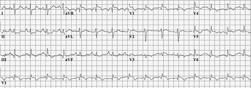
Image Source: Wikimedia Commons (2010). Pulm_embolism, Licensed under a creative commons licence. Available for free at: https://commons.wikimedia.org/wiki/File:Pulm_embolism.jpg (Accessed: 5 June 2022).
- Imaging
- Chest x-ray (CXR)– useful to exclude alternative diagnoses, and a normal CXR is also a prerequisite for interpreting a ventilation/perfusion (V/Q) scan.
- CT pulmonary angiogram (CTPA) – the main imaging modality of choice to look for filling defects associated with large or subsegmental PEs.
- Ultrasound doppler – not a direct diagnostic test for PE, but sometimes used to identify lower limb DVTs in patients where ionising radiation is contraindicated (i.e., in pregnancy). If a VTE elsewhere is identified, and the patient has the classical signs and symptoms, treatment can be started empirically.
- Ventilation/perfusion (V/Q) scanning – imaging using radioisotopes to identify any areas of mismatch in ventilation and perfusion of the lungs.
- Transthoracic echocardiogram – not part of the formal diagnostic pathway, but can be used as adjunctive imaging to identify features of right heart strain, such as right ventricular dilatation and a poorly compressible internal vena cava (IVC) (3).

Image Source: Wikimedia Commons (2007). Pulmonary embolism CTPA, Licensed under a creative commons licence. Available for free at: https://commons.wikimedia.org/wiki/File:Pulmonary_embolism_CTPA.JPEG (Accessed: 5 June 2022).

Differential diagnoses
Pulmonary embolism is a life-threatening diagnosis which is important not to miss. There is a broad differential for both chest pain and shortness of breath, and symptoms and signs can be shared with a multitude of other conditions. We will focus mainly on other diagnoses; it would be important to differentiate from PE due to comparable clinical urgency.
Acute coronary syndrome
Acute coronary syndrome is a term that encompasses patients presenting with unstable angina, non-ST-elevation and ST-elevation myocardial infarction. Classical symptoms include chest pain or tightness, shortness of breath, diaphoresis, nausea, vomiting and dizziness. PE shares a lot of similar features, although generally has a greater association with hypoxia and tachycardia, and the nature of the chest pain is classically described as more pleuritic. ECG would be a mainstay of differentiating between the two diagnoses – if this raised significant clinical suspicion of a myocardial infarction, the patient would need to be taken for urgent cardiac catheterisation. Blood tests can help guide diagnosis, but caution should be used when relying on biochemical markers alone, as troponin and d-dimer can be deceptively raised in both conditions.
Pneumothorax
Primary, secondary and tension pneumothoraces can all present with sudden onset pleuritic chest pain and shortness of breath. A past medical history of pre-existing airways disease may point towards a diagnosis of secondary pneumothorax, but overall, few features in the history alone can differentiate between pneumothorax and pulmonary embolism. On examination, the most notable feature would be reduced breath sounds and hyperresonance to percussion on the side of a pneumothorax, as well as tracheal deviation in the case of a tension pneumothorax. Comparably, air entry should sound normal in a PE. In many cases, definitive treatment will involve chest drain insertion, so early access to chest radiograph imaging for diagnosis is essential.
Pericarditis
Pericarditis classically presents with chest pain relieved on leaning forwards, shortness of breath and sometimes dizziness. Similarly to a PE, it can present in a young population in the absence of a clear trigger. Features that raise suspicion of pericarditis would include a clear history of recent viral infection, co-existing inflammatory disorders such as rheumatoid arthritis or a fever at presentation itself. ECG features also include widespread saddle-shaped ST-elevation. It is important to keep in the differential as the life-threatening complication of pericardial effusion can occur.
Aortic dissection
Aortic dissection is a medical emergency that causes sudden onset, severe, tearing chest pain that often radiates to the back. Classical features on examination include a wide pulse pressure and radial-radial or radial-femoral delay. Definitive diagnosis can only be made by CT aortogram imaging, so a high index of suspicion must be maintained when reviewing any patient presenting with chest pain, particularly patients with a past medical history of aortic aneurysm or hypertension.
Other disorders
As shortness of breath, pleuritic chest pain, cough and haemoptysis are common presentations associated with a range of clinical conditions, the differential must be kept broad and guided by findings on history and examination. Any respiratory disorder such as pneumonia, lung cancer or pleural effusion could potentially be a mimic of a PE. Equally, musculoskeletal causes of chest pain, such as costochondritis or Tietze’s syndrome, can commonly produce pain of a similar nature, but these must remain a diagnosis of exclusion (4).
Management
The first step of managing an acute PE involves a similar approach to any other acute medical scenario by assessing the patient in an ABCDE fashion, identifying and treating features such as hypoxia and hypotension. Once the patient has been stabilised, it is important to risk stratify patients into low, intermediate and high-risk categories. The Pulmonary Embolism Severity Index (PESI) and, later, the simplified PESI (sPESI) have been developed to help clearly delineate risk by calculating 30-day mortality. This should be calculated for any patient presenting with a confirmed PE, unless there is clear evidence of haemodynamic compromise on presentation, in which case a medical emergency algorithm approach should be taken to proceed immediately to thrombolysis. An sPESI score of one or more indicates the patient is at least intermediate risk and should be managed as an inpatient. If the sPESI score is zero, in some scenarios, the patient can be managed on an ambulatory basis (5).
High-risk features in PE:
- Haemodynamic instability – persistent hypotension <90mmHg or a systolic drop of ≥40mmHg lasting for 15 or more minutes, not caused by new-onset arrhythmia, hypovolaemia or sepsis.
- sPESI score of ≥1
- Features of right ventricular dysfunction on TTE or CTPA
- Elevation of cardiac troponin levels
Table 3 – sPESI score.
| Criteria | Score |
|---|---|
| Age >80 | 1 |
| History of cancer | 1 |
| History of chronic cardiopulmonary disease | 1 |
| Heart rate ≥ 110bpm | 1 |
| Systolic blood pressure <100mmHg | 1 |
| O2 saturation <90% | 1 |
Table 3. Source: Jimenez D. Simplified PESI (Pulmonary Embolism Severity Index) – MDCalc [Internet]. Mdcalc.com. Available from: https://www.mdcalc.com/simplified-pesi-pulmonary-embolism-severity-index#use-cases
Pharmacological Management
Anticoagulation
The mainstay of treatment for PE is early anticoagulation. When there is a high clinical suspicion, anticoagulation should be started even prior to confirmation of PE on diagnostic tests. Most commonly, the anticoagulant of choice is once-daily subcutaneous low molecular weight heparin (LMWH) or fondaparinux at a weight-adjusted therapeutic dose. Alternatively, there is also now good evidence for oral anticoagulation with a non-vitamin K oral anticoagulant (NOAC) agent, such as apixaban or rivaroxaban. In the case of poor renal function or significant bleeding risk, unfractionated heparin can be used, although this is associated with a higher risk of heparin-induced thrombocytopenia.
Warfarin monotherapy is not used in the acute setting, as it normally requires several days to reach the required therapeutic range for international normalised ratio (INR). When it is being used as a long-term anticoagulation agent, concurrent use of a LMWH must be maintained until the INR is within the therapeutic range of 2.0-3.0. Following the acute period, all patients will need a period of long-term anticoagulation to prevent recurrence. Most patients can be converted to NOAC treatment, but for patients with active malignancy, guidelines recommend remaining on LMWH, which has been found to be significantly more efficacious at reducing recurrence of VTE than oral agents (6).
| Clinical scenario | Recommended long-term anticoagulation |
|---|---|
| Provoked PE (defined as having a transient major risk factor such as major surgery, recent trauma or confined to hospital bed for ≥3 days with an acute illness) | 3 months |
| Unprovoked or recurrent PE | Consider continuing anticoagulation beyond 3 months based on risk/benefit profile. |
| PE in a patient with non-transient risk factors such as active cancer or antiphospholipid antibody syndrome | Continue anticoagulation for at least 6 months and consider lifelong. |
Table 4 – Recommendations for anticoagulation in PE
Reperfusion treatment
- Systemic thrombolysis – for patients with features of haemodynamic compromise as defined above (‘massive PE’), intravenous thrombolysis can be undertaken with agents such as recombinant tissue-type plasminogen activator (alteplase) or streptokinase, in the absence of major bleeding risk factors.
- Catheter-directed thrombolysis – there is growing use of targeted thrombolysis therapy and mechanical thrombectomy using percutaneous approach via pulmonary artery catheterisation.
- Surgical embolectomy – open embolectomy of the pulmonary artery clots requiring cardio-pulmonary bypass (7).
Supportive management
- Respiratory support is often required in PE, ranging from nasal cannulae and simple face mask oxygen support to invasive ventilation and, rarely, extracorporeal membrane oxygenation (ECMO).
- Haemodynamic support – intravenous fluids, and in some cases, vasopressor and inotropic support.
- Multidisciplinary pulmonary embolism response teams (PERTs) – there is a growing use of MDTs comprising members from various disciplines, including cardiology, haematology and interventional radiology, amongst others, to make clinical decisions on optimal PE management on an individual basis.
Reperfusion treatment
- Systemic thrombolysis – for patients with features of haemodynamic compromise as defined above (‘massive PE’), intravenous thrombolysis can be undertaken with agents such as recombinant tissue-type plasminogen activator (alteplase) or streptokinase, in the absence of major bleeding risk factors.
- Catheter-directed thrombolysis – there is growing use of targeted thrombolysis therapy and mechanical thrombectomy using percutaneous approach via pulmonary artery catheterisation.
- Surgical embolectomy – open embolectomy of the pulmonary artery clots requiring cardio-pulmonary bypass (7).
Supportive management
- Respiratory support is often required in PE, ranging from nasal cannulae and simple face mask oxygen support to invasive ventilation and, rarely, extracorporeal membrane oxygenation (ECMO).
- Haemodynamic support – intravenous fluids, and in some cases, vasopressor and inotropic support.
- Multidisciplinary pulmonary embolism response teams (PERTs) – there is a growing use of MDTs comprising members from various disciplines, including cardiology, haematology and interventional radiology, amongst others, to make clinical decisions on optimal PE management on an individual basis.
Internal vena cava filters
In patients where anticoagulation in PE is absolutely contraindicated, insertion of an internal vena cava (IVC) filter can be considered. Theoretically, these prevent recurrent PEs from occurring by mechanically blocking the travel of potential venous thromboemboli into the proximal circulation. Whilst they do decrease risk of recurrent PE, they are actually associated with a significantly increased risk of DVT, likely due to instrumentation in the absence of anticoagulation, and thus have no mortality benefit overall.
References
- Pathophysiology | Background information | Pulmonary embolism | CKS | NICE [Internet]. Cks.nice.org.uk. 2022 [cited 4 March 2022]. Available from: https://cks.nice.org.uk/topics/pulmonary-embolism/background-information/pathophysiology/
- Hall T. PACES for the MRCP. Saint Louis: Elsevier Health Sciences UK; 2014.
- Elyas A. [Internet]. Escardio.org. 2022 [cited 4 April 2022]. Available from: https://www.escardio.org/static-file/Escardio/Subspecialty/EACVI/Education/Teaching%20courses/2019/Use%20of%20echocardiography%20in%20diagnosis%20of%20pulmonary%20embolism.pdf
- Thompson B, Kabhrel C, Pena C. UpToDate [Internet]. Uptodate.com. 2022 [cited 4 June 2022]. Available from: https://www.uptodate.com/contents/clinical-presentation-evaluation-and-diagnosis-of-the-nonpregnant-adult-with-suspected-acute-pulmonary-embolism#H746822394
- Tapson V, Weinberg A. UpToDate [Internet]. Uptodate.com. 2022 [cited 4 April 2022]. Available from: https://www.uptodate.com/contents/treatment-prognosis-and-follow-up-of-acute-pulmonary-embolism-in-adults
- Provoked versus unprovoked pulmonary embolism (PE) or deep vein thrombosis (DVT) – General Practice Notebook [Internet]. Gpnotebook.com. 2018 [cited 4 April 2022]. Available from: https://gpnotebook.com/en-gb/simplepage.cfm?ID=x20120725074001799563
- Konstantinides S, Meyer G, Becattini C, Bueno H, Geersing G, Harjola V et al. 2019 ESC Guidelines for the diagnosis and management of acute pulmonary embolism developed in collaboration with the European Respiratory Society (ERS). European Heart Journal. 2019;41(4):543-603.
Complete Guide to Clinical Genetics ST3 Application and Recruitment (2027)
Written by Medibuddy. Last updated: October 2026. Clinical Genetics is

Complete Guide to Clinical Radiology ST3 Application and Recruitment (2027)
Recruitment paused: NHS England is not running Clinical Radiology ST3+ recru

Complete Guide to Paediatric Surgery ST3 Application and Recruitment in United Kingdom (2026)
Written by Medibuddy. Last updated: October 2026. If you’re planning


Take your subscriptions with you
Our mobile app allows you to access your interview and exam question banks wherever you are.

