


 Close
Close
 17th January 2024
Clinical scoring tools
Wells’ score for PE
A risk stratification tool that determines recommended further investigations when used in the presence of clinical suspicion of PE. A score of >4 indicates that PE is likely and the clinician should proceed directly to CT pulmonary angiogram (CTPA), whilst a score of 4 or less suggests PE is less likely and that a d-dimer blood test should first be performed. If the d-dimer result is then negative, PE can be ruled out, whilst a positive d-dimer result would indicate further investigation +/- treatment is required.
Table 1 – Wells’ score for PE.
| Criteria | Points if present |
|---|---|
| Clinical signs and symptoms of DVT | 3 |
| PE is the number one diagnosis OR equally likely | 3 |
| Heart rate >100 | 1.5 |
| Immobilisation for at least 3 days OR surgery in the previous 4 weeks | 1.5 |
| Previous, objectively diagnosed PE or DVT | 1.5 |
| Haemoptysis | 1 |
| Malignancy within 6 months of treatment OR palliative | 1 |
Pulmonary Embolism rule-out criteria (PERC score)
The PERC score is strictly a rule-out criteria in patients who have a low pre-test probability of PE. When used in the correct patient group, a PERC score of 0 indicates the clinician can safely rule out a PE without the need for further investigations. Scoring even a single point means the PERC rule cannot be used to rule out PE, but importantly, it cannot be used to make a positive diagnosis in itself.
Table 2 – PERC rules.
| Age ≥ 50 | 1 |
| Heart rate ≥ 100 bpm | 1 |
| Oxygen saturations on room air <95% | 1 |
| Unilateral leg swelling | 1 |
| Haemoptysis | 1 |
| Recent surgery or trauma requiring general anaesthesia ≤ 4 weeks ago | 1 |
| Prior PE or DVT | 1 |
| Hormone use | 1 |
Table 1 Source: Wells’ Criteria for DVT – MDCalc [Internet]. Available from: https://www.mdcalc.com/wells-criteria-dvt
Table 2 Source: PERC Rule for Pulmonary Embolism – MDCalc [Internet]. Available from: https://www.mdcalc.com/perc-rule-pulmonary-embolism
Investigations
- Blood tests
- Full blood count (FBC)
- Clotting screen
- Urea and electrolytes (U&Es) – important in particular when considering whether the patient is fit for a CTPA with contrast
- D-dimer – only to be used in patients with a low pre-test probability
- Troponin – to help risk stratify
- Arterial blood gas sampling – look for hypoxia and hypocapnia
- Electrocardiogram (ECG) – features include:
- Sinus tachycardia
- Right bundle branch block (RBBB)
- S1Q3T3 – Deep S-waves in lead I, pathological Q-waves in lead III, inverted T-waves in lead III
- T-wave inversion in V1-V3 (right ventricular strain pattern)
- P pulmonale
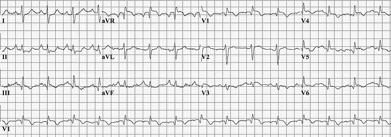
Image Source: Wikimedia Commons (2010). Pulm_embolism, Licensed under a creative commons licence. Available for free at: https://commons.wikimedia.org/wiki/File:Pulm_embolism.jpg (Accessed: 5 June 2022).
- Imaging
- Chest x-ray (CXR)– useful to exclude alternative diagnoses, and a normal CXR is also a prerequisite for interpreting a ventilation/perfusion (V/Q) scan.
- CT pulmonary angiogram (CTPA) – the main imaging modality of choice to look for filling defects associated with large or subsegmental PEs.
- Ultrasound doppler – not a direct diagnostic test for PE, but sometimes used to identify lower limb DVTs in patients where ionising radiation is contraindicated (i.e., in pregnancy). If a VTE elsewhere is identified, and the patient has the classical signs and symptoms, treatment can be started empirically.
- Ventilation/perfusion (V/Q) scanning – imaging using radioisotopes to identify any areas of mismatch in ventilation and perfusion of the lungs.
- Transthoracic echocardiogram – not part of the formal diagnostic pathway, but can be used as adjunctive imaging to identify features of right heart strain, such as right ventricular dilatation and a poorly compressible internal vena cava (IVC) (3).
A CTPA demonstrating a saddle pulmonary embolus with thrombus visualised below in pulmonary arteries bilaterally. Myat A, Ahsan A
Image Source: Wikimedia Commons (2007). Pulmonary embolism CTPA, Licensed under a creative commons licence. Available for free at: https://commons.wikimedia.org/wiki/File:Pulmonary_embolism_CTPA.JPEG (Accessed: 5 June 2022).
- Imaging
- Chest x-ray (CXR)– useful to exclude alternative diagnoses, and a normal CXR is also a prerequisite for interpreting a ventilation/perfusion (V/Q) scan.
- CT pulmonary angiogram (CTPA) – the main imaging modality of choice to look for filling defects associated with large or subsegmental PEs.
- Ultrasound doppler – not a direct diagnostic test for PE, but sometimes used to identify lower limb DVTs in patients where ionising radiation is contraindicated (i.e., in pregnancy). If a VTE elsewhere is identified, and the patient has the classical signs and symptoms, treatment can be started empirically.
- Ventilation/perfusion (V/Q) scanning – imaging using radioisotopes to identify any areas of mismatch in ventilation and perfusion of the lungs.
- Transthoracic echocardiogram – not part of the formal diagnostic pathway, but can be used as adjunctive imaging to identify features of right heart strain, such as right ventricular dilatation and a poorly compressible internal vena cava (IVC) (3).

Image Source: Wikimedia Commons (2007). Pulmonary embolism CTPA, Licensed under a creative commons licence. Available for free at: https://commons.wikimedia.org/wiki/File:Pulmonary_embolism_CTPA.JPEG (Accessed: 5 June 2022).
Complete Guide to Clinical Genetics ST3 Application and Recruitment (2027)
Written by Medibuddy. Last updated: October 2026. Clinical Genetics is

Complete Guide to Clinical Radiology ST3 Application and Recruitment (2027)
Recruitment paused: NHS England is not running Clinical Radiology ST3+ recru

Complete Guide to Paediatric Surgery ST3 Application and Recruitment in United Kingdom (2026)
Written by Medibuddy. Last updated: October 2026. If you’re planning


Take your subscriptions with you
Our mobile app allows you to access your interview and exam question banks wherever you are.

