


 Close
Close
 10th July 2023
Miscellaneous
Skin in Systemic Disease
Respiratory
Sarcoidosis
Skin manifestations are seen in a quarter of those with systemic sarcoid, sometimes in the absence of systemic disease. Cutaneous manifestations include erythema nodosum, lupus pernio (see image below), scar sarcoid and scarring alopecia.

Lupus pernio
Image Source: Wikimedia Commons (2011). Lupus pernio 01, Licensed under a creative commons licence. Available for free at: https://commons.wikimedia.org/wiki/File:Lupus_pernio_01.jpg (Accessed on: 16 July 2022).

Tuberculosis
Tuberculosis is associated with scrofuloderma (breakdown of skin overlying TB focus), lupus vulgaris (50%), verrucosa cutis, scarring alopecia, and gumma.
Metabolic Disorders
The Porphyrias
The classic skin manifestations of porphyrias are blister formation, photosensitivity, scarring with milia and hypertrichosis.
| Type | Description |
|---|---|
| Porphyria cutanea tarda |
i.e., alcohol, oestrogens, hepatitis C, iron, and lead
|
| Acute intermittent porphyria |
|
| Congenital erythropoietic porphyria |
|
Table 1: Types of porphyrias
Diabetes
Diabetes can often present with a wide range of cutaneous signs, such as necrobiosis lipoidica, granuloma annulare, diabetic rubeosis, diabetic dermopathy, candidiasis, vitiligo, and neuropathic foot ulcers.
- Diabetic dermopathy presents with dark red papules that eventually become atrophic and is associated with a long duration of diabetes mellitus.
- Necrobiosis lipoidica (waxy yellow lesions) mainly affects the shins and is associated with telangiectasia
- Granuloma annulare present as papules with central depression.
Acanthosis nigricans
Often described as dark, thick, velvety patches that present in skin folds, namely the underarms, neck, and groin. It often indicates an underlying health concern, such as internal malignancy, acromegaly, Cushing syndrome, obesity, insulin resistant diabetes or hypothyroidism. It can also be seen in those that take the oral contraceptive pill. Acanthosis nigricans can be familial.
Thyroid disease
| Hypothyroidism |
|
| Hyperthyroidism |
|
Table 2: Thyroid disease and its cutaneous manifestations
Rheumatology
Rheumatoid arthritis
Specific cutaneous changes include:
- Rheumatoid nodules
- Vasculitis causing changes such as nail fold infarcts and ulcers
- Pyoderma gangrenosum
Dermatomyositis
Skin manifestations include the classic heliotrope rash around the eyes, Gottron papules (red plaques on the extensor surfaces of finger joints), Gottron sign (erythema over knees and elbows), dilated nail fold capillaries, ragged cuticles, and nail fold infarcts. Please note that when a patient presents with dermatomyositis, this warrants an investigation for an underlying malignancy, namely bronchial, breast and ovarian.
Lupus Erythematosus
- Discoid lupus erythematosus is associated with scaly erythematous plaques with follicular plugging on sun-exposed areas of the skin. They tend to heal with scarring.
- Systemic lupus erythematosus (SLE) is commonly associated with cutaneous manifestations such as a malar rash (facial butterfly rash with purple hue), photosensitivity, vasculitis, Raynaud’s phenomenon, alopecia, and oropharyngeal ulceration.
Paraneoplastic Cutaneous Manifestations
Dermatological signs can be seen in various types of malignancy, with some being more common in certain types of neoplasia.
| Malignancy | Cutaneous manifestation |
|---|---|
| Gastrointestinal adenocarcinoma | Acanthosis nigricans
Acanthosis palmaris |
| Bronchial carcinoma | Erythema gyratum repens (wood grain pattern)
Acanthosis palmaris (tripe palms) Clubbing |
| Lymphoma | Acquired ichthyosis
Erythroderma (also seen in leukaemia) Generalised pruritus |
| Glucagonoma | Necrolytic migratory erythema |
| Pancreatic carcinoma | Migratory thrombophlebitis |
| Myeloproliferative tumours | Pyoderma gangrenosum
Sweet’s syndrome (febrile neutrophilic dermatoses) Herpes zoster |
| Oesophageal carcinoma | Tylosis |
| T cell lymphoma | Mycosis Fungoides |
Table 3: Types of cutaneous manifestations in internal malignancies
Pyoderma Gangrenosum (PG)
PG presents clinically as painful, deep red necrotic ulcerating lesions with a violaceous border that mainly affects the lower extremities. Underlying medical disease is noted in approximately 50% of these patients, namely inflammatory bowel disease, rheumatoid arthritis, SLE and myeloid leukaemia. PG can characteristically cause peristomal complications in inflammatory bowel disease. Treatment involves oral steroid supplementation. PG is not typically associated with diabetes.
| System | Medical Conditions associated with PG |
|---|---|
| Gastroenterology | Ulcerative colitis
Crohn’s colitis |
| Hepatology | Chronic active hepatitis
Primary biliary cirrhosis Sclerosing cholangitis |
| Rheumatology | Rheumatoid arthritis
Ankylosing spondylitis |
| Haematology | Lymphoma
Leukaemia Myeloproliferative disorders |
| Other | Diabetes mellitus
Thyroid disease Sarcoidosis Wegener’s granulomatosis Other malignancies |
Table 4: Medical conditions associated with PG

Pyoderma gangrenosum
Image Source: Wikimedia Commons (2008). Crohnie Pyoderma gangrenosum, Licensed under a creative commons licence. Available for free at: https://commons.wikimedia.org/wiki/File:Crohnie_Pyoderma_gangrenosum.jpg (Accessed on: 16 July 2022).

Gastrointestinal disorders
Hepatology
| Viral hepatitis | Common skin symptoms associated with hepatitis include jaundice, generalised pruritus, acute urticaria, transient exanthems, erythema multiforme, and erythema nodosum.
Skin conditions associated with hepatitis B and C include cryoglobulinemia types II and III (cutaneous vasculitis), lichen planus, cutaneous squamous cell carcinomas, papular acrodermatitis and dermatomyositis. |
| Liver cirrhosis
Portal hypertension |
Common skin symptoms associated with liver cirrhosis include xerosis, pruritus, nodular prurigo, ecchymosis, petechiae, purpura, palmar erythema, spider naevi, onycholysis, leukonychia, loss of pubic and armpit hair, and caput medusae. |
Table 5: Cutaneous manifestations of liver disease
Malnutrition
Please refer to the topic ‘Nutrition’ in the Gastroenterology section for more detail.
Inherited Skin Disease
There are a multitude of genetic conditions that have associated skin manifestations (see table).
| Condition | Description |
|---|---|
| Ataxia Telangiectasia | Autosomal recessive
Mucocutaneous telangiectasia Increased risk of lymphoma and leukaemia |
| Cowden Syndrome | Trichilemmomas
Warty hyperplasia of mucosal surfaces Associated with breast and thyroid carcinoma |
| Darier disease | Autosomal dominant
Scaly crusted papules in a seborrhoeic distribution ATP2A2 |
| Gardner syndrome | Epidermal cysts, lipomas, and fibromas
Associated with colonic carcinoma |
| Gorlin syndrome | Basal cell naevus syndrome
Multiple basal cell carcinomas Associated with brain tumours and ovarian cancer |
| Hereditary haemorrhagic telangiectasia | Autosomal dominant
20% spontaneous Diagnostic criteria include telangiectasias, visceral bleeding, epistaxis, AV malformations, and first-degree relative involvement. |
| Howel-Evans syndrome | Tylosis
Associated with oesophageal carcinoma |
| LEOPARD syndrome | L – lentigines (multiple brown-black spots on the skin)
E – electrocardiographic (ECG) conduction defects O – ocular hypertelorism P – pulmonary stenosis A – abnormalities of genitals R – retarded growth resulting in short stature D – deafness or hearing loss due to inner ear malfunction |
| Mild inherited ichthyosis | Autosomal dominant
Incomplete penetrance |
| Neurofibromatosis | 6 or more café-au-lait macules
Axillary freckling Neurofibromas Associated with malignant schwannomas and astrocytoma |
| Peutz-Jeghers syndrome | Mucocutaneous pigmentation (especially in the perioral region)
Associated with gastrointestinal malignancy |
| Sturge-Weber syndrome | Port wine stain
Associated with vascular meningeal malformation and epilepsy |
| Tuberous sclerosis | Angiofibromas
Periungual fibromas Shagreen patches Ash leaf macules Associated with sarcomas and rhabdomyomas |
| Von hippel Lindau syndrome | Café-au-lait macules
Haemangiomas Associated with vascular tumours, phaeochromocytomas, renal and pancreatic carcinoma |
| Wiskott-Aldrich syndrome | X-linked inheritance
Eczema Immunodeficiency Increased risk of lymphoma and leukaemia |
Table 6: Inherited conditions with dermatological manifestations.
Hair and Nail Disorders
Hair Disorders
Hypertrichosis and Hirsutism
Excessive hair growth can be androgen-independent (hypertrichosis) or androgen-dependent (hirsutism – excess hair growth in a male pattern that occurs in women).
| Causes of Hypertrichosis | Causes of Hirsutism |
|---|---|
| Hereditary: Congenital hypertrichosis lanuginosa Epidermolysis bullosa Hurler syndromeAcquired: Acquired hypertrichosis lanuginosa Thyroid disease Medication: diazoxide, minoxidil, ciclosporin, streptomycin Malnutrition Anorexia nervosaHereditary/Acquired: Porphyria, e.g. PCT |
Polycystic ovary syndrome
Ovarian tumours |
Table 7: Causes of hypertrichosis and hirsutism
Treatment options can include physical hair removal or medication such as the combined oral contraceptive pill (that contains anti-androgenic cyproterone acetate), spironolactone, finasteride and topical eflornithine.
Scarring alopecia
Scarring alopecia is hair loss with destruction of hair follicles. Causes include ichthyosis, tuberculosis, syphilis, burns, radiotherapy, kerion, lichen planus, discoid lupus, morphoea, sarcoidosis and cicatricial pemphigoid.
Non scarring alopecia
Non scarring alopecia is hair loss with preservation of hair follicles. Alopecia areata often presents as the classic ‘exclamation mark’ hair. It is often associated with nail dystrophy, cataracts, and other autoimmune diseases such as vitiligo, autoimmune thyroid disease, pernicious anaemia, and Addison’s disease. 50% of patients have hair growth within 1 year. First line treatment includes topical corticosteroids.
Other causes of non-scarring alopecia include male pattern (androgenic) alopecia, hypopituitary state, thyroid disease, hypoparathyroidism, pregnancy, iron deficiency anaemia, telogen effluvium, trichotillomania, and chronic illness. Medications that can lead to non-scarring alopecia include retinoids, anticoagulants, anti-mitotic agents, oral contraceptive pill, carbimazole, thiouracil and lithium.
Trichotillomania
Also known as a hair pulling disorder leading to alopecia. It affects 4% of the general population and is more commonly seen in females. It is associated with obsessive-compulsive disorder and is triggered by stress and anxiety. There have been studies that have identified a genetic linkage.
Nail Disorders
Nail changes associated with skin disorders
- Psoriasis: nail changes include onycholysis, nail pitting, subungual hyperkeratosis, pustule, and occasional loss of the nail.
- Fungal: fungal infection signs include thickening of the nail, onycholysis and discolouration. Treatment: oral terbinafine.
- Bacterial: usually secondary to staphylococcal infections. Pseudomonas infections give the nail a green/black discolouration and are often treated with oral ciprofloxacin.
- Lichen planus: nail changes occur in up to 10% of patients. Changes include thinning of the nail plate and longitudinal linear depressions/ ridging. Occasionally patients present with pterygium.
- Alopecia areata: pitting, thickening, and ridging of the nail – known as sandpaper nail
- Dermatitis: coarse pits, cross ridging and onycholysis may be seen
Nail changes associated with systemic disease
- Koilonychia: thin, brittle spoon shaped/concave nails. Associated with iron deficiency anaemia.
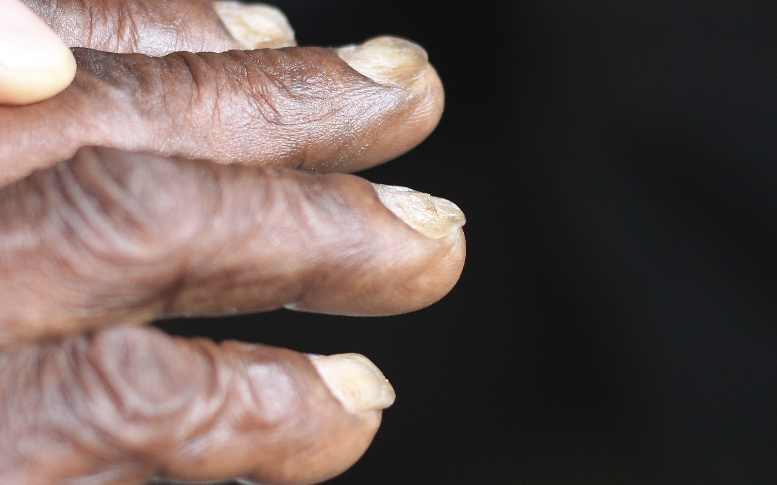
Koilonychia
Image Source: Wikimedia Commons (2018). Koilonychia iron deficiency anemia, Licensed under a creative commons licence. Available for free at: https://commons.wikimedia.org/wiki/File:Koilonychia_iron_deficiency_anemia.jpg (Accessed on: 16 July 2022).

- Yellow nail syndrome: excessively curved, yellow nails. Often associated with recurrent pleural effusions, chronic bronchitis, bronchiectasis, congenital lymphedema, nephrotic syndrome, and hypothyroidism.
- Nail-patella syndrome: loss of the ulnar half of the nail, usually affecting the thumbnail. Associated with small patellae, bony spines over the posterior iliac crests, renal abnormalities, over extension of the joints and laxity of the skin.
- Beau’s lines: transverse depressions in the nail due to temporary arrest in growth. They usually occur after illness or infection.

Beau’s Lines
Image Source: Wikimedia Commons (2014). Beau’s line on left, middle fingernail, Licensed under a creative commons licence. Available for free at: https://commons.wikimedia.org/wiki/File:Beau%27s_line_on_left,_middle_fingernail.jpg (Accessed on: 16 July 2022).

Onycholysis
Onycholysis is when the nail plate separates/lifts away from the underlying nail bed. Causes include dermatological (psoriasis, dermatitis), infective (fungal), vascular (Raynaud’s), metabolic (hypo/euthyroid), and traumatic (such as through regular manicures).
Sebaceous and Sweat Gland Disorders
Please refer to the Dermatology topic ‘Inflammatory Disorders’, where the topics acne vulgaris and acne rosacea are covered.
Hyperhidrosis
Hyperhidrosis is excessive sweating. Sweat is produced by the eccrine glands of the skin. There are two types of sweat glands – apocrine (open into hair follicles) and eccrine (widely distributed, more serous fluid expulsion). A dysfunction in the eccrine glands often leads to hyperhidrosis Causes include climate changes, anxiety, spicy food, stroke, malignancy, diabetes, menopause, hyperthyroidism, alcohol, caffeine and many more.
Treatment options:
First line: topical aluminium chloride
Iontophoresis
Botulinum toxin – axilla
Endoscopic transthoracic sympathectomy
Anhidrosis
Anhidrosis or lack of sweating can be associated with systemic diseases such as Sjogren’s syndrome, Fabry’s disease, burns, psoriasis and disorders of the central nervous system.
Hidradenitis Suppurativa (HS)
A skin condition that primarily affects the apocrine glands of the groin and axillae. It is characterised by persistent or recurrent boil-like nodules and abscesses that culminate in a purulent discharge, sinuses, and scarring. Risk factors include obesity, cigarette smoking, family history (30%), African ethnicity, Crohn’s disease, and medications such as lithium and sirolimus. The lesions often present as comedones and painful papules that eventually progress into draining sinuses and scarring. Complications include pyogenic granulomas, secondary infections, lymphoedema and squamous cell carcinomas. Management involves lifestyle measures. Medical options include topical benzoyl peroxide, topical clindamycin, anti-androgen therapies, systemic steroids, ciclosporins and surgical drainage. The treatment of HS can often be challenging for clinicians and a multidisciplinary team approach is often needed.
Complete Guide to Clinical Genetics ST3 Application and Recruitment (2027)
Written by Medibuddy. Last updated: October 2026. Clinical Genetics is

Complete Guide to Clinical Radiology ST3 Application and Recruitment (2027)
Recruitment paused: NHS England is not running Clinical Radiology ST3+ recru

Complete Guide to Paediatric Surgery ST3 Application and Recruitment in United Kingdom (2026)
Written by Medibuddy. Last updated: October 2026. If you’re planning


Take your subscriptions with you
Our mobile app allows you to access your interview and exam question banks wherever you are.

