


 Close
Close
 19th February 2024
Sympathetic nervous system
Main structure
The preganglionic neurons of the sympathetic nervous system arise within the thoracolumbar section of the spinal cord. These can be found within the lateral horns of the spinal cord within spinal levels T1-L2. The sympathetic fibres will exit the spinal cord with the spinal nerves. However, they will quickly diverge from the spinal nerve as the white rami communicans and synapses onto the sympathetic chain.
The sympathetic chain consists of 3 separate structures: 2 paravertebral ganglion chains that run alongside the spinal cord and a central prevertebral ganglion located anterior to the aorta. The paravertebral chains extend along the entire length of the spinal cord. The prevertebral ganglion is closely associated with the origin of the major blood vessels within the abdominal aorta.
The preganglionic sympathetic neurons will follow one of the following paths once it has entered the sympathetic chain:
- Ascend and synapse with a ganglion at a higher vertebral level
- It is usual for sympathetic fibres to leave the T1-T5 levels as they supply the cervical ganglion
- Three main cervical ganglion exist – Superior, middle and inferior
- Inferior ganglion can be merged with the first thoracic ganglion to form the stellate ganglion.
- Descend and synapse with a ganglion at a lower vertebral level
- Usual for fibres from T5-L2 as it supplies the lumbosacral ganglions
- Synapse within the same vertebral level
- Fibres will leave the chain as a grey ramus communicans and merge with the spinal nerve’s anterior rami to arrive at its target organ.
- Travel without synapsing towards its target organ
- This usually occurs with the T5 -L2 preganglionic fibres as they travel towards the prevertebral ganglion to contribute towards the splanchnic nerves.
Once the sympathetic fibre synapses within the sympathetic chain, the postganglionic nerve fibres will leave and join the course of the peripheral nerves to reach their end-organ targets. These include the sweat glands, smooth muscle walls, erector pili muscles, respiratory lining, cardiac tissues, GI tract, urinary tracts, ciliary muscles within the eye and the adrenal glands.
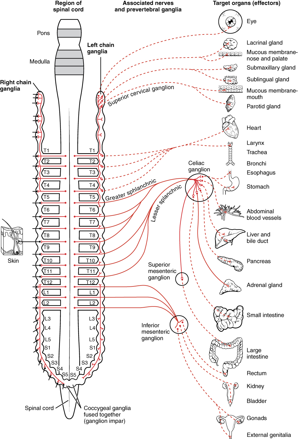 Image Source: Wikimedia Commons (2013). 1501 Connections of the Sympathetic Nervous System, Licensed under a creative commons licence. Available for free at: https://upload.wikimedia.org/wikipedia/commons/f/fd/1501_Connections_of_the_Sympathetic_Nervous_System.jpg (Accessed: 9 January 2024).
Complete Guide to Clinical Genetics ST3 Application and Recruitment (2027)
Written by Medibuddy. Last updated: October 2026. Clinical Genetics is

Complete Guide to Clinical Radiology ST3 Application and Recruitment (2027)
Recruitment paused: NHS England is not running Clinical Radiology ST3+ recru

Complete Guide to Paediatric Surgery ST3 Application and Recruitment in United Kingdom (2026)
Written by Medibuddy. Last updated: October 2026. If you’re planning


Take your subscriptions with you
Our mobile app allows you to access your interview and exam question banks wherever you are.

