


 Close
Close
 19th February 2024
Spinal cord
Clinical anatomy
The spinal cord is a continuation of the medulla oblongata after it exits the foramen magnum. It is housed within the vertebral canal of the spinal cord and continues down to the level of L2. The spinal cord contains a cervical and lumbar enlargement to accommodate the formation of the brachial plexus and the lumbosacral plexus, respectively. At the level of L1, the spinal cord tapers off into a conical structure known as the conus medullaris before dividing into the final spinal nerves as the cauda equina at L2. After the cord divides into the cauda equina, the extension of the pia and dura mater travels inferiorly to form the filum terminale, providing longitudinal anchoring for the spinal cord. The spinal cord is covered with a similar lining to the cerebral cortex: the dura, subarachnoid, and pia mater. CSF flows within the subarachnoid space between the subarachnoid and pia mater.
Transverse sections of the spinal cord reveal the internal morphology of the spinal cord. The grey matter of the spinal cord is located centrally as an H-shaped structure and can be divided into several laminae based on its function. The anterior (ventral) horn contains the motor nerve cells, while the posterior (dorsal) horn contains the sensory nerve cells. Within levels T1-L2, the spinal cord also contains a pair of lateral horns within the spinal cord grey matter, which contains neurons responsible for sympathetic innervation and reflex pathways. The white matter of the spinal cord surrounds the grey matter, which houses the main ascending and descending tracts of the nervous system. The white matter tracts are divided into three sections: anterior (ventral) funiculus, lateral funiculus and posterior (dorsal) funiculus.
The posterior column contains the primary sensory tract, the dorsal column-medial lemniscus system. This column receives proprioception, fine touch and two-point discrimination sensation and transmits this to the sensory cortex.
The spinothalamic tract is the other major sensory tract within the spinal cord. The ventral spinothalamic tract transmits crude touch and pressure sensation, while the lateral spinothalamic tract transmits pain and temperature sensation. It is important to note that sensory fibres of both spinothalamic tracts will cross the midline at each level of the spinal accord before ascending the spinal cord.
The ventral column holds the corticospinal tract. This column carries motor fibres from the brain’s motor cortex down to the muscles of the body, allowing voluntary muscle movement. Extrapyramidal tracts that are responsible for the maintenance of posture, tone and reflexes are also found within the ventral column. This includes the rubrospinal tract, vestibulospinal tract, reticulospinal tract and tectospinal tract.
Sensory nerve fibres enter the spinal cord via the dorsal root ganglion into the posterior spinal cord. The motor fibres exit the spinal cord via the ventral root through the anterior spinal cord. The motor and sensory roots meet at the intervertebral foramen to form the spinal nerve, and there are 31 pairs of spinal nerves for each corresponding spinal level. Each spinal root then divides into the ventral and dorsal rami. The spinal nerve’s dorsal rami typically innervates the back’s skin and muscles. The ventral rami of the spinal nerves will form the bulk of the peripheral nervous system, and this will be discussed in a further section.
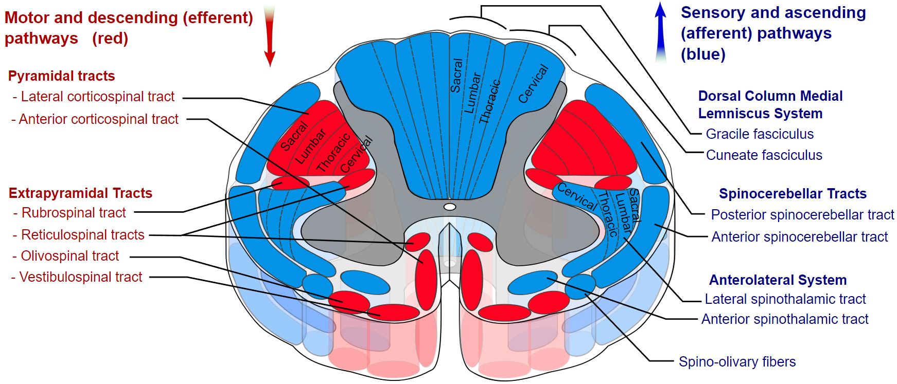 Image Source: Wikimedia Commons (2019). Spinal cord tracts – English, Licensed under a creative commons licence. Available for free at: https://upload.wikimedia.org/wikipedia/commons/b/b2/Spinal_cord_tracts_-_English.svg (Accessed: 9 January 2024).
Vasculature
The main blood supply of the spinal cord originates from the anterior and posterior spinal arteries. The anterior spinal artery originates from the anastomosis of the two vertebral arteries and supplies the anterior 2/3rd of the spinal cord. The posterior spinal artery arises from the posterior inferior cerebellar and vertebral arteries.
Complete Guide to Clinical Genetics ST3 Application and Recruitment (2027)
Written by Medibuddy. Last updated: October 2026. Clinical Genetics is

Complete Guide to Clinical Radiology ST3 Application and Recruitment (2027)
Recruitment paused: NHS England is not running Clinical Radiology ST3+ recru

Complete Guide to Paediatric Surgery ST3 Application and Recruitment in United Kingdom (2026)
Written by Medibuddy. Last updated: October 2026. If you’re planning


Take your subscriptions with you
Our mobile app allows you to access your interview and exam question banks wherever you are.

