


 Close
Close
 19th February 2024
The cerebellum and the brainstem
Cerebellum
The cerebellum is located within the posterior fossa and is inferior to the occipital lobe. It consists of 2 cerebellar hemispheres connected by the cerebellar vermis. The cerebellum communicates with the remainder of the cerebral cortex and brainstem via three main peduncles: superior cerebellar peduncle, middle cerebellar peduncle, and inferior cerebellar peduncle. Each cerebellar hemisphere controls the ipsilateral half of the body. The cerebellum’s primary function is to allow the control and coordination of complex movement within the extremities, along with the maintenance of normal muscle tone and ocular movements. Proprioceptive information is also processed within the cerebellum to allow control of balance.
The vascular supply to the cerebellum is via the basilar and vertebral arteries. The basilar artery supplies the superior and anterior aspects of the cerebellum via the superior cerebellar artery and anterior inferior cerebellar artery, respectively. The superior cerebellar peduncle is supplied by the superior cerebellar artery, while the superior and anterior inferior cerebellar artery supply the middle cerebellar peduncle. The vertebral artery supplies the posterior aspect of the cerebellum and inferior cerebellar peduncle via the posterior inferior cerebellar artery.
The cerebellar veins will drain via the transverse and superior petrosal sinuses.

Image Source: Wikimedia Commons (2015). Cerebellum arteries en, Licensed under a creative commons licence. Available for free at: https://upload.wikimedia.org/wikipedia/commons/d/d9/Cerebellum_arteries_en.svg (Accessed: 9 January 2024).
Brainstem
The brainstem consists of 3 separate structures: the midbrain, pons and medulla. The brainstem holds the nuclei of several cranial nerves, sensory and motor tracts, and reflex centres.
The midbrain, or mesencephalon, is between the cerebral cortex, pons and cerebellum. The midbrain houses the oculomotor (CNIII) and the trochlear nerve (CNIV) nuclei. The Edinger-Westphal nucleus is also located within the midbrain between the oculomotor nucleus and the Sylvius aqueduct and acts as the parasympathetic innervation to the iris and ciliary muscle. Other structures within the midbrain include the red nucleus, ventral tegmental area, periaqueductal grey area and the substantia nigra. There are also complex reticular networks that exist within the midbrain that are responsible for visceral functions, movement control and maintenance of consciousness/wakefulness. The reticular formation forms three columns, including the raphe nucleus (serotonergic nucleus), gigantocellular reticular nuclei (movement control) and parvocellular reticular nuclei (expiration control). Lastly, the midbrain also has ascending and descending pathways, including the spinothalamic, corticospinal, and rubrospinal tract.
The midbrain receives an extensive blood supply, with the basilar artery branches supplying the anterior aspects of the midbrain, the posterior cerebral artery supplying the lateral aspects, and the superior cerebellar artery supplying the posterior aspects.
 Image Source: Wikimedia Commons (2009). Midbrainsection, Licensed under a creative commons licence. Available for free at: https://upload.wikimedia.org/wikipedia/commons/c/cb/Midbrainsection.svg (Accessed: 9 January 2024).
The pons are inferior to the midbrain and medulla oblongata. The pons serve as the site of origin for several cranial nerve nuclei, including the trigeminal nerve (CN V), abducens nerve (CN VI), facial nerve (CN VII) and the vestibulocochlear nerve (CN VIII). Many ascending and descending tracts also pass through the pons, including the corticospinal, corticobulbar, spinothalamic and medial lemniscus tracts. The vasculature to the pons predominantly arises from the pontine artery of the basilar artery. Small contributions arise from the anterior inferior cerebellar and superior cerebellar arteries.

Image Source: Wikimedia Commons (2016). Pons section at facial colliculus, Licensed under a creative commons licence. Available for free at: https://upload.wikimedia.org/wikipedia/commons/1/13/Pons_section_at_facial_colliculus.png (Accessed: 9 January 2024).
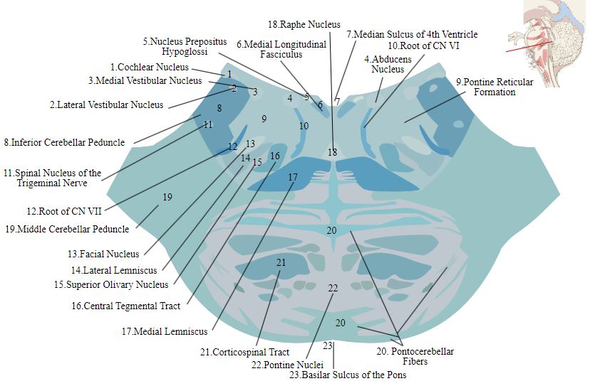
Image Source: Wikimedia Commons (2011). Lower pons horizontal KB, Licensed under a creative commons licence. Available for free at: https://upload.wikimedia.org/wikipedia/commons/d/dc/Lower_pons_horizontal_KB.svg (Accessed: 9 January 2024).
The medulla oblongata is the inferior-most portion of the brainstem. The ventral portions of the medulla house the pyramids and olives. The pyramids house the descending motor tracts of the brain, and the decussation of the motor fibres at this level forms the anterior and lateral lemniscus tracts of the spinal cord. The olivary nuclei allow cerebellar motor function and sound perception. It also houses the nuclei for the glossopharyngeal nerve (CN IX), vagus nerve (CN X), accessory nucleus nerve (CN XI) and the hypoglossal nerve (CN XII). Lastly, the medulla also houses the cardiovascular and respiratory regulatory centres within the ventral medulla. The vascular supply to the medulla is complex, with blood supplies originating from the anterior spinal artery, vertebral artery, posterior spinal artery and the posterior inferior cerebellar artery.
 Image Source: Wikimedia Commons (2013). Sobo 1909 665, Licensed under a creative commons licence. Available for free at: https://upload.wikimedia.org/wikipedia/commons/7/79/Sobo_1909_665.png (Accessed: 9 January 2024).
Ocular system
The visual pathway begins within the retinal layer of the eye. The fibres of the retina unite at the optic disc to form the optic nerve. The optic nerve is surrounded by the dura mater as a direct extension of the intracranial dura mater. It exits via the orbital foramen and enters the cranium via the optic canal into the middle cranial fossa. The optic nerves will unite above and behind the pituitary stalk to form the optic chiasm. At the optic chiasm, the nasal fibres from the right eye will unite with the temporal fibres of the left eye to form the left optic tract. Each optic tract continues posteriorly through the lateral geniculate body to the genico-calcarine tracts within the parietal and temporal lobes. It terminates within the primary visual cortex in the occipital lobe. A small proportion of fibres will pass through the superior colliculus and Edinger-Westphal nucleus to provide parasympathetic innervation to the eye.
The visual and parasympathetic system drives the pupillary reflex. The optic nerve controls the afferent limb, and its impulses are directed towards the pretectal nucleus and, subsequently, the Edinger-Westphal nucleus bilaterally. The oculomotor nerve controls the efferent limb as parasympathetic fibres leave the Edinger-Westphal nucleus into the ciliary ganglion and the oculomotor nerve before terminating at the pupillary sphincter muscle.
Complete Guide to Clinical Genetics ST3 Application and Recruitment (2027)
Written by Medibuddy. Last updated: October 2026. Clinical Genetics is

Complete Guide to Clinical Radiology ST3 Application and Recruitment (2027)
Recruitment paused: NHS England is not running Clinical Radiology ST3+ recru

Complete Guide to Paediatric Surgery ST3 Application and Recruitment in United Kingdom (2026)
Written by Medibuddy. Last updated: October 2026. If you’re planning


Take your subscriptions with you
Our mobile app allows you to access your interview and exam question banks wherever you are.

