


 Close
Close
 19th February 2024
Peripheral nervous system
Brachial plexus
Root, trunk, division and cord
The brachial plexus provides the motor, sensory and autonomic innervation to the upper limb, including the scapular region. The C5-T1 spinal nerves will contribute towards the brachial plexus. The anterior (ventral) rami of these spinal nerves will form the roots of the brachial plexus. These spinal nerves will pass along the border of the anterior and medial scalene muscles to enter the base of the neck.
Once at the base of the neck, the roots will combine to form the brachial plexus trunks. Three trunks exist:
- Superior trunk – C5-C6 roots
- Middle trunk – C7 root
- Inferior trunk – C8-T1 roots
The brachial plexus trunks will then divide into the brachial plexus divisions at the neck’s posterior triangle. Each trunk will divide into an anterior and posterior division, resulting in 6 nerve fibres passing into the axillae
Each division of the brachial plexus will combine within the axillae to form 3 cords, and the nomenclature of the cord depends on the nerve fibre’s relation to the axillary artery. The three cords consist of the following:
- Lateral cord – Anterior division of the superior and middle trunk
- Posterior cord – Posterior divisions of the superior, middle and medial trunk
- Medial cord – Anterior division of the inferior trunk
The cords will then continue down the upper limb and give rise to the major nerves of the upper limb.
Major branches
The first nerve to exit the brachial plexus arises from the root of the brachial plexus. The dorsal scapular nerve arises directly from the C5 spinal nerve and innervates the levator scapulae, rhomboid major and rhomboid minor muscles. The long thoracic nerve will arise from the C5, C6 and C7 spinal nerves and innervates the serratus anterior muscle.
The superior trunk of the brachial plexus gives rise to the suprascapular nerve and nerve to subclavius. The suprascapular nerve innervates the supraspinatus and infraspinatus muscles. There are no branches from the middle and inferior trunks. There are also no branches arising from the division of the brachial plexus.
The branches of the cords of the brachial plexus are as follows:
- Lateral cord
- Lateral pectoral nerve (C5-C7 roots) – Innervates the pectoralis major
- Middle cord
- Medial pectoral nerve (C8-T1 roots) – Innervates the pectoralis major and pectoralis minor
- Medial brachial cutaneous nerve (C8-T1 roots) – Innervate medial side of the arm
- Medial antebrachial cutaneous nerve (C8-T1 roots) – Innervates the medial side of the forearm.
- Posterior cord
- Upper subscapular nerve (C5-6 roots) – Innervates the subscapularis muscle
- Middle subscapular nerve (C6-8 roots) – Innervates the latissimus dorsi muscle
- Lower subscapular nerve (C5-6 roots) – Innervates the subscapularis and teres major muscles.
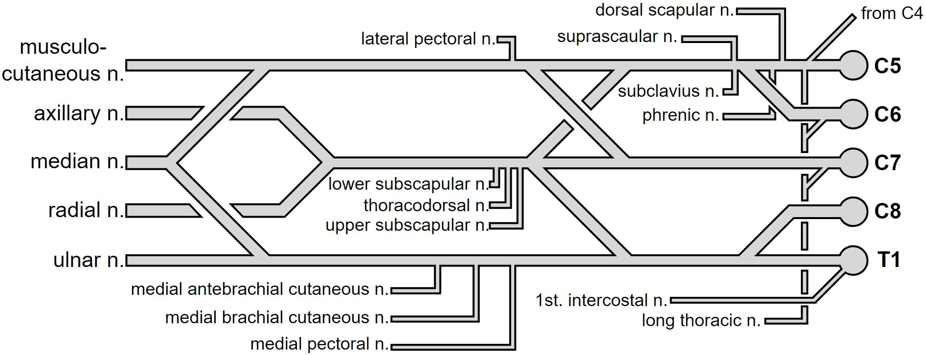 Image Source: Wikimedia Commons (2007). Brachial plexus, Licensed under a creative commons licence. Available for free at: https://upload.wikimedia.org/wikipedia/commons/7/73/Brachial_plexus.svg (Accessed: 9 January 2024).
The cords of the brachial plexus will terminate as the five major nerves within the upper extremities. These are the musculocutaneous, axillary, ulnar, radial and median nerves.
Musculocutaneous nerve
The musculocutaneous nerve is the terminal branch of the lateral cord and consists of the C5, C6 and C7 nerve roots. It provides motor innervation to the coracobrachialis, brachialis and biceps brachialis. As the nerve emerges lateral to the biceps within the forearm, it provides sensory innervation to the lateral forearm as the lateral cutaneous nerve. Injuries to this nerve are relatively uncommon due to its protected location in the axillae but can still be damaged iatrogenically or via stabbing injuries to the axillae.
Axillary nerve
The axillary nerve is a continuation of the posterior cord and comprises the C5 and C6 roots. The nerve passes posterior to the axillary artery, exits along the inferior border of the subscapularis, and passes medial to the surgical neck of the humerus. It will divide into its 3 terminal branches past this point:
- Posterior terminal branch: Motor innervation to the posterior deltoid and teres major + sensory innervation of the skin over the inferior deltoid (as supplied by the upper lateral cutaneous nerve of the arm).
- Anterior terminal branch – Motor innervation to the anterior deltoid muscle.
- Articular branch – Supplies the glenohumeral joint.
Injury to the axillary nerve commonly occurs with proximal humeral or shoulder injuries, including shoulder surgery, shoulder dislocations and proximal humeral fractures.
Ulnar nerve
The ulnar nerve is the continuation of the middle cord and contains the C8 and T1 root fibres. The ulnar nerve does not branch within the arm. As it courses through the forearm, it gives off three branches:
- Muscular branch – Innervates the flexor carpi ulnaris and the medial half of flexor digitorum profundus.
- Palmar cutaneous branch – Supplies the medial 1/3rd of the palm
- Dorsal cutaneous branch – Supplies the medial 1/3rd of the dorsum of the hand and medial 1/3rd of digits.
The ulnar nerve traverses superficial to the flexor retinaculum and enters via the Guyon’s canal. It terminates as the ulnar nerve divides into a superficial and deep branch of the ulnar nerve. The superficial branch of the ulnar nerve innervates the palmaris brevis along with sensory innervation to the palmar surface of the medial half of the fourth digit and the fifth digit. The deep branch of the ulnar nerve innervates the muscles within the hypothenar eminence and the interosseous muscles. Ulnar nerve injuries can occur with injuries to the elbow at the level of the medial epicondyle, along with compression along the cubital tunnel. It can also be damaged at the level of the wrist.
Median nerve
The median nerve is derived from the anterior divisions of the medial and lateral cords and contains the roots from the C6-T1 spinal nerve. No branches arise within the arm. Within the forearm, the anterior interosseous and palmar cutaneous nerves arise from the median nerve. The anterior interosseous nerve provides motor innervation to the deep flexors of the anterior compartment of the forearm. The palmar cutaneous branch innervates the lateral aspect of the palm. The median nerve will provide direct innervation to the superficial and middle flexors of the forearm.
The median nerve continues into the palm via the carpal tunnel and divides into its terminal branches. The recurrent branch of the median nerve will innervate the thenar muscles. The palmar digital branch will provide sensory innervation of the palmar surface and fingertips of the lateral three-and-a-half digits. Damage to the median nerve can occur via injury to the nerve at the carpal tunnel level or the level of humeral condyles.
Radial nerve
The radial nerve is the continuation of the posterior cord of the brachial plexus, which contains C5-T1 spinal roots. As it passes down the arm, it courses along the radial groove and supplies the triceps muscle. Three sensory branches of the radial nerve arise in the arm:
- The lower lateral cutaneous nerve of the arm – Innervates the lateral aspect of the arm inferior to the deltoid insertion
- Posterior cutaneous nerve of the arm – Innervates the posterior surface of the arm
- Posterior cutaneous nerve of the forearm – Innervates the skin along the middle of the forearm.
It terminates as it divides into two branches, the deep and superficial branches. The deep branch of the radial nerve provides motor innervation to the posterior compartment of the forearm. It collectively allows extension of the forearm and fingers along with the supination of the forearm. The superficial branch of the radial nerve provides sensory innervation to the dorsal surface of the lateral three-and-a-half digits of the hand. Injuries to the radial nerve can occur within the axillae, radial groove during a humeral fracture, within the forearm or during injuries to the radial head.
Reference
Moore, K. L., Dalley, A. F., & Agur, A. M. R. (2014). Upper Limb. In Moore’s Clinically Orientated Anatomy (7th ed.). China: Lippincott.
Okwumabua E, Thompson JH. Anatomy, Shoulder and Upper Limb, Nerves. [Updated 2021 Jul 28]. In: StatPearls [Internet]. Treasure Island (FL): StatPearls Publishing; 2022 Jan-. Available from: https://www.ncbi.nlm.nih.gov/books/NBK526056/
Polcaro L, Charlick M, Daly DT. Anatomy, Head and Neck, Brachial Plexus. [Updated 2021 Aug 25]. In: StatPearls [Internet]. Treasure Island (FL): StatPearls Publishing; 2022 Jan-. Available from: https://www.ncbi.nlm.nih.gov/books/NBK531473/
Lumbosacral plexus
Lumbar plexus
The lumbar plexus originates from the anterior rami of the L1-L4 spinal nerves, and they will combine to contribute towards six major nerves. The six nerves that arise from the lumbar plexus include:
- Iliohypogastric nerve
- Contains the T12 and L1 spinal roots. It provides sensory innervation to the lateral gluteal region.
- Motor innervation is provided to the internal oblique and transverse abdominis muscle.
- Ilioinguinal nerve
- Contains nerve roots from the L1 spinal nerve.
- It provides motor innervation to the transverse abdominis and internal obliques.
- Sensory innervation is provided to the skin of the anteromedial thigh. Sensation is also supplied to the root of the penis and anterior scrotum in males, while it supplies the mons pubis and labia majora in females.
- Genitofemoral nerve
- Contains the L1 and L2 nerve roots.
- Provides motor function to the cremasteric muscle.
- The genital branch provides sensory innervation to the anterior scrotum in males and the mons pubis and labia majora in females. Its femoral branch provides sensory innervation to the anterior thigh.
- Lateral cutaneous nerve of the thigh
- Contains the L2 and L3 spinal roots.
- Provides sensation to the anterior and lateral thigh down to the knee.
- Obturator nerve
- Contains the L2, L3 and L4 spinal roots.
- Motor innervation to the medial thigh (obturator externus, adductor longus, adductor brevis, adductor magnus, gracilis)
- Sensory innervation to the medial thigh
- Femoral nerve
- Contains the L2, L3 and L4 spinal roots
- Motor innervation to the anterior thigh (iliacus, pectineus, sartorius and quadriceps femoris)
- Sensory innervation to the anterior thigh and medial leg.
Sacral plexus
The sacral plexus is the network of nerves that supplies the lower limbs, predominantly arising from the S1-S4 spinal nerves. It also receives contributions from the L4 and L5 spinal nerves. Five major nerves arise from the sacral plexus and leave the pelvic wall via the greater sciatic foramen. The following major nerves are derived from the sacral plexus:
- Superior gluteal nerve
- Contains the L4, L5 and S1 spinal roots.
- Motor innervation to the gluteus minimus, gluteus medius and the tensor fascia lata
- Inferior gluteal nerve
- Contains the L5, S1 and S2 spinal roots
- Motor innervation to the gluteus maximus.
- Posterior femoral cutaneous nerve of the thigh
- Contains the S1, S2 and S3 spinal roots
- Provides sensory innervation to the posterior surface of the thigh, leg and the perineum.
- Pudendal nerve
- Contains the S2, S3 and S4 spinal roots
- Motor innervation to the perineal muscles, urethral sphincter, anal sphincter and levator ani.
- Sensory innervation is provided to the penis, clitoris and most of the perineal skin.
- Sciatic nerve
- Contains the L4, L5, S1, S2 and S3 spinal roots
- Motor innervation to the posterior compartment of this thigh (biceps femoris, semimembranosus, semitendinosus and the ischial portion of the adductor magnus)
- It divides further into two major branches after passing the popliteal fossa
- Tibial nerve
- Motor innervation to the posterior compartment of the leg and some intrinsic muscles of the foot
- Sensory innervation to the posterolateral leg, lateral foot and sole of the foot.
- Common peroneal/fibular nerve
- Motor innervation to the anterior and lateral compartments of the leg and intrinsic muscles of the foot
- Sensory innervation to the lateral aspect of the leg and the dorsum of the foot.
- Tibial nerve

Image Source: Wikimedia Commons (2010). Lumbar plexus, Licensed under a creative commons licence. Available for free at: https://upload.wikimedia.org/wikipedia/commons/7/73/Lumbar_plexus.svg (Accessed: 9 January 2024).
Reference
Moore, K. L., Dalley, A. F., & Agur, A. M. R. (2014). Lower Limb. In Moore’s Clinically Orientated Anatomy (7th ed.). China: Lippincott.
Spence KT, Forro SD. Anatomy, Bony Pelvis and Lower Limb, Nerves. [Updated 2021 Jul 31]. In: StatPearls [Internet]. Treasure Island (FL): StatPearls Publishing; 2022 Jan-. Available from: https://www.ncbi.nlm.nih.gov/books/NBK532304/
Singh O, Al Khalili Y. Anatomy, Back, Lumbar Plexus. [Updated 2021 Aug 11]. In: StatPearls [Internet]. Treasure Island (FL): StatPearls Publishing; 2022 Jan-. Available from: https://www.ncbi.nlm.nih.gov/books/NBK545137/
Complete Guide to Clinical Genetics ST3 Application and Recruitment (2027)
Written by Medibuddy. Last updated: October 2026. Clinical Genetics is

Complete Guide to Clinical Radiology ST3 Application and Recruitment (2027)
Recruitment paused: NHS England is not running Clinical Radiology ST3+ recru

Complete Guide to Paediatric Surgery ST3 Application and Recruitment in United Kingdom (2026)
Written by Medibuddy. Last updated: October 2026. If you’re planning


Take your subscriptions with you
Our mobile app allows you to access your interview and exam question banks wherever you are.

