


 Close
Close
 7th June 2023
Stroke
Classification
Stroke is defined by the World Health Organisation as a ‘clinical syndrome of vascular origin, with rapidly developing signs of focal or global cerebral dysfunction, lasting for over 24 hours or leading to death’.
85% of strokes are ischaemic. These are due to an interruption in the blood supply to a particular area of the brain. Ischaemic strokes can be caused by the following:
- Cardioembolism – most commonly due to atrial fibrillation
- Large vessel thrombosis
- Small vessel disease
- Hypoperfusion – for example, due to systemic hypotension during a period of critical illness
- Venous sinus thrombosis
The remaining 15% are haemorrhagic due to the rupture of a blood vessel or vascular structure in the brain. This can be due to the following:
- Intracerebral haemorrhage – bleeding within the brain, which can be further divided into:
- Intraparenchymal – within the brain tissue
- Intraventricular – within the ventricles of the brain
- Subarachnoid haemorrhage
Ischaemic Stroke
It is helpful with ischaemic strokes to consider the arterial blood supply to the brain as this determines how ischaemic strokes are subdivided in the Bamford classification, according to which part of the cerebral blood supply is affected.
Circle of Willis
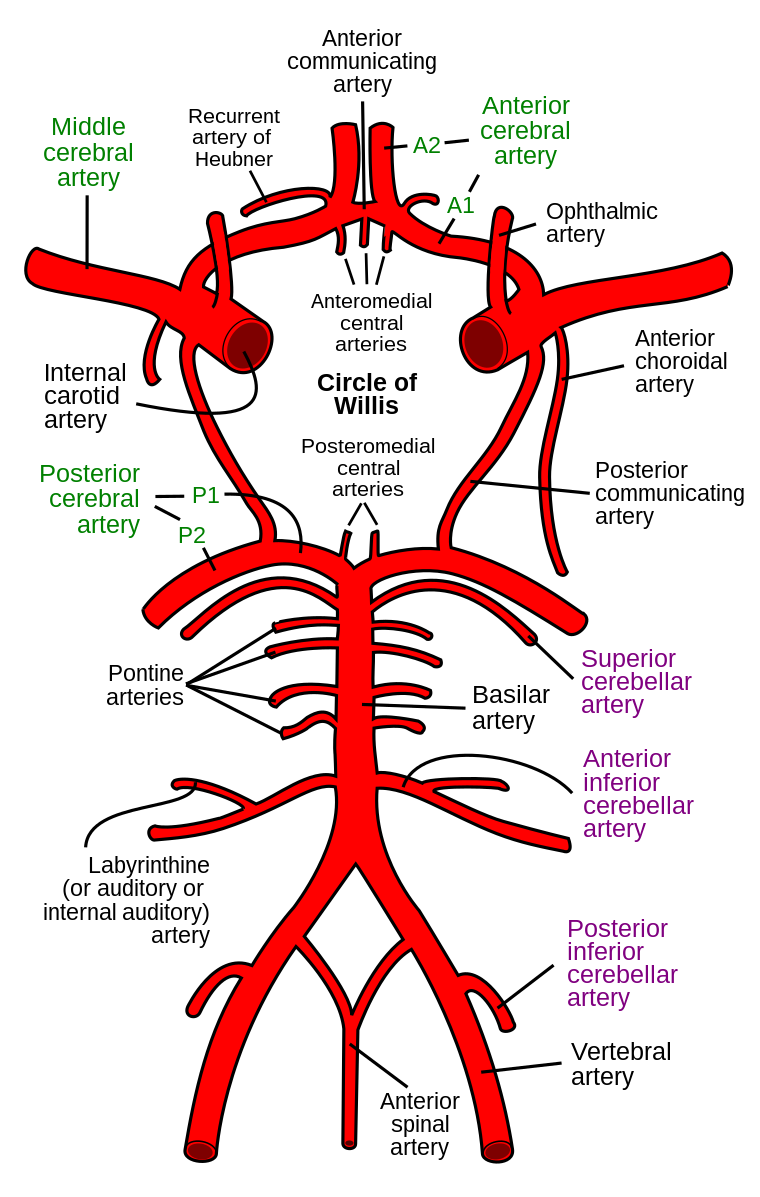
Diagram of the Circle of Willis.
Image Source: Wikimedia Commons (2007). Circle of Willis en, Licensed under a creative commons licence. Available for free at: https://commons.wikimedia.org/wiki/File:Circle_of_Willis_en.svg (Accessed on: 11 January 2023).

Branches of the anterior and posterior circulations of the brain form a ring-like structure known as the circle of Willis. This connects the anterior and posterior circulation and, in the event of an ischaemic lesion, will provide collateral blood flow to maintain cerebral perfusion.
The anterior circulation is formed by the internal carotid artery dividing into the anterior and middle cerebral arteries. The anterior communicating artery connects the anterior cerebral arteries. The posterior circulation is formed by the vertebral artery forming the basilar artery, giving rise to the pontine and posterior cerebral arteries. These then connect to the anterior part of the circulation via the posterior communicating arteries.
Bamford Classification
This divides strokes into four subtypes by their clinical presentation and depending on which part of the circulation is affected.
| Type of Stroke | Diagnosis | |
|---|---|---|
| TACS (Total anterior circulation stroke) |
Large cortical stroke Anterior/middle cerebral arteries |
All three of:
|
| PACS (Partial anterior circulation stroke) |
Large cortical stroke Anterior/middle cerebral arteries |
Any two of:
|
| POCS (Posterior circulation syndrome) |
Posterior circulation syndrome | Any one of:
|
| LACS (Lacunar stroke) |
Subcortical stroke Caused by small vessel disease No higher cerebral dysfunction |
5 syndromes:
|
Investigation
Investigations for stroke can be divided into those to confirm the diagnosis of stroke and those to identify the cause of the stroke, which will affect secondary prevention measures.
Initial investigation in the emergency setting is most commonly with a non-contrast CT brain, which aims to rule out a bleed and look for stroke mimics, such as tumours, etc. There may be early signs of stroke, such as a hyperdense vessel, loss of insular ribbon and loss of grey-white differentiation, but the absence of these signs does not rule out a stroke. The changes on CT associated with stroke can take hours or days to become apparent, and it is common for a person presenting with an acute stroke to initially have a completely normal CT scan. MRI with diffusion-weighted imaging (DWI) is very sensitive at picking up acute ischaemia. Changes are apparent within approximately forty minutes but start to fade after ten days. It is important to note, however, that about ten percent of strokes are negative on MRI with DWI; therefore, stroke remains a clinical diagnosis.
Investigations to look for a cause include a 48-hour tape or Holter monitor for atrial fibrillation as a cause of cardioembolic stroke, cholesterol and glucose levels to look for risk factors or hypercholesterolaemia and diabetes and an echocardiogram to look for a cardiac thrombus. Carotid dopplers are indicated for anterior circulation strokes, and if symptomatic carotid artery stenosis >50% on the affected side is found, the patient should be referred for carotid endarterectomy.
In cases of young stroke or where a cause cannot be identified (cryptogenic stroke), one can consider screening for thrombophilia (e.g. antiphospholipid syndrome, factor V Leiden) as well as rarer causes such as a patent foramen ovale with a bubble echocardiogram.
Management
The acute management of ischaemic strokes consists of intravenous thrombolysis with alteplase (tissue plasminogen activator) and/or clot retrieval (mechanical thrombectomy) in suitable patients. Thrombolysis is indicated within four and a half hours of symptom onset. There is a risk of causing bleeding, including intracerebral haemorrhage, which can be life-threatening. The risk increases with increasing age and with time from symptom onset. Contraindications include being on a therapeutic anticoagulant and recent bleeding, although the relative risks and benefits are considered on an individual basis.
Mechanical thrombectomy is indicated within six hours of symptom onset and is considered for patients with a significant level of impairment from their stroke and good premorbid function.
High-dose antiplatelets (usually aspirin) are given if a patient is not suitable for either thrombolysis or thrombectomy and continued for two weeks before switching to a maintenance dose. Longer term, the mainstays of treatment are antiplatelet therapy, statins, blood pressure lowering drugs, and management of other risk factors. Patients should be anticoagulated if atrial fibrillation is present and referred for carotid endarterectomy if significant carotid artery stenosis is found.
Transient Ischaemic Attack
Transient ischaemic attack (TIA) is ‘an acute loss of focal cerebral or ocular function with symptoms lasting less than 24 hours and which is thought to be due to inadequate cerebral or ocular blood supply as a result of low blood flow, thrombosis or embolism associated with diseases of the blood vessels, heart, or blood’. Patients are at higher risk for having further vascular events, especially in the first few weeks and therefore require urgent assessment by a stroke specialist. Treatment is with antiplatelet therapy and treatment of vascular risk factors such as hypertension, hypercholesterolaemia, for example.
Haemorrhagic Stroke
Intracerebral haemorrhage
Intracerebral haemorrhage accounts for approximately 15% of strokes and causes include hypertension, therapeutic anticoagulants, increasing age, recreational drug use (e.g. cocaine), vascular anomalies, venous sinus thrombosis and brain tumours.
Symptoms can be gradual or sudden in onset and are related either to the part of the brain involved or to raised intracranial pressure, such as nausea, vomiting, headache, drowsiness, and seizures. Investigation is with non-contrast CT or MRI brain.
Conservative management consists of stopping the bleeding by reversing anticoagulants and blood pressure control. Haemorrhages in the basal ganglia and thalamus are generally managed conservatively as there is no evidence that surgery improves outcomes. Surgical treatment is considered for patients with haemorrhage in particular locations, such as cerebellar and lobar, as well as for complications, such as hydrocephalus.
Subarachnoid Haemorrhage
Subarachnoid haemorrhage is bleeding into the subarachnoid space between the arachnoid membrane and the pia mater. It causes sudden onset of severe headache (known as ‘thunderclap headache’), decreased consciousness, focal neurology and meningism (nuchal rigidity, photophobia). It is most often due to rupture of an intracranial aneurysm (Berry aneurysm) which can be sporadic or occur secondary to genetic conditions such as autosomal dominant polycystic kidney disease, Ehlers-Danlos syndrome, Marfan syndrome and neurofibromatosis.
Diagnosis is from a CT brain scan. If this is negative, cerebrospinal fluid (CSF) should be examined for xanthochromia (discolouration of the CSF due to lysis of blood).
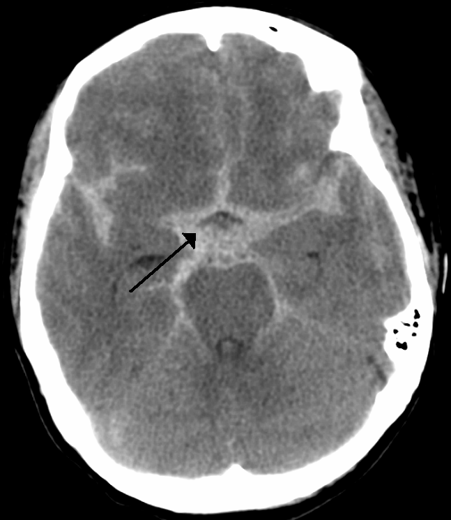
Subarachnoid haemorrhage on CT head.
Image Source: Wikimedia Commons (2010). Circle of Willis en, Licensed under a creative commons licence. Available for free at: https://commons.wikimedia.org/wiki/File:SubarachnoidP.png (Accessed on: 11 January 2023).

Cerebral Venous Sinus Thrombosis
This is a rare cause of stroke, produced by a clot in the dural venous sinuses, resulting in obstruction of blood draining out of the brain, causing venous congestion, and raised intracranial pressure. Clinical features include headache, vomiting, papilloedema, focal neurological signs and cranial nerve palsies.
Diagnosis is by CT venography, and treatment is with anticoagulation, initially with heparin which is later converted to warfarin.
Complete Guide to Clinical Genetics ST3 Application and Recruitment (2027)
Written by Medibuddy. Last updated: October 2026. Clinical Genetics is

Complete Guide to Clinical Radiology ST3 Application and Recruitment (2027)
Recruitment paused: NHS England is not running Clinical Radiology ST3+ recru

Complete Guide to Paediatric Surgery ST3 Application and Recruitment in United Kingdom (2026)
Written by Medibuddy. Last updated: October 2026. If you’re planning


Take your subscriptions with you
Our mobile app allows you to access your interview and exam question banks wherever you are.

