


 Close
Close
 20th January 2024
Osteoarthritis
Background
Epidemiology
Osteoarthritis (OA) is the most common form of joint disease, and is thought to be a leading cause for pain and disability worldwide. It has an estimated overall prevalence of 24% and 11% for knee and hip OA respectively, in the general adult population.
Pathophysiology
OA is a clinical syndrome characterised by joint pain, which causes varying degrees of functional limitation and a reduced quality of life. It is a disorder of synovial joints, which occurs when joint damage caused by repeated excessive loading or injury triggers a repair process, culminating in structural changes to the joint. These structural changes can lead to characteristic features of OA, including localised loss of cartilage, remodelling of adjacent bone and formation of osteophytes, and inflammation of the synovial membranes. Periarticular damage can also lead to capsular stretching, muscle weakness and ligament laxity. Any synovial joint can be affected, but the most commonly affected peripheral joints are the knees, followed by hips and small joints of the hands.
Causes and risk factors
The exact cause of OA is unknown, but it is thought to result from an interaction between genetic, biological and biomechanical factors. Heritability of hand, hip and knee OA is estimated at around 40-60%. Increasing age is the strongest risk factor for developing OA, particularly after the age of 50 years, and is more common in females than in men. Other risk factors include obesity, high bone mineral density and low oestrogen levels. Biomechanical factors such as joint injury, occupational and recreational joint stresses, articular congenital deformities, history of inflammatory arthropathy, joint laxity and reduced muscle strength can all contribute to an increased risk in the development of OA.
Definitions
OA can be classified as either primary or secondary:
- Primary (idiopathic): no history of preceding joint injury
- Localised: affecting hands, knee, hip or foot
- Generalised: affecting hands and another joint
- Secondary: occurs following a pre-existing insult to the joint, e.g. congenital abnormality, trauma, inflammatory arthropathies, repetitive physical or occupational strain.
References
- Pereira D, Peleteiro B, Araujo J, et al. The effect of osteoarthritis definition on prevalence and incidence estimates: a systematic review. Osteoarthritis Cartilage 2011;19:1270–85.
- Conaghan PG, Dickson J, Grant RL. Care and management of osteoarthritis in adults: summary of NICE guidance. Bmj. 2008 Feb 28;336(7642):502-3.
- National Institute for Health and Care Excellence (Great Britain).Osteoarthritis: care and management. National Institute for Health and Care Excellence (NICE); 2014
Presentation
Background
There is large variability in clinical presentation of OA between individuals and different joints affected in the same person. The diagnosis of OA is largely clinical and can be made in patients with suggestive features without the need for further investigations. The most common symptoms are joint pain and stiffness, although patients often present with functional impairment. The knees, hips and small joints of the hands, namely the proximal interphalangeal (PIP) and distal interphalangeal (DIP) joints, and the spine (particularly lumbar and cervical regions) are most commonly affected. OA is an unlikely diagnosis in patients with ankle or wrist symptoms, and other differentials should be considered.
Symptoms
- Joint pain:
- Exacerbated by activity/weight-bearing
- Relieved by rest (except in advanced disease where rest and night pain can occur)
- Typically, only one or a few joints are affected at any given point
- Develops over months or years
- Joint stiffness:
- Occurs usually in the morning for less than 30 minutes or after rest
- Typically resolves in a few minutes with motion of the joint
- Functional impairment
Signs
- Bony swelling and joint deformity (due to osteophytes)
- Joint effusion (can occur in the knee)
- Reduced range of movement in the joint
- Pain at extremes of joint movement
- Tenderness/warmth around the joint
- Joint instability
- Muscle weakness or wasting around affected joint
- Crepitus – grating/crunching sound, commonly felt on passive or active movement of joint, caused by friction between bone and cartilage
- Absence of systemic features, e.g. rash, fever
The specific presentation of OA depends on the affected joint, and a detailed history and examination can reveal the following features:
| Hand OA | |
|---|---|
| History | Examination |
| Pain can radiate towards the thumb or wrist and distal forearm. Often exacerbated by strong grip | Typically affects the first carpometacarpal (CMC) joint at the base of the thumb, the DIP joints and the PIP joints |
| Reduced grip or pinch strength, which can cause difficulties with tasks such as opening jars, turning keys, fastening buttons, writing, etc | Fixed flexion deformity of CMC joint, with hyperextension of the distal joints |
| Wasting of thenar muscles | |
| Heberden’s nodes: bony nodules on the DIP joints
Bouchard’s nodes: bony nodules on the PIP joints |
|
| Pain/warmth/redness/swelling of affected joints | |
| Ulnar or radial deviation at affected joints | |
| Painful mucous-filled cysts on the dorsum of the finger | |
| “Squaring” at the base of the thumb caused by partial dislocation of CMC joint | |
Table 1: Possible clinical features of hand osteoarthritis
Adapted from: National Institute for Health and Care Excellence (NICE) Clinical Knowledge Summaries: Osteoarthritis [Internet]. Cks.nice.org.uk. 2021 [cited 12 August 2022]. Available from: https://cks.nice.org.uk/topics/osteoarthritis/diagnosis/diagnosis/

Image Source: Wikimedia Commons (2006). Heberden-Arthrose, Licensed under a creative commons licence. Available for free at: https://commons.wikimedia.org/wiki/File:Heberden-Arthrose.JPG#globalusage (Accessed: 24 June 2021).
| Hip OA | |
|---|---|
| History | Examination |
| Pain felt in the groin and anterior/lateral thigh. Can also be referred to the knee or the testicle on the affected side in males | Painful restriction of internal rotation with hip flexed |
| Difficulty walking, climbing stairs, driving, painful sexual intercourse | Antalgic gait
Trendelenburg gait in advanced disease – towards affected hip with reduced time weight-bearing on that side; pelvis tilting down on unaffected side due to wasting and weakness of gluteal and anterior thigh muscles |
| Shortening of the lower limb | |
Table 2: Possible clinical features of hip osteoarthritis
Adapted from: National Institute for Health and Care Excellence (NICE) Clinical Knowledge Summaries: Osteoarthritis [Internet]. Cks.nice.org.uk. 2021 [cited 12 August 2022]. Available from: https://cks.nice.org.uk/topics/osteoarthritis/diagnosis/diagnosis/
| Knee OA | |
|---|---|
| History | Examination |
| Usually, bilateral pain, felt in and around the knee. Unilateral symptoms usually secondary to pre-existing trauma or disease | Varus (bow-legged) or valgus (knock-knee) deformities |
| Difficulty walking, particularly when going down the stairs if patellofemoral involvement | Locking (inability to straighten knee due to loose meniscal cartilage in the joint |
| Progressive pain on sitting, relieved by standing | Crepitus or joint line tenderness |
| Sensation of knee ‘giving way’ due to weak quadriceps and altered load-bearing | Small to moderate effusions |
| Bony swelling around knee in advanced disease | |
| Antalgic gait – towards affected hip with reduced time weight-bearing on that side; pelvis is in neutral position | |
Table 3: Possible clinical features of knee osteoarthritis
Adapted from: National Institute for Health and Care Excellence (NICE) Clinical Knowledge Summaries: Osteoarthritis [Internet]. Cks.nice.org.uk. 2021 [cited 12 August 2022]. Available from: https://cks.nice.org.uk/topics/osteoarthritis/diagnosis/diagnosis/
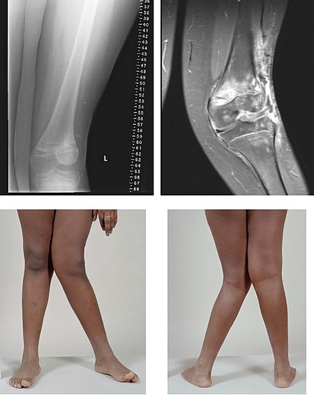
Image Source: Wikimedia Commons (2007). Valgus, Licensed under a creative commons licence. Available for free at: https://commons.wikimedia.org/wiki/File:Valgus.jpg (Accessed: 24 June 2021).
References
- Conaghan PG, Dickson J, Grant RL. Care and management of osteoarthritis in adults: summary of NICE guidance. Bmj. 2008 Feb 28;336(7642):502-3.
- National Institute for Health and Care Excellence (Great Britain).Osteoarthritis: care and management. National Institute for Health and Care Excellence (NICE); 2014
- Arden N, Blanco F, Cooper C, Guermazi A, Hayashi D, Hunter D, Javaid MK, Rannou F, Roemer F, Reginster JY. Atlas of osteoarthritis. Springer Healthcare Limited; 2014.
- Osteoarthritis | Health topics A to Z | CKS | NICE [Internet]. Cks.nice.org.uk. 2021 [cited 12 August 2022]. Available from: https://cks.nice.org.uk/topics/osteoarthritis/diagnosis/diagnosis/
Diagnosis
Background
NICE guidelines recommend suspecting a diagnosis of OA if the following features are present and alternative diagnoses have been excluded:
- Age 45 years or over
- Activity-related joint pain
- Absence of morning joint-related stiffness or morning stiffness lasting no longer than 30 minutes
- Functional impairment
Investigations
OA is a clinical diagnosis; therefore, investigations should be ordered if there is a suspicion of alternative diagnoses, such as inflammatory arthritis, based on initial history and clinical examination.
OA is a clinical diagnosis; therefore, investigations should be ordered if there is a suspicion of alternative diagnoses, such as inflammatory arthritis, based on initial history and clinical examination.
Blood tests
- Serum CRP and ESR: levels would be normal in OA
- Rheumatoid factor and anti-cyclic citrullinated peptide (anti-CCP): can be requested if there is diagnostic uncertainty, or suspicion of co-existing rheumatoid arthritis with OA
- FBC, U&Es and LFTs: may be checked prior to commencing non-steroidal anti-inflammatory drugs (NSAIDS)
Imaging
Routine radiography of the affected joints is not usually required for confirmation of diagnosis of OA. Plain X-rays can be considered if the diagnosis is uncertain to exclude alternative causes for pain and when there are unexpected changes in symptoms or rapid progression in a patient with known OA. However, radiological findings are thought to correlate poorly with symptoms and functional impairment. Typical x-ray findings in OA are:
- Loss or narrowing of joint space (indicates severity of cartilage damage)
- Osteophyte formation (formation of new bone at joint margins)
- Subarticular sclerosis
- Subchondral cysts

Image Source: Wikimedia Commons (2017). Severe (Tönnis grade 3) osteoarthritis of the hip, Licensed under a creative commons licence. Available for free at: https://commons.wikimedia.org/wiki/File:Severe_(Tönnis_grade_3)_osteoarthritis_of_the_hip.jpg (Accessed: 24 June 2021).
Kellgren-Lawrence radiographic classification of osteoarthritis
The Kellgren-Lawrence system is frequently used in epidemiological studies to demonstrate the grading of radiological findings in OA:
- Grade 0 (none): no radiological findings of osteoarthritis
- Grade 1 (doubtful): doubtful joint space narrowing and possible osteophytic lipping
- Grade 2 (minimal): definite osteophytes and possible joint space narrowing
- Grade 3 (moderate): moderate multiple osteophytes, definite narrowing of joint space and some sclerosis, and possible deformity of bone ends
- Grade 4 (severe): large osteophytes, marked narrowing of joint space, severe sclerosis, and definite deformity of bone ends.
Although MRI is more sensitive in investigating changes associated with OA, it is not indicated for the diagnosis of simple OA. Therefore, the use of MRI is limited to excluding other causes for hip or knee pain, such as bone tumours or avascular necrosis. It should also be considered in cases of spinal OA with the presence of neurological deficits to rule out nerve involvement. Ultrasound (US) and computed tomography (CT) scans can be used to investigate alternative diagnoses – US is more useful for imaging soft tissues, whereas bone is best visualised on MRI or CT. In cases where diagnoses such as septic arthritis or crystal arthropathies are suspected, patients may need to undergo a joint aspiration.
Classification
American College of Rheumatology criteria for the classification and reporting of osteoarthritis of the knee
Clinical and laboratory diagnostic criteria:
- Knee pain plus at least 5 of the following 9 criteria: age >50 years; stiffness <30 minutes; crepitus; bony tenderness; bony enlargement; no palpable warmth; erythrocyte sedimentation rate (ESR) <40 mm/hour; rheumatoid factor <1.40; synovial fluid signs of OA.
Clinical and radiographic diagnostic criteria:
- Knee pain plus osteophytes, plus at least 1 of the following 3 criteria: age >50 years; stiffness <30 minutes; crepitus.
American College of Rheumatology criteria for the classification and reporting of osteoarthritis of the hand
Clinical criteria:
- Hard tissue enlargement involving at least 2 of 10 selected joints, swelling in <3 metacarpophalangeal (MCP) joints, and hard tissue enlargement of at least 2 distal interphalangeal (DIP) joints.
- If the patient has <2 enlarged DIP joints, then deformity of at least 1 of the 10 selected joints is necessary to classify the symptoms as being due to OA.
American College of Rheumatology criteria for the classification and reporting of osteoarthritis of the hip
Clinical and laboratory diagnostic criteria:
- Hip pain is present plus either 1) hip internal rotation ≥15°; pain present on internal rotation of the hip; morning stiffness of the hip for ≤60 minutes; age >50 years, or 2) hip internal rotation <15°; ESR ≤45 mm/hour. If no ESR was obtained, hip flexion ≤115° is substituted (sensitivity 86%, specificity 75%)
Clinical and radiographic diagnostic criteria:
- Hip pain plus 2 of the following 3 radiographic criteria: osteophytes (femoral or acetabular); joint space narrowing (superior, axial, and/or medial); ESR <20 mm/hour (sensitivity 89%, specificity 91%).
References
- Wesseling J, Bierma-Zeinstra SM, Kloppenburg M, Meijer R, Bijlsma JW. Worsening of pain and function over 5 years in individuals with ‘early’ OA is related to structural damage: data from the Osteoarthritis Initiative and CHECK (Cohort Hip & Cohort Knee) study. Annals of the Rheumatic Diseases. 2015 Feb 1;74(2):347-53
- Kellgren JH, Lawrence JS. Radiological assessment of osteo-arthrosis. Ann Rheum Dis. 1957 Dec;16(4):494-502.
- Altman R, Asch E, Bloch D, Bole G, Borenstein D, Brandt K, Christy W, Cooke TD, Greenwald R, Hochberg M, Howell D. Development of criteria for the classification and reporting of osteoarthritis: classification of osteoarthritis of the knee. Arthritis & Rheumatism: Official Journal of the American College of Rheumatology. 1986 Aug;29(8):1039-49.
- Hayashi D, Roemer FW, Jarraya M, Guermazi A. Imaging of osteoarthritis. Geriatric imaging. 2013:93-121.
- Conaghan PG, Dickson J, Grant RL. Care and management of osteoarthritis in adults: summary of NICE guidance. Bmj. 2008 Feb 28;336(7642):502-3.
- National Institute for Health and Care Excellence (Great Britain).Osteoarthritis: care and management. National Institute for Health and Care Excellence (NICE); 2014
Differential diagnoses
Background
OA is a clinical diagnosis; however in the presence of atypical features, further investigations are required to distinguish between other rheumatological conditions which can present similarly. It is vital to exclude the other conditions discussed below, as they have different pathological processes and subsequent management pathways. However, it is worth being aware that there can often be overlap between the features of different conditions and that they can co-exist in the same patient.
Rheumatoid arthritis (RA)
A number of features can be used to distinguish between RA and OA, especially the number and distribution of affected joints. RA tends to cause a symmetrical small-joint polyarthritis. The joints most commonly affected are the metacarpophalangeal (MCP) joints in the hands, with sparing of the distal interphalangeal joints (DIPS). Patients with RA generally experience morning stiffness for longer than 30 minutes, which can be relieved with activity and can often present with systemic symptoms such as fever and fatigue. In terms of investigations, CRP and ESR are raised in RA, rheumatoid factor and anti-CCP are positive, and characteristic features can be seen on imaging, such as loss of joint space, erosions, soft tissue swelling and soft bones (osteopenia). Nevertheless, it can be difficult to distinguish between OA and inflammatory arthropathies, and the two conditions can often co-exist.
Psoriatic arthritis
Psoriatic arthritis often presents with asymmetric joint involvement, mostly affecting the DIPs. However, in certain types of psoriatic arthritis where only DIPS are affected, it can be difficult to differentiate from OA. A diagnosis of psoriatic arthritis can be suspected if there are psoriatic skin changes; however, these can often either be absent or manifest many years after arthritic symptoms.
Bursitis
Greater trochanteric bursitis in the hip and pes anserine bursitis in the knee can also present with localised pain in the hip and knee respectively. However, in these conditions, there is often local tenderness on examination, which is usually absent in OA. Local anaesthetic and corticosteroid injections can be used to treat bursitis.
Gout and pseudogout
Gout and pseudogout usually present with an acute monoarthritis; however, they can both mimic an acute exacerbation of OA. A joint affected by gout or pseudogout is usually warm, erythematous and tender to palpation. Joint fluid analysis can be used to distinguish between the conditions. The most commonly affected joint in gout is the first metatarsophalangeal joint. Gout will be suspected in the presence of sodium monourate crystals present in the synovium. The serum uric acid level would also be raised (> 450 µmol/l). Pseudogout usually occurs in the wrist and knee but can also involve MCPs. Pyrophosphate crystals would be found on joint fluid analysis, and x-rays would demonstrate features such as chondrocalcinosis.
Avascular necrosis (AVN)
AVN presents sub-acutely, usually in the hip and knee joints. Patients often have risk factors such as corticosteroid use. There may be localised bony tenderness in the affected joint. AVN can be distinguished from OA with MRI imaging, which is the most sensitive test for AVN.
Internal derangements (e.g., meniscal tears)
Meniscal tears tend to present acutely and can cause hugely debilitating symptoms. Patients usually have a history of preceding trauma, although this can be minor. Joint locking can occur in both conditions; however, MRI can be useful in distinguishing between them.
Malignancy
Malignancy, including bony metastases, can present with persistent and severe bone pain unrelated to activity, joint pain, rapid progression of symptoms, unexplained swelling or potentially neurological features. Systemic features such as weight loss and night sweats may also be reported. Patients who present with these red-flag symptoms are required to be investigated further with suitable imaging modalities.
Septic arthritis
Sudden onset of a red, hot, swollen joint associated with systemic features such as fever can trigger suspicion of septic arthritis, especially in the presence of risk factors such as prior joint damage, joint surgery, presence of joint prosthesis or immunodeficiency. Joint fluid analysis and aspiration with microscopy and gram staining can be used to identify organisms in septic arthritis.
Other differentials to consider
Other alternative diagnoses according to affected joints:
Hands
- Ganglion
- De Quervain’s tenosynovitis
- Trigger thumb
Hips
- Greater trochanteric pain syndrome
- Meralgia paraesthetica
- Sciatica
- Spinal stenosis
Knees
- Patellofemoral pain
- Anterior cruciate ligament tear
References
- Wenham CY, Grainger AJ, Conaghan PG. The role of imaging modalities in the diagnosis, differential diagnosis and clinical assessment of peripheral joint osteoarthritis. Osteoarthritis and Cartilage. 2014 Oct 1;22(10):1692-702.
- Conaghan PG, Dickson J, Grant RL. Care and management of osteoarthritis in adults: summary of NICE guidance. Bmj. 2008 Feb 28;336(7642):502-3.
- Graham DJ, Campen D, Hui R, Spence M, Cheetham C, Levy G, Shoor S, Ray WA. Risk of acute myocardial infarction and sudden cardiac death in patients treated with cyclo-oxygenase 2 selective and non-selective non-steroidal anti-inflammatory drugs: nested case-control study. The Lancet. 2005 Feb 5;365(9458):475-81.
- National Institute for Health and Care Excellence (Great Britain).Osteoarthritis: care and management. National Institute for Health and Care Excellence (NICE); 2014
- Doherty M, Abhishek A, Hunter D. Clinical manifestations and diagnosis of osteoarthritis. UpToDate [Internet]. 2017:1-30.
Management
The main treatment goal in OA is symptomatic control of joint pain and stiffness, which in turn can improve function. NICE recommends a holistic management strategy for OA, formulated in partnership with the patient and tailored to their individual needs, considering their preferences and comorbidities. The management of OA can be divided into non-pharmacological approaches, pharmacological therapies and surgery.
Non-pharmacological management
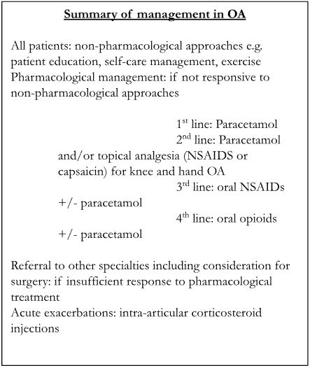
The first step in the management of OA is non-pharmacological approaches. All patients should be given access to appropriate information regarding their condition in a way that they can best understand. This can be offered through verbal advice, websites (e.g. Versus Arthritis, www.versusarthritis.org, Arthritis and Musculoskeletal Alliance ARMA, www.arma.uk.net) or NHS information leaflets. Weight loss should be recommended to those who are overweight, and advice should be given on local muscle-strengthening exercises and aerobic fitness training to protect the joints. Examples of helpful exercises are balance exercises, tai chi, yoga and water-based exercise. Additional advice on appropriate footwear, use of walking sticks, and alternative methods of pain management, for example, heat or cold packs and transcutaneous electrical nerve stimulation (TENS) can be suggested. No evidence has been reported in the literature for alternative therapies, such as the use of glucosamine or acupuncture. Patients can also be offered support for their condition and its associated psychological impact.
Pharmacological management
If non-pharmacological approaches do not achieve adequate symptom control, pharmacological management strategies are required, although these can often be used on an as-required basis in some patients.
Patients are initially advised to use simple analgesia such as paracetamol and topical non-steroidal anti-inflammatory drugs (NSAIDs), especially in hand or knee involvement, provided there are no contraindications to treatment. Potential adverse effects of paracetamol include hepatic toxicity, although this is rare at low doses. Patients using topical ibuprofen should be advised against the risk of severe burns in the presence of naked flames. If these drugs fail to produce sufficient symptomatic control, oral NSAIDs such as ibuprofen or COX 2 inhibitors such as etoricoxib and celecoxib can be used in conjunction with a proton pump inhibitor. Care should be taken, especially when prescribing these drugs in the elderly or those with renal, cardiac or gastrointestinal problems. NSAIDs and COX-2 inhibitors should be used at the lowest effective dose for the shortest period of time possible. Selective COX-2 inhibitors are associated with reduced risk of gastrointestinal side effects compared with NSAIDs, but carry a similar risk of renal toxicity. A careful selection of drugs and adequate patient education are required to maximise the risk-to-benefit ratio. If oral NSAIDs are being used, treatment with topical NSAIDs should be stopped. If the above treatment is ineffective, opioids such as codeine can be used if there are no contraindications. Topical capsaicin can be considered, especially for hand or knee involvement. In patients with acute exacerbations of OA in the knee or hip with moderate to severe pain, intra-articular corticosteroid injections can be considered as adjuncts to treatment.
Referral to other specialties
As part of the management strategy for OA, patients should be reviewed regularly to reinforce education and self-help advice and assess response to treatment. For patients who do not experience symptom resolution despite optimal self-management strategies and pharmacological therapies, referrals can be made to other members of the multidisciplinary team, including physiotherapists, occupational therapists, podiatrists, orthopaedic surgeons or pain clinics, depending on clinical scenario.
Surgery
Prior to referral for surgery, patients must have been offered non-pharmacological and pharmacological therapies. Surgical therapy should be considered for those patients who experience symptoms with a substantial impact on their function and quality of life and who are refractory to non-surgical management. Patient factors such as age, sex, smoking status, and BMI should not be considered as barriers to referral for surgery. Surgical treatment usually involves joint replacement or fusion; for patients with knee osteoarthritis with a history of mechanical locking, arthroscopic lavage and debridement may be considered. Complications associated with surgery include infection, aseptic loosening, pain and dislocation.
Adapted from: NICE guidelines: Osteoarthritis: care and management. National Institute for Health and Care Excellence (NICE); 2014
References
- Conaghan PG, Dickson J, Grant RL. Care and management of osteoarthritis in adults: summary of NICE guidance. Bmj. 2008 Feb 28;336(7642):502-3.
- Graham DJ, Campen D, Hui R, Spence M, Cheetham C, Levy G, Shoor S, Ray WA. Risk of acute myocardial infarction and sudden cardiac death in patients treated with cyclo-oxygenase 2 selective and non-selective non-steroidal anti-inflammatory drugs: nested case-control study. The Lancet. 2005 Feb 5;365(9458):475-81.
- National Institute for Health and Care Excellence (Great Britain). Osteoporosis: assessing the risk of fragility fracture. National Institute for Health and Care Excellence (NICE); 2017
Complete Guide to Clinical Genetics ST3 Application and Recruitment (2027)
Written by Medibuddy. Last updated: October 2026. Clinical Genetics is

Complete Guide to Clinical Radiology ST3 Application and Recruitment (2027)
Recruitment paused: NHS England is not running Clinical Radiology ST3+ recru

Complete Guide to Paediatric Surgery ST3 Application and Recruitment in United Kingdom (2026)
Written by Medibuddy. Last updated: October 2026. If you’re planning


Take your subscriptions with you
Our mobile app allows you to access your interview and exam question banks wherever you are.

