


 Close
Close
 31st July 2023
Other GI Tract Pathology
Congenital Abnormalities of the GI tract
Meckel’s Diverticulum
Meckel’s diverticulum are congenital diverticulum due to embryonic remnants of the vitelline duct in the distal ileum, made up of gastric and pancreatic tissue. The remnant tissue may secrete gastric acid, leading to ulceration and bleeding, and it is an important differential for occult GI bleeding in adults.
The majority of those with this condition are asymptomatic. Those with symptoms typically present early in life to paediatrics (<2 years), but symptoms can potentially arise at any age. Clinical features may include fresh PR bleeding (or melaena) and those of bowel obstruction. Rarely, it can present with severe right iliac fossa pain and mimic acute appendicitis. Other complications include diverticulitis, perforation and the formation of a Littre hernia.
The rule of 2s may be used as a memory aid to remember some clinical features:
- 2% of the population affected
- 2 inches in length
- 2 feet proximal to the ileocaecal valve
- 2 types of ectopic tissue
- 2 years old – peak age for presentation
- 2:1 male to female ratio
The diagnosis is usually made with a radionuclide scan (technetium-99m pertechnetate) which will accumulate in ectopic gastric tissue. In asymptomatic patients, it is often found incidentally during laparoscopy or laparotomy.
Patients with a symptomatic Meckel’s diverticulum should be referred to the general surgery team for resection. For those with an incidental Meckel’s diverticulum, the decision regarding whether to operate is made depending on age, comorbidities and size of diverticulum.

A surgical specimen of Meckel’s diverticulum.
Image Source: Wikimedia Commons (2010). Meckel’s Diverticulum AFIP, Licensed under a creative commons licence. Available for free at: https://commons.wikimedia.org/wiki/File:Meckel%27s_Diverticulum_AFIP.jpg (Accessed: 12 May 2022).

Gilbert’s Syndrome
Gilbert’s syndrome is an autosomal recessive* condition causing decreased activity of UDP glucuronosyltransferase activity, causing defective bilirubin conjugation to glucuronic acid. The prevalence is likely 1-2% in the general population.
Some patients will never have symptoms, whereas others may be jaundiced during periods of stress, such as intercurrent illness, intense exercise, fasting or pregnancy. Patients may have a family history of jaundice. Bloods show an unconjugated hyperbilirubinaemia without any other significant abnormalities. As the additional serum bilirubin is not conjugated, there will be no urobilinogen increase in the urine.
The differential for an unconjugated hyperbilirubinaemia include causes of haemolytic anaemia (e.g., autoimmune, haemoglobinopathies, red blood cell enzyme disorders, red blood cell membrane disorders) and Crigler-Najjar syndrome. Reticulocytes and other markers of haemolysis should always be checked.
To confirm the diagnosis, bilirubin may be checked after periods of prolonged fasting or after nicotinic acid administration, which increases osmotic fragility of red blood cells, causing a higher proportion to break down than usual.
Gilbert’s is a benign condition, and no specific management is required aside from patient education.
* some rarer variants appear to be inherited in an autosomal dominant pattern
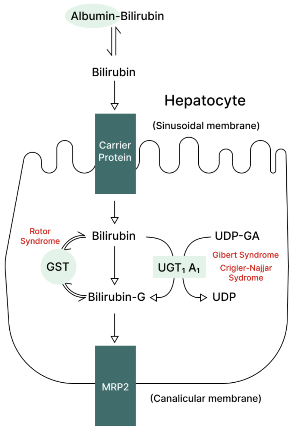
Metabolic pathway for bilirubin in the hepatocyte. Gilbert’s syndrome and Crigler-Najjar syndrome are associated with decreased UDPGT activity; therefore, bilirubin is not conjugated.
Crigler Najjar Syndrome
Crigler Najjar syndrome is an autosomal dominant condition causing absent activity of UDP glucuronyl transferase activity. In contrast to the above, the condition presents in infancy with jaundice, with or without CNS signs (secondary to kernicterus).
Bloods will again show an unconjugated hyperbilirubinaemia. The disease is life-threatening and the only cure is an urgent liver transplant.
Dubin-Johnson Syndrome
Dubin-Johnson syndrome is an autosomal recessive disorder causing a conjugated hyperbilirubinaemia. Typically, patients present in adolescence with intermittent jaundice and right upper quadrant pain. It often worsens during pregnancy.
It is due to a defect in the canalicular multispecific organic anion transporter (cMOAT) protein, leading to defective hepatocyte excretion of conjugated bilirubin. Therefore, unlike the two conditions above, urobilinogen will be raised in the urine. Liver biopsy shows a characteristic pigmented, ‘black’ liver.
No treatment is required.
Anaemia
Iron Deficiency Anaemia
Anaemia is not a disease in its own right, and an underlying cause should always be sought. Further information regarding anaemia more generally can be found in the ‘Haematology’ section.
Patients may have clinical features of anaemia in general, such as fatigue, dyspnoea, pallor and tachycardia. Those with chronic iron deficiency may develop atrophic glossitis, angular stomatitis and/or koilonychia.
Iron deficiency is the most common cause of anaemia, and patients will have a microcytosis along with their low haemoglobin levels. MCV, MCH and MCHC are all reduced. The exception is when patients have coexistent folate and/or B12 deficiency, as this will lead to a dimorphic red cell population with both microcytes and macrocytes circulating. In these instances, the red cell distribution width (RCDW) would be high.
The differential for a microcytic anaemia includes thalassaemia, sideroblastic anaemia and lead poisoning. These can be distinguished from iron deficiency anaemia with haematinics (serum ferritin, iron and transferrin saturation) and a blood film. The film in iron deficiency anaemia reveals hypochromic, microcytic cells, and there may also be target cells and pencil poikilocytes.
Patients with chronic bleeding typically present with iron deficiency anaemia, and as such, 1 in 6 menstruating women have the condition. New onset iron deficiency anaemia in all men, and all women after the menopause, should be assumed to be from a GI tract lesion until proven otherwise, unless they have overt non-gastrointestinal bleeding. Other causes include malabsorption (e.g., coeliac disease), dietary deficiency and hookworm infection.
NICE guidelines state that all men and postmenopausal women with iron deficiency anaemia should be referred to gastroenterology, where they will undergo both upper and lower GI endoscopy. A coeliac screen should also be sent. If patients are over 60, or over 50 and have rectal bleeding, the referral should be under a 2-week wait pathway. Premenopausal women may also be referred to the gastroenterology team if they have bowel symptoms, a strong family history of GI malignancy or resistant disease.
If the above investigations do not reveal pathology and a cause is yet to be found, the small bowel should be imaged if the anaemia is transfusion dependent (otherwise, the diagnostic yield is very low). This can be done with video capsule endoscopy. Finally, angiography of the coeliac and mesenteric vessels may be required.
Along with managing the underlying cause, the iron deficiency itself can be treated with iron supplementation. Oral iron is adequate for most patients, e.g., ferrous sulphate or ferrous fumarate. This should be continued until haemoglobin returns to normal range, and for at least 3 months after this to allow stores to be replenished. Intravenous iron is required in a small proportion, such as those with chronic kidney disease. These patients should be iron replete prior to a trial of erythropoiesis stimulating agents.
The most common reason that patients fail to respond to iron replacement is compliance. Side effects include constipation and GI irritation; advise that these typically settle down with time. Troublesome adverse effects can be minimised if iron supplements are taken with or after food, or if dose frequency is reduced to alternate days.
Monitoring is important, and haemoglobin levels should be rechecked within 4 weeks of commencing iron supplementation, and periodically after this.

Algorithm for the management of iron deficiency anaemia, as suggested by the British Society of Gastroenterology. IDA is iron deficiency anaemia; IRT is iron replacement therapy; OGD is oesophago-gastroduodenoscopy.
Pernicious Anaemia
Describes autoimmune destruction of gastric parietal cells that secrete intrinsic factor, reducing vitamin B12 absorption. B12 is key in the erythropoiesis and in myelination of neurons, leading to the clinical features. Pernicious anaemia is the most common cause of B12 deficiency; the differential includes dietary deficiency (especially seen in vegans), disease of the terminal ileum (e.g., Crohn’s disease) or previous gastrectomy.
Pernicious anaemia is strongly associated with other autoimmune conditions; thus, a thorough past medical history and family history is required. The associations, clinical features and investigations are summarised in Table 1. The condition is rare in those <40 years old. Diagnosis is usually made with positive autoantibodies following clinical suspicion.
| Associations | Clinical features | Investigations |
|---|---|---|
| Autoimmune thyroid disease | Those of anaemia: lethargy, weakness, dyspnoea | FBC: anaemia, macrocytosis, possibly reduced white cell count and platelets |
| Vitiligo | ‘Lemon tinge’ to skin due to pallor from anaemia, and mild jaundice from haemolysis | Blood film: hypersegmented neutrophils Marrow: megaloblasts |
| Addison’s disease | Neurological: peripheral neuropathy, paraesthesia | B12 levels may be low. Also, check folate levels – if both are deficient, give B12 first to avoid precipitating subacute combined degeneration of the cord |
| Rheumatoid arthritis | Psychiatric: depression, dementia, irritability | Intrinsic factor antibodies: not particularly sensitive (50%) but highly specific |
| Type 1 diabetes mellitus | Those of subacute combined degeneration of the cord if untreated: ataxia, weakness and paraesthesia. Extensor plantars with absent knee and ankle jerks | Parietal cell antibodies: sensitive (90%) but not particularly specific |
| Blood group A | Increased risk of gastric cancer | (Schilling tests involved administering radiolabelled B12 and IF, then measuring urinary B12 levels, but is no longer used in the UK) |
Table 1: Associations, clinical features and investigations for pernicious anaemia.
Management involves B12 replacement in the form of intramuscular hydroxocobalamin. If there are no neurological features, 3 injections per week are given for the first 2 weeks, followed by injections once every 3 months. The 1mg doses are given more frequently if neurological features are present; contact the haematology team for an appropriate regimen.

Schematic showing normal absorption of vitamin B12 from the diet. In pernicious anaemia, there is reduced or absent intrinsic factor secretion; therefore, much less vitamin B12 is absorbed at the terminal ileum.
Vascular Disorders of the GI Tract
Acute Mesenteric Ischaemia
Describes sudden-onset ischaemia of the small bowel secondary to hypoperfusion of the superior mesenteric artery (SMA) or its branches. The hypoperfusion can be secondary to arterial thrombosis, arterial embolism or non-occlusive ischaemia due to poor cardiac output.
The predisposing factors, and signs and symptoms, of all syndromes of ischaemia to the lower gastrointestinal tract are similar and are summarised in Table 1. However, the management and prognosis vary greatly; therefore, it is important to differentiate the various types.
| Risk factors | Clinical features |
|---|---|
| Atrial fibrillation (always suspect ischaemia to the lower GI tract if AF + abdominal pain) | Abdominal pain: particularly severe in acute mesenteric ischaemia, with pain out of proportion to the physical examination findings. |
| Cardiovascular risk factors: hypertension, smoking, hyperlipidaemia, diabetes mellitus | Per rectal bleeding |
| Cocaine use | Fever |
| Increasing age | Diarrhoea |
| Other causes of embolism: infective endocarditis, mural thrombus, malignancy | Lactic acidosis: therefore, a blood gas is imperative in all cases of acute abdomen |
Table 2: Common risk factors and clinical features to both mesenteric ischaemia and ischaemic colitis
The sudden-onset, severe pain is usually constant, central within the abdomen and responds poorly to analgesia. Typically, there are no other abdominal signs. Along with a raised serum lactate, there may be an increased serum amylase and leucocytosis.
The diagnosis is usually made on CT abdomen and pelvis with contrast, though this should not delay laparotomy if there is high suspicion for the condition. The CT may reveal gas in the intestinal wall or portal vein.
The general surgery team will typically organise an emergency laparotomy, and the prognosis is especially poor if the operation is delayed. The bowel is assessed for viability and the necrotic bowel is removed. Endovascular thrombolysis or thrombectomy may be an option in a minority of patients. Patients will usually remain on long-term anticoagulation.
Complications include peritonitis, with or without sepsis, due to bacterial translocation across the necrotic bowel. Therefore, broad-spectrum antibiotics are usually commenced, with a regimen that will also target anaerobic bacteria (e.g., ceftriaxone + metronidazole). Mortality rate is high, estimated at around 50%.

Acute bowel ischaemia leading to necrosis
Image Source: Wikimedia Commons (2008). Intestinal Obstruction, Licensed under a creative commons licence. Available for free at: https://commons.wikimedia.org/wiki/File:Intestinal_Obstruction.JPG (Accessed: 28 June 2023).

Chronic Mesenteric Ischaemia
Also known as intestinal angina. Usually a consequence of atherosclerotic plaques in the large mesenteric vessels, leading to colicky, post-prandial abdominal pain. Typically, the pain commences around thirty minutes after eating and lasts approximately one hour. Other symptoms include weight loss (due to ‘fear’ of eating and/or malabsorption), nausea & vomiting and PR bleeding. Examination may reveal bruits of the mesenteric vasculature.
Patients will almost always have atheromatous disease elsewhere, thus enquire about features of cardiovascular disease, cerebrovascular disease, peripheral arterial occlusive disease and renovascular disease.
Diagnosis is usually made with CT or MR angiography, which will reveal atherosclerosis. Duplex ultrasound of the mesenteric arteries may aid in analysis of blood flow through a blockage in real time.
Conservative approaches target the underlying causes of atherosclerosis. Surgical options include angioplasty + stenting, or bypassing the blocked vessel with an open procedure.
Ischaemic Colitis
Describes compromised blood flow to the large bowel. Severity can range from mild ischaemia to severe gangrenous colitis. Most commonly occurs at ‘watershed’ areas, such as the splenic flexure (Griffiths point), which is located at the border of the territory supplied by the superior and inferior mesenteric arteries.
The most common symptoms are left iliac fossa pain and haematochezia.
Abdominal XR may reveal ‘thumbprinting’ due to submucosal oedema. CT is usually diagnostic, with bowel walls thickening a common feature.
Management is conservative in the first instance and involves fluids and antibiotics. A small proportion of patients will require bowel resection with stoma formation.
Complications in severe ischaemic colitis include ongoing haemorrhage, bacterial contamination causing peritonitis and perforation.

Watershed areas of the colon.
Image Source: Wikimedia Commons (2008). Colonic blood supply, Licensed under a creative commons licence. Available for free at: https://commons.wikimedia.org/wiki/File:Colonic_blood_supply.svg (Accessed: 28 June 2023).

Ischaemic Hepatitis
A cause of acute liver failure secondary to insufficient perfusion to the liver. This is frequently due to systemic hypotension, though it can rarely be caused by a clot in the hepatic artery or significant activation of the renin-angiotensin system, which results in splanchnic vasoconstriction. The condition is often termed ‘shock liver’ and is commonly a result of septic, hypovolaemic, cardiogenic or other types of shock. Therefore, it is very likely that there will be concurrent damage to other end organs that are sensitive to ischaemia, such as the kidneys and brain.
The patient may present with jaundice and features of hepatic encephalopathy, such as confusion and asterixis. However, the diagnosis is usually made on the basis of a marked increase in aminotransferase levels after an inciting event. The differential for a very high ALT level (>1000 IU/L) includes paracetamol overdose and acute viral hepatitis, and these should be excluded.
Treating the underlying cause of the ischaemia is the mainstay of management, and critical care support may be required to restore and maintain adequate blood pressure.
The typical histopathological findings of the liver at post-mortem are centrilobular necrosis with normal periportal hepatocytes.
Budd-Chiari Syndrome
The eponymous name for hepatic vein occlusion. Typically caused by a thrombus but can also be secondary to infiltration by a tumour.
The classic triad includes right upper quadrant pain, ascites and hepatomegaly. The clinical features are summarised in Table 3.
| Clinical feature | Additional notes |
|---|---|
| Abdominal pain | Typically sudden onset and severe. Usually in the right upper quadrant but can be epigastric. |
| Liver enlargement | The hepatomegaly is usually tender. The differential for tender hepatomegaly includes right-sided heart failure and constrictive pericarditis, which similarly lead to hepatic congestion. |
| Ascites | May present as abdominal distension. Serum-ascites albumin gradient (SAAG) would reveal a transudate (>11g/L). |
| Portal hypertension | If left unchecked, can lead to complications such as acute variceal bleeding. |
| Hepatocyte damage | Leads to a transaminitis, with raised ALT and AST. Can rarely lead to fulminant, acute, subacute or chronic liver failure, or can cause decompensation of existing chronic liver disease. Liver cirrhosis is a possibility with ongoing hepatocyte destruction and subsequent fibrosis. |
Table 3: Features of Budd-Chiari syndrome
Budd-Chiari syndrome is exceptionally rare in the absence of a procoagulant condition, and its presence should trigger investigation for the cause of hypercoagulability. A rarer alternative is the direct extension of a tumour mass into the hepatic vein. The causes are shown in Table 2.
| Hypercoagulable state | Tumour infiltration |
|---|---|
| Polycythaemia rubra vera or other primary myeloproliferative disorders | Kidney tumour |
| Thrombophilia: factor V Leiden, protein C or S deficiency, antithrombin III deficiency | Hepatic tumour |
| Pregnancy | Adrenal tumour |
| Combined oral contraceptive pill | |
| Paroxysmal nocturnal haemoglobinuria | |
| Antiphospholipid syndrome |
Table 4: Causes of Budd-Chiari syndrome
If Budd-Chiari syndrome is suspected, the initial radiological investigation is typically ultrasound with doppler flow studies, which is highly sensitive for hepatic vein occlusion. An alternative is a CT with contrast, for which hypertrophy of the caudate lobe is a characteristic sign (highly specific but only seen in ~50% of cases).
The management is variable and will typically be made by the hepatology team. Lifelong anticoagulation is often required, though this may be contraindicated with concurrent oesophageal varices. The occlusion may need to be bypassed with a surgical shunt or a transjugular intrahepatic portosystemic shunt (TIPSS). If fulminant liver failure is present, the patient may be eligible for a liver transplant.
Complete Guide to Clinical Genetics ST3 Application and Recruitment (2027)
Written by Medibuddy. Last updated: October 2026. Clinical Genetics is

Complete Guide to Clinical Radiology ST3 Application and Recruitment (2027)
Recruitment paused: NHS England is not running Clinical Radiology ST3+ recru

Complete Guide to Paediatric Surgery ST3 Application and Recruitment in United Kingdom (2026)
Written by Medibuddy. Last updated: October 2026. If you’re planning


Take your subscriptions with you
Our mobile app allows you to access your interview and exam question banks wherever you are.

