


 Close
Close
 31st July 2023
Congenital Abnormalities of the GI tract
Meckel’s Diverticulum
Meckel’s diverticulum are congenital diverticulum due to embryonic remnants of the vitelline duct in the distal ileum, made up of gastric and pancreatic tissue. The remnant tissue may secrete gastric acid, leading to ulceration and bleeding, and it is an important differential for occult GI bleeding in adults.
The majority of those with this condition are asymptomatic. Those with symptoms typically present early in life to paediatrics (<2 years), but symptoms can potentially arise at any age. Clinical features may include fresh PR bleeding (or melaena) and those of bowel obstruction. Rarely, it can present with severe right iliac fossa pain and mimic acute appendicitis. Other complications include diverticulitis, perforation and the formation of a Littre hernia.
The rule of 2s may be used as a memory aid to remember some clinical features:
- 2% of the population affected
- 2 inches in length
- 2 feet proximal to the ileocaecal valve
- 2 types of ectopic tissue
- 2 years old – peak age for presentation
- 2:1 male to female ratio
The diagnosis is usually made with a radionuclide scan (technetium-99m pertechnetate) which will accumulate in ectopic gastric tissue. In asymptomatic patients, it is often found incidentally during laparoscopy or laparotomy.
Patients with a symptomatic Meckel’s diverticulum should be referred to the general surgery team for resection. For those with an incidental Meckel’s diverticulum, the decision regarding whether to operate is made depending on age, comorbidities and size of diverticulum.

A surgical specimen of Meckel’s diverticulum.
Image Source: Wikimedia Commons (2010). Meckel’s Diverticulum AFIP, Licensed under a creative commons licence. Available for free at: https://commons.wikimedia.org/wiki/File:Meckel%27s_Diverticulum_AFIP.jpg (Accessed: 12 May 2022).

Gilbert’s Syndrome
Gilbert’s syndrome is an autosomal recessive* condition causing decreased activity of UDP glucuronosyltransferase activity, causing defective bilirubin conjugation to glucuronic acid. The prevalence is likely 1-2% in the general population.
Some patients will never have symptoms, whereas others may be jaundiced during periods of stress, such as intercurrent illness, intense exercise, fasting or pregnancy. Patients may have a family history of jaundice. Bloods show an unconjugated hyperbilirubinaemia without any other significant abnormalities. As the additional serum bilirubin is not conjugated, there will be no urobilinogen increase in the urine.
The differential for an unconjugated hyperbilirubinaemia include causes of haemolytic anaemia (e.g., autoimmune, haemoglobinopathies, red blood cell enzyme disorders, red blood cell membrane disorders) and Crigler-Najjar syndrome. Reticulocytes and other markers of haemolysis should always be checked.
To confirm the diagnosis, bilirubin may be checked after periods of prolonged fasting or after nicotinic acid administration, which increases osmotic fragility of red blood cells, causing a higher proportion to break down than usual.
Gilbert’s is a benign condition, and no specific management is required aside from patient education.
* some rarer variants appear to be inherited in an autosomal dominant pattern
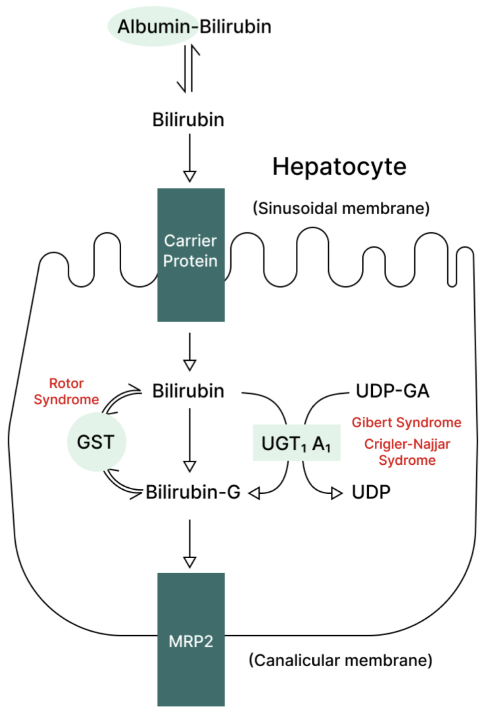
Metabolic pathway for bilirubin in the hepatocyte. Gilbert’s syndrome and Crigler-Najjar syndrome are associated with decreased UDPGT activity; therefore, bilirubin is not conjugated.
Crigler Najjar Syndrome
Crigler Najjar syndrome is an autosomal dominant condition causing absent activity of UDP glucuronyl transferase activity. In contrast to the above, the condition presents in infancy with jaundice, with or without CNS signs (secondary to kernicterus).
Bloods will again show an unconjugated hyperbilirubinaemia. The disease is life-threatening and the only cure is an urgent liver transplant.
Dubin-Johnson Syndrome
Dubin-Johnson syndrome is an autosomal recessive disorder causing a conjugated hyperbilirubinaemia. Typically, patients present in adolescence with intermittent jaundice and right upper quadrant pain. It often worsens during pregnancy.
It is due to a defect in the canalicular multispecific organic anion transporter (cMOAT) protein, leading to defective hepatocyte excretion of conjugated bilirubin. Therefore, unlike the two conditions above, urobilinogen will be raised in the urine. Liver biopsy shows a characteristic pigmented, ‘black’ liver.
No treatment is required.
Complete Guide to Clinical Genetics ST3 Application and Recruitment (2027)
Written by Medibuddy. Last updated: October 2026. Clinical Genetics is

Complete Guide to Clinical Radiology ST3 Application and Recruitment (2027)
Recruitment paused: NHS England is not running Clinical Radiology ST3+ recru

Complete Guide to Paediatric Surgery ST3 Application and Recruitment in United Kingdom (2026)
Written by Medibuddy. Last updated: October 2026. If you’re planning


Take your subscriptions with you
Our mobile app allows you to access your interview and exam question banks wherever you are.

