


 Close
Close
 10th January 2024
Pleural Effusion
Background
Epidemiology
Pleural effusions are a common pathology seen in respiratory medicine, with approximately 200,000 new cases in the UK each year with over 50 underlying aetiologies spanning a breadth of medical specialities. Pleural effusions can largely be categorised into 2 forms: transudative and exudative. The most common underlying cause of a transudative pleural effusion is heart failure, whereas the most common cause of an exudative effusion is pneumonia.
Pathophysiology
In normal physiological circumstances, the volume of pleural fluid is carefully governed by a balance between liquid into the pleural space (via the parietal pleura) and liquid outflow (via the parietal and visceral pleura). A small amount of fluid (estimated at 10-20mL, or 0.3mL/kg) is always present within the space to allow effective ventilation of the lungs. Several mechanisms help maintain this tightly controlled volume: Starling forces at the visceral pleura, lymphatic drainage at the parietal pleura and “electrolyte-coupled liquid absorption” via the mesothelium on both visceral and parietal pleura.
Pleural effusions result from either increased pleural fluid production or impaired absorption via a) a change in the transpleural pressure balance and subsequently Starling’s forces, b) impairment of the lymphatic system or c) change in mesothelial permeability.
Transudates
Transudative pleural effusions result from increases in the systemic capillary pressure in the setting of congestive cardiac failure. This increased capillary pressure changes hydrostatic and oncotic pressures across the pleura, thus altering the normal equilibrium seen, i.e., Starling’s forces. This alteration results in pleural fluid production exceeding its absorption and resultant accumulation. As the mesothelium remains largely unaltered, protein and cell leakage into the fluid is minimal.
Transudative pleural effusions secondary to chronic liver disease and nephrotic syndrome also result from a change in Starling’s forces at the capillary membrane, but in these cases, it is a reduction in oncotic pressure from hypoalbuminaemia. This drives the balance in favour of hydrostatic pressure, and subsequently, fluid is moved into the pleural space.
Exudates
Exudative pleural effusions are either a result of impaired protein and cell removal from the pleural space (via the lymphatic system) or from increased permeability of the mesothelial membrane to water and small solutes. This results in fluid accumulation that is high in cell number and protein within the pleural space.
Specifically, in parapneumonic exudative effusions (the most common form of exudative pleural effusion) it is the direct result of inflammatory-mediating cytokines and cells interacting with the mesothelial membrane, which increase its permeability to cells and protein. At times, this inflammatory-mediated damage to the mesothelium can be so significant that bacteria can enter the pleural space, resulting in the formation of an empyema (pleural infection).
Presentation
Pleural effusion presentation can be variable due to several changeable factors (size of effusion, how quickly the effusion accumulated, its underlying cause), but the overwhelmingly common symptom is “shortness of breath.” A detailed history of the shortness of breath should be undertaken to determine character, speed of onset, duration and how limiting the symptomology is on activities of daily living. Given there is a significant range of pathologies that pleural effusions can be associated with, there is also variety in the associated symptoms.
Important associated symptoms to consider would include presence of –
- Constitutional symptoms (fever, night sweats, weight loss),
- Cough and sputum production
- Recent travel history
- Past medical history (including specifically if any current or previous cancer diagnosis),
- Occupational history
- Smoking history.
Signs
On clinical examination, reduced chest expansion may be noted, either unilaterally or bilaterally, dependent on the underlying effusion aetiology. The typical finding described is “stony” dull percussion over the area of the pleural effusion with associated reduced breath sounds. Bronchial breathing can also be present just superior to the uppermost point of the effusion. In massive pleural effusion tracheal deviation away from the side of effusion can also be seen.
In addition to the effusion itself, clinical examination can give clues to the underlying aetiology:
| Clinical Sign | Possible associated aetiology |
|---|---|
| Clubbing, cachexia and tar staining | Bronchial carcinoma |
| Productive cough, fever | Pneumonia/ empyema |
| Peripheral oedema, raised JVP and displaced apex beat | Congestive cardiac failure |
| Multiple spider naevi (>5), palmar erythema, jaundice | Hepatic failure |
| AV fistula, renal transplant | Renal failure |
| Swan neck deformity, Boutonniere deformity | Rheumatoid arthritis |
Table 1: Clinical features with their associated underlying aetiology.
Table 1: Table adapted from –
Bhatnagar R, Maskell N. The modern diagnosis and management of pleural effusions. BMJ. 2015;351(September).
Saguil A, Wyrick K, Hallgren J. Diagnostic approach to pleural effusion. Am Fam Physician. 2014;90(2):99–104.
Diagnosis
A diagnosis of pleural effusion should be considered in any patient presenting to hospital with shortness of breath. The combination of history and thorough physical examination revealing findings, as detailed above, should alert the clinician to the possibility of an underlying pleural effusion.
Chest x-ray
Plain posteroanterior (PA) chest x-rays are the first-line imaging modality used in suspected pleural effusion and become abnormal with around 200mL of pleural fluid accumulation. The fluid results in blunting of the costophrenic angle with associated development of the meniscus. If there is air associated with the pleural effusion, called a hydropneumothorax, a straight line at the air-fluid interface will be seen, rather than a meniscus.

Large L-sided unilateral pleural effusion in a female patient.
Image Source: Wikimedia Commons (2022). Pleural Effusion, Licensed under a creative commons licence. Available for free at: https://commons.wikimedia.org/wiki/Category:Pleural_effusion (Accessed on: 17 May 2022).
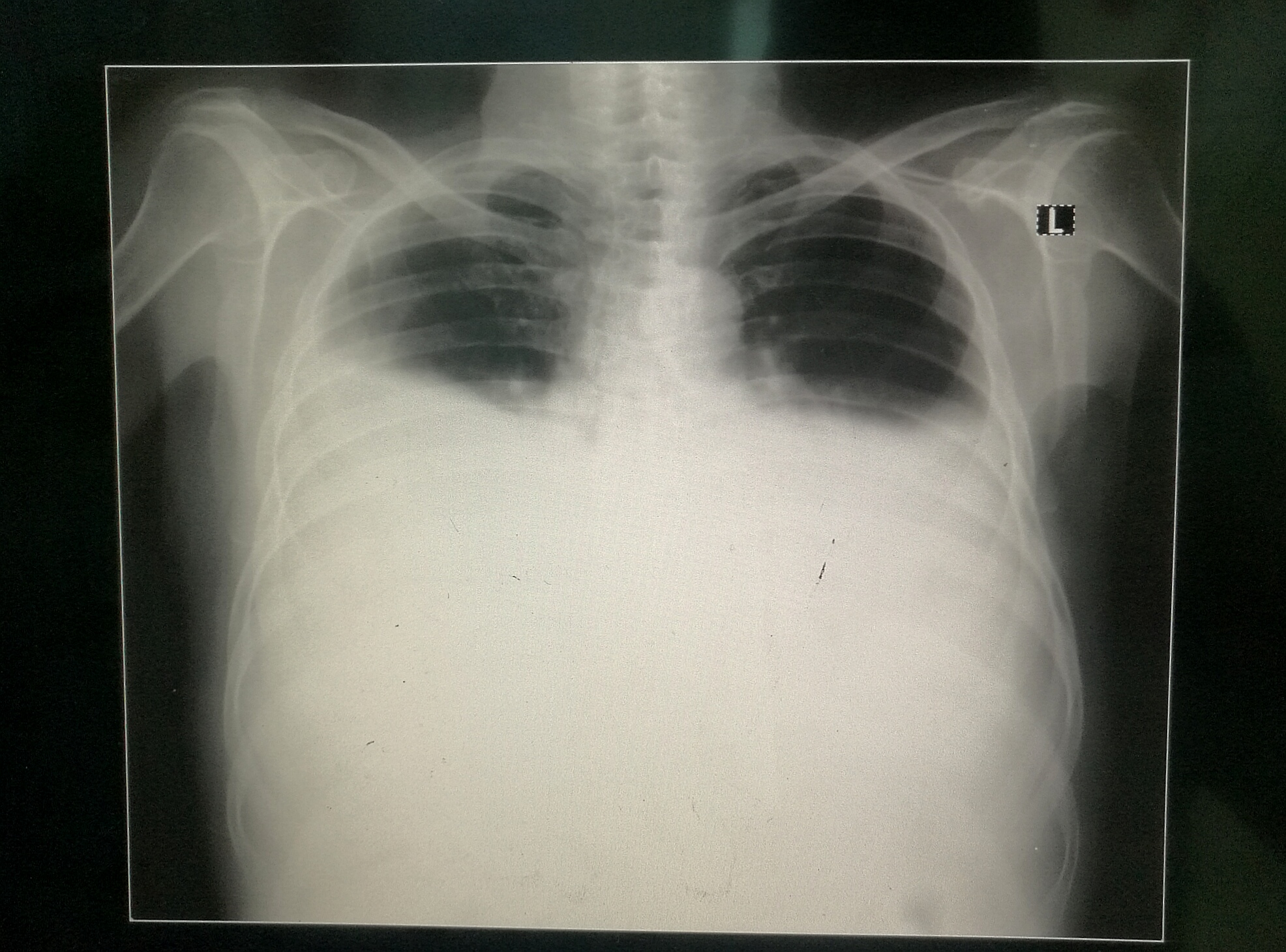
Large bilateral pleural effusions.
Image Source: Wikimedia Commons (2017). Bilateral Pleural Effusion, Licensed under a creative commons licence. Available for free at: https://upload.wikimedia.org/wikipedia/commons/3/34/Bilateral_Pleural_Effusion.jpg (Accessed on: 17 May 2022).
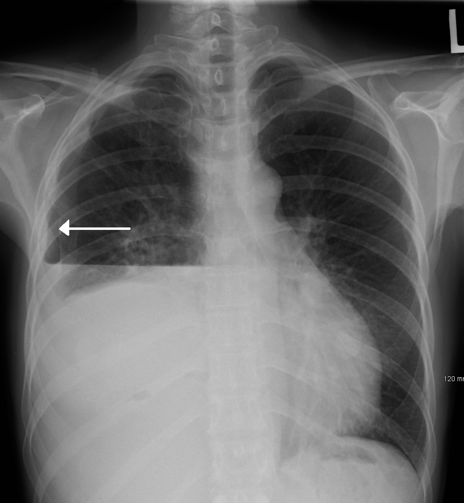
R-sided hydropneumothorax in a male patient. Key difference to note compared to a pleural effusion without associated pneumothorax is the straight line caused by the air-fluid interface.
Image Source: Wikimedia Commons (2017). Hydropneumothorax, Licensed under a creative commons licence. Available for free at: https://upload.wikimedia.org/wikipedia/commons/7/7d/HydropneumoX.png (Accessed on: 17 May 2022).
Ultrasound
Bedside pleural ultrasound can be used to identify pleural effusions and be utilised to identify a site for diagnostic aspiration of pleural fluid. It is particularly useful in the diagnosis of small pleural effusions, which may not be evident on a plain radiograph. In addition to its uses for ultrasound-guided diagnostic aspiration of pleural fluid, ultrasound can be used to determine additional characteristics of the fluid, e.g. septations, which may be suggestive of an underlying malignant or infective effusion.
Diagnostic Aspiration
Diagnostic aspiration is the key diagnostic test in evaluating a unilateral pleural effusion. British Thoracic Guidelines (BTS) state this should be a bedside direct ultrasound-guided aspiration of fluid in the most lateral position possible to reduce associated complications. A 50mL sample of fluid is obtained and sent for:
- Protein
- Lactate dehydrogenase
- Gram staining
- Microbiology culture
- Cytology
- pH (if an infected pleural effusion is suspected)
On aspiration, pleural fluid can take on many appearances. “Normal” pleural fluid is straw-coloured and inodorous. However, in the setting of pathology, many different appearances of pleural fluid can be noted, including blood-strained, frank blood, purulent (suggesting empyema), milky (suggesting chylothorax) or bile-stained.

Diagnostic algorithm for pleural effusion once identified.
Flowchart adapted from guidance available at – Maskell N. British Thoracic Society pleural disease guidelines – 2010 update. Thorax. 2010;65(8):667–9.
Pleural Effusion Fluid Analysis
The key distinction pleural fluid analysis attempts to achieve is exudative vs. transudative pleural effusions. This distinction allows a narrowing of the differential diagnosis (see below).
Pleural fluid protein (g/L) measurement is the key to differentiating between transudates and exudates. Pleural fluid protein >30g/L is considered exudative and protein <30g/L is considered transudative. However, the closer the level is to 30g/L, the less accurate this distinction becomes. In order to achieve a distinction between transudate and exudate in this setting we employ Light’s Criteria:
- Pleural fluid protein to serum protein ratio >0.5
- Pleural fluid LDH to serum LDH ratio >0.6
- Pleural LDH >0.67 x upper limit of normal for serum LDH
If any one of these criteria are met, an exudative pleural effusion diagnosis can be made. Lactate dehydrogenase is utilised in Light’s Criteria as it is a non-specific marker of inflammation – a key component of exudative pleural effusion pathology.
Microscopy, Culture and Sensitivity
Microscopy, culture and sensitivity (M, C + S) is the gold-standard test that is sent with all diagnostic pleural aspirations as per BTS guidelines. TB cultures can be sent in addition if this is suspected in order to look for acid-fast bacilli.
Cell differential is widely reported to be of limited diagnostic benefit as, in the majority of cases, the differential diagnosis remains wide. Pleural fluid can be predominantly lymphocytic, neutrophilic or eosinophilic.
- Lymphocytic. Defined as >50% of pleural fluid cells being lymphocytes. In this instance differentials to consider would be tuberculosis (TB) and malignancy. If a significantly raised lymphocyte count is noted (>80%), this should prompt the clinician to think about TB, as well as rheumatological conditions: rheumatoid arthritis (RA) and sarcoidosis.
- Neutrophilic. This is seen in acute processes and, most commonly, in parapneumonic effusions.
- Eosinophilic. Defined as >10% of pleural fluid cells, and the differential remains wide, but the most common cause is pneumothorax (air in the pleural space) or haemothorax (blood in the pleural space). Rarer conditions, such as eosinophilic granulomatosis with polyangiitis (EGPA), can cause eosinophil-predominant pleural effusions.
Specific Diagnostic Tests of Pleural Effusion
- Amylase. A raised amylase within pleural fluid is highly sensitive for pancreatic disease, oesophageal rupture or malignancy. The presence of a raised pleural fluid amylase (defined as any pleural amylase level above the limit of normal) narrows the differential significantly to these conditions.
- Triglycerides. High levels of pleural fluid triglyceride concentrations >110mg/dL are diagnostic of chylothorax.
- Cholesterol. Significantly raised cholesterol (>250mg/dL) aids with diagnosis of a pseudochylothorax.
- pH. pH can be utilised in suspected non-purulent pleural infections (empyema). Normal pleural pH is around 7.60, and pleural fluid acidosis is defined as a pH <7.30 and can be seen in a range of causes: malignancy, TB and connective tissue disease. A pleural fluid pH of <7.2 though is highly suggestive of an empyema and should be treated as per pleural infection guidelines (see “Management” below).
- Cytology. Cytological analysis can be utilised in patients with pleural effusions suspected to be secondary to malignancy and has a sensitivity of around 60%. The presence of malignant cells within a pleural fluid sample is the gold standard diagnostic test for malignant pleural effusions.
Differential Diagnosis
The aetiology of pleural effusion is wide, with over 50 aetiologies possible spanning multiple medical specialities. A thorough clinical history, examination and diagnostic approach helps narrow the differential diagnosis. The table below is by no means exhaustive but incorporates important differentials to know for the MRCP exams.
| Aetiology | Transudate or Exudate | Other pleural fluid characteristics | Clinical clues |
|---|---|---|---|
| Cardiovascular | |||
| Heart Failure | Transudate | Straw-coloured (pale yellow). Typically bilateral. Occasionally, lymphocytosis is noted on cell differential of pleural fluid. |
Pulmonary or peripheral oedema Raised pro-brain natriuretic peptide (pro-BNP) level. |
| Gastrointestinal | |||
| Liver cirrhosis | Transudate | Straw-coloured. | Signs of chronic liver disease. Ascites. History of alcohol excess/ viral hepatitis/ non-alcoholic fatty liver disease (NAFLD). |
| Nephrotic syndrome | Transudate | Straw-coloured. | Generalised anasarca. Proteinuria/ haematuria. |
| Pancreatitis | Exudate | Pleural fluid amylase above the limit of normal reference range. | Epigastric tenderness. Raised serum amylase. History of common underlying cause of pancreatitis: gallstones/ alcohol excess. |
| Genitourinary | |||
| Meigs syndrome | Transudate | Straw-coloured. | History of ovarian fibroma. High serum Ca-125. |
| Oncology | |||
| Malignancy | Exudate* (* exception – Meigs syndrome) |
Positive cytology | History of underlying malignancy (lung most common, followed by lymphoma). Weight loss. History of smoking/ tar staining/ clubbing. |
| Respiratory | |||
| Parapneumonic effusion | Exudate | Turbid appearance. Neutrophil-predominance. pH of fluid normal. |
Associated fever/ productive cough. Consolidative changes on chest x-ray. |
| Tuberculosis | Exudate | Lymphocytosis. Acid-fast bacilli (but visualised in <5%). Very high protein (>50g/L) and LDH levels. |
Travel history. Weight loss/ fevers/ haemoptysis. Chest x-ray or thoracic imaging suggestive of TB. Positive sputum for AAFBs. |
| Malignant mesothelioma | Exudate | Abnormal mesothelial cells with positive cytogenetic analysis. | Asbestos exposure previously. |
| Yellow nail syndrome | Exudate | Clear pleural fluid. Lymphocytosis. |
Dystrophic changes to nails. Recurrent infections. |
| Rheumatology | |||
| Rheumatoid arthritis | Exudate | Very low pleural fluid glucose level (typically <1.6mmol/L). High cholesterol. Low pH <7.30. Milky appearance if pseudochylothorax. |
Rheumatoid arthritis signs on peripheral examination: swan neck deformities, Z-thumb, for example. |
| SLE | Exudative | Can be unilateral or bilateral. No distinctive biochemical features noted. |
Multi-system disorder with evidence of photosensitive rash/ arthropathy/ Raynaud’s, for example. |
| Other | |||
| Chylothorax | Exudative | Milky appearance. High triglycerides >110mg/dL. Low cholesterol. Absent cholesterol crystals. |
History of preceding trauma or thoracic surgery. |
| Pseudochylothorax | Exudative | Milky appearance. High cholesterol and cholesterol crystals present. Normal triglycerides. |
Rheumatoid arthritis. High serum cholesterol. |
| Haemothorax | Exudative | Frank blood appearance. | History of trauma. On anticoagulation. |
Table 2: Differential diagnosis of pleural effusion with key points relating to pleural effusion and clinical presentation characteristics.
Table adapted from – Bhatnagar R, Maskell N. The modern diagnosis and management of pleural effusions. BMJ. 2015;351(September). Saguil A, Wyrick K, Hallgren J. Diagnostic approach to pleural effusion. Am Fam Physician. 2014;90(2):99–104. UpToDate. Diagnostic evaluation of a pleural effusion in adults: Initial testing [Internet]. 2022 [cited 2022 May 17]. Available from: https://www.uptodate.com/contents/diagnostic-evaluation-of-a-pleural-effusion-in-adults-initial-testing
Empyema
An important differential diagnosis of pleural effusion is an empyema, meaning infection of the pleural fluid. The gold standard diagnostic test for empyema is the growth of an organism from the aspirated pleural fluid, but it can also be suggested by a low pleural fluid pH <7.2. Pleural infections are seen more commonly at the extremes of age: in the very young and the very old, and as such, has a high associated mortality of 20% in the UK.
The causative organisms vary depending on whether the pleural infection was community-acquired or hospital-acquired in origin. In the community, gram-positive aerobic organisms such as Streptococcus and Staphylococci are the most commonly seen organisms, but the incidence of gram-negative isolates from these patients is increasing. In the hospital setting, the most common organism seen is Staphylococcal aureus (S aureus), with a high proportion of patients growing methicillin-resistant staphylococcal aureus (MRSA). Gram-negative organisms such as E coli, Enterobacter and Pseudomonas species are seen in higher proportion in hospital-acquired cases when compared to the community.
| Setting | Common Organisms |
|---|---|
| Community-acquired | Streptococcus spp. (52%)
Staphylococcus aureus |
| Hospital-acquired | Staphylococci spp
Gram negative anaerobes e.g. Escherichia coli, Pseudomonas aeruginosa. |
Table 3: Common causative organisms of empyema.
Table adapted from – Maskell N. British Thoracic Society pleural disease guidelines – 2010 update. Thorax. 2010;65(8):667–9.
Strange C. Management and prognosis of parapneumonic pleural effusion and empyema in adults [Internet]. UpToDate. 2021 [cited 2022 May 30]. Available from: https://www.uptodate.com/contents/management-and-prognosis-of-parapneumonic-pleural-effusion-and-empyema-in-adults
Empyema, or pleural infection, should be suspected in any patient who has remained persistently unwell with high inflammatory markers and temperatures despite appropriate antimicrobial treatment. Signs and symptoms consistent with sepsis can develop if a pleural infection remains untreated. The gold standard diagnostic test for pleural infection is pleural fluid culture, and would be the main way to differentiate from the other causes of pleural effusion detailed above, alongside a pleural pH level.
Haemothorax
Haemothorax refers to the presence of frank blood in the pleural space most commonly secondary to thoracic trauma. Its appearance on a plain chest x-ray is identical to a standard pleural effusion, but clinical history, pleural ultrasound features and diagnostic aspiration can help differentiate it.
Haemothorax tends to develop in the context of polytrauma, both in the young and old, and these patients may have evidence of haemodynamic instability from bleeding: hypotension or tachycardia. In addition to radiological features, a haemothorax may be suggested by a significant serum haemoglobin drop. Radiologically, it appears identical to all other causes of pleural effusion on chest x-ray, but on pleural ultrasound, it is a cause of a loculated pleural effusion (other causes being malignancy and pleural infection). The key to diagnosis, though, is on diagnostic aspiration with identification of pleural fluid: blood haematocrit ratio >50%.
If identified, haemothorax should be managed urgently (see management).
Management
Given there is a wide range of aetiologies resulting in the formation of a pleural effusion, there are also a variety of management options available to treat them. The mainstay of treatment is related to treating the underlying condition once identified, with or without drainage of the effusion. For example, pleural effusions secondary to cardiac failure should initially be treated with optimising treatment for the underlying cardiac disease. There are 3 specialised circumstances where management is altered: pleural infection, haemothorax and malignant pleural effusions.
The majority of bilateral pleural effusions can be treated by managing the underlying condition: optimising medical management of cardiac failure, consideration of transjugular intrahepatic portosystemic shunt (TIPS) in liver cirrhosis or consideration of haemofiltration in renal failure.
Management of Unilateral Pleural Effusion
Unilateral effusions, once a cause has been identified, can be managed conservatively in the majority of cases as long as they remain uncomplicated (absence of organisation and septation features on pleural ultrasound) and asymptomatic. Following a diagnostic aspiration, in this setting of the asymptomatic patient with pleural effusion, a repeat chest x-ray can be requested to follow up in 6 weeks’ time to assess for resolution. However, if a patient is symptomatic, there is a general consensus that drainage should be carried out under ultrasound guidance. The only exceptions relate to asymptomatic pleural infections or haemothoraces, which should be drained regardless.
Drainage of a pleural effusion can happen in one of two ways:
- Therapeutic thoracentesis (also referred to as therapeutic aspiration). This procedure, performed under ultrasound guidance, simultaneously allows samples for diagnostic purposes as well as removal of a larger volume of pleural fluid to provide symptomatic benefit. This procedure leaves some residual pleural fluid, which can be utilised if the original samples are non-diagnostic in more invasive procedures.
- Intercostal drain insertion. Ultrasound (US) guided drain insertion allows complete drainage of pleural fluid as well as diagnostic sampling. The advantage of complete drainage means attempts at pleurodesis can be made, e.g. talc pleurodesis to prevent reaccumulating fluid. This can be utilised in patients who already have a known cause for their pleural effusion or in patients who would be unsuitable for further invasive investigation of the underlying cause (poor performance status, non-ambulatory).
When pleural fluid is drained, BTS recommends no more than 1.5L of fluid removal at once due to the risk of re-expansion pulmonary oedema. The development of this condition can be suggested by the development of a cough and chest pain, followed by significant respiratory deterioration, instability and potentially collapse.
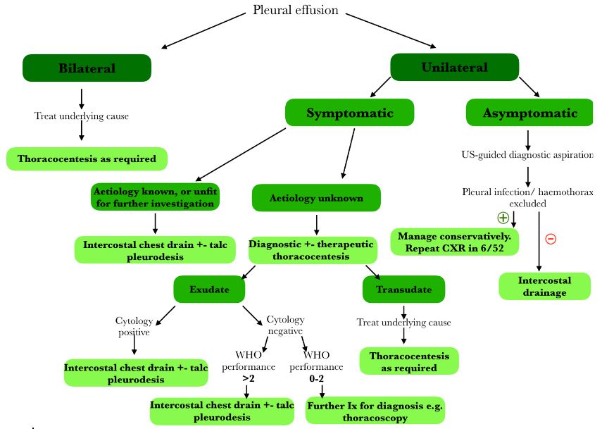
Summary of Management of Pleural Effusion.
Flowchart adapted from guidance available at – Maskell N. British Thoracic Society pleural disease guidelines – 2010 update. Thorax. 2010;65(8):667–9.
Li D, Ajmal S, Tufail M, Panchal RK. Modern-day management of a unilateral pleural effusion. Clin Med J R Coll Physicians London. 2021;21(6):E561–6.
Refractory Non-Malignant Pleural Effusions
Non-malignant pleural effusions that are refractory to management of the underlying cause should be considered for intercostal drainage to reduce symptomatic burden on these patients. Patients should receive optimal medical management of the underlying medical problem, ideally for 72 hours before proceeding to invasive procedures.
Management of Malignant Pleural Effusion
The presence of malignant cells in pleural fluid is diagnostic of metastatic cancer, most commonly secondary to an underlying bronchial malignancy. Management options of malignancy-associated pleural effusions are largely determined by the patient’s performance status, as per the WHO performance status categories, alongside the primary tumour, symptomatology and the predicted degree of lung re-expansion following removal of pleural fluid.
- Performance status 0. Able to carry out all normal activities without restriction.
- Performance status 1. Restricted in strenuous activity but ambulatory and able to carry out light work.
- Performance status 2. Ambulatory and capable of all self-care, but unable to carry out any work activities; up and about >50% of waking hours.
- Performance status 3. Symptomatic and chair/bed-bound>50% of the day.
- Performance status 4. Cannot carry out any self-care; confined to bed/chair.
Patients who are asymptomatic of their malignant pleural effusion should not undergo drainage routinely and should be observed in the outpatient setting. Drainage of asymptomatic pleural effusions does not prevent re-accumulation and progression of the effusion; hence, the observation method is recommended in these patients.
Patients who are symptomatic at presentation with a known malignant pleural effusion can undergo one of several options: therapeutic pleural aspiration, intercostal drainage without pleurodesis or intercostal drainage with pleurodesis. Therapeutic aspiration is not recommended for routine use in known malignant pleural effusions due to the high rate of re-accumulation but can be utilised in patients with very poor prognosis (<1 month). As such, those patients whose prognosis is >1 month should undergo intercostal drainage with the aim of pleurodesis once all pleural fluid has been removed. A chest x-ray should be completed prior to talc pleurodesis to confirm full reinflation of the lung and absence of pleural fluid.

Management of the Malignant Pleural Effusion.
Flowchart adapted from guidance available at – Maskell N. British Thoracic Society pleural disease guidelines – 2010 update. Thorax. 2010;65(8):667–9.
Heffner JE. Management of malignant pleural effusions [Internet]. UpToDate. 2022 [cited 2022 May 30]. Available from: https://www.uptodate.com/contents/management-of-malignant-pleural-effusions
Pleurodesis
Pleurodesis refers to a procedure that sticks the pleura to the chest wall, with the aim of preventing re-accumulation of pleural fluid. In the case of malignant pleural effusions, BTS recommends sterile talc pleurodesis once drainage and re-expansion of the lung has been confirmed. Sterile talc pleurodesis is quoted as being between “81% and 100% effective”.
Trapped Lung
Trapped lung, otherwise known as nonexpendable lung, is seen more commonly in patients with malignant pleural effusions. It refers to the inability of the lung to fully re-expand, and radiologically, the previous fluid is replaced with air in the pleural space on a chest x-ray. It can be suggested clinically by failure to improve symptomatology despite satisfactory drainage of the pleural effusion. In this situation, pleurodesis is of no benefit as visceral and parietal pleura do not come back together. In addition, recurrent thoracentesis does not improve the “trapped lung”, so repeated thoracentesis is not recommended. In patients who are significantly symptomatic with this condition, an indwelling pleural catheter (IPC) can be placed to assist with control of symptoms.
Recurrent symptomatic malignant pleural effusions
In patients who have recurrent symptomatic pleural effusions that reaccumulate despite attempts at pleurodesis, then a tunnelled pleural catheter can be considered to help alleviate symptoms associated with pleural fluid accumulation. It is particularly important for those patients with “trapped lung,” as above, but can also be utilised in patients without this phenomenon.
Management of Pleural Infection/ Empyema
Pleural infections are an ever-increasing complication of pleural fluid accumulation, particularly seen in the very young and very old populations or in those considered to be immunosuppressed. Management of pleural infections focuses on removing the source alongside intravenous antibiotics, guided by the causative agent and its sensitivities.
Pleural infections can be identified in a number of ways, and the way they are identified or suspected are important for their management strategy. The key to diagnosis is the diagnostic pleural aspiration once a pleural effusion has been identified:
- Frankly purulent pleural fluid on aspiration. Any fluid during a diagnostic aspiration which appears to be frank pus or is significantly turbid/cloudy should be considered to be a pleural infection, and immediate intercostal drainage should be completed.
- Low pH pleural fluid <7.2. In those patients who have features suggestive of possible pleural infection (high inflammatory markers, vulnerable population demographic, recurrent fevers despite antibiotics) but not a frankly purulent aspiration, pleural pH should be completed. A pH <7.2 is highly suggestive of pleural infection, and proceeding to intercostal drainage is recommended.
- Gram stain or culture-positive pleural fluid. The presence of organisms identified within pleural fluid should prompt discussion with respiratory physicians regarding the need for drainage. In some situations, these can be managed by antibiotics alone, but this is assessed on a case-by-case basis.
- Loculated pleural effusions identified on pleural USS. Locules on pleural ultrasound are suggestive of organisation of the pleural fluid and are commonly seen in pleural infection but also in haemothorax and malignant pleural effusions. It is this appearance on ultrasound that allows clinicians to refer to it as a “complicated” pleural effusion. Drainage of loculated pleural effusion is recommended due to the potential for individual locules to become infected in isolation.
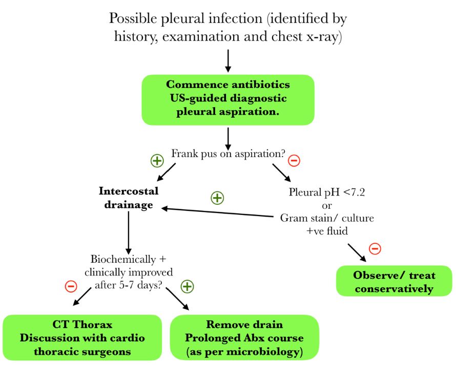
Management of the possible pleural infection (empyema).
Flowchart adapted from guidance available at – Maskell N. British Thoracic Society pleural disease guidelines – 2010 update. Thorax. 2010;65(8):667–9.
Strange C. Management and prognosis of parapneumonic pleural effusion and empyema in adults [Internet]. UpToDate. 2021 [cited 2022 May 30]. Available from: https://www.uptodate.com/contents/management-and-prognosis-of-parapneumonic-pleural-effusion-and-empyema-in-adults
In addition to drainage of the pleural infection, all patients should receive antibiotics. The antibiotics each patient gets will be dependent on several factors: the organisms identified on gram staining/ culture, the sensitivity profile of these organisms, whether the pleural infection was community or hospital-acquired and local resistance patterns.
Pleural infections have the potential to be caused by aerobic and anaerobic organisms, gram-positive and gram-negative organisms, meaning broad-spectrum cover may be needed in those patients who do not grow a specific organism from pleural sampling. Penicillin-based regimes remain the preferred choice due to both the good penetration into the pleural space and the broad-spectrum of cover they provide. Aminoglycosides, e.g. gentamicin, should be avoided due to their poor penetration into the pleural space. All antimicrobial therapy for pleural infection should be discussed with local microbiology teams to allow local resistance patterns to be accounted for. Initially, intravenous antibiotics are recommended, but these can be stepped down to the oral route once biochemical and clinical improvement is noted. Duration of treatment will be guided by local microbiology input, but it is recommended they are continued for at least 3 weeks.
| Type of Pleural Infective | Recommended First-Line Antibiotics | Penicillin-allergy |
|---|---|---|
| S. pneumoniae infection S. milleria infection |
Penicillin-based therapy, e.g. amoxicillin. Or third-generation cephalosporin + metronidazole. |
Carbapenem-based monotherapy, e.g. meropenem. |
| Unknown organism. Community-acquired pleural infection |
Penicillin + beta-lactamase inhibitor e.g. co-amoxiclav or amoxicillin + metronidazole combination. Or third-generation cephalosporin + metronidazole. |
Clindamycin Or, Clindamycin + ciprofloxacin/ cephalosporin. |
| Unknown organism. Hospital-acquired pleural infection. |
Penicillin + beta-lactamase inhibitor with good anaerobic cover and anti-pseudomonal cover e.g. piperacillin-tazobactam. MRSA cover, e.g. vancomycin or teicoplanin, until results available. |
Clindamycin + ciprofloxacin/ cephalosporin. Or antipseudomonal carbapenem e.g. meropenem. MRSA cover, e.g. vancomycin or teicoplanin, until results available. |
Table 4: Common causative organisms of empyema.
Table adapted from – Maskell N. British Thoracic Society pleural disease guidelines – 2010 update. Thorax. 2010;65(8):667–9.
Strange C. Management and prognosis of parapneumonic pleural effusion and empyema in adults [Internet]. UpToDate. 2021 [cited 2022 May 30]. Available from: https://www.uptodate.com/contents/management-and-prognosis-of-parapneumonic-pleural-effusion-and-empyema-in-adults
In patients who do not respond to drainage and antibiotic therapy after a 5-7-day treatment period, a CT thorax and referral to cardiothoracic surgeons for consideration of surgical management of pleural infection should be completed. A CT thorax allows further evaluation of pathology and planning of subsequent surgical procedures. The type of surgical procedure selected will be dependent on many factors, guided by the expertise of cardiothoracic colleagues.
Management of Haemothorax
The essential management of haemothorax is intercostal drainage, alongside appropriate resuscitation such as intravenous fluid and red blood cell replacement as required. In the case of haemothorax, large-bore chest drains should be used in order to allow the passage of clots. If there is persistent bleeding or haemodynamic stability, an urgent referral to cardiothoracic surgery for consideration of urgent thoracotomy should be completed.
References
- Beaudoin S, Gonzalez A V. Evaluation of the patient with pleural effusion. Cmaj. 2018;190(10):E291–5.
- Bhatnagar R, Maskell N. The modern diagnosis and management of pleural effusions. BMJ. 2015;351(September).
- BMJ Best Practice. Empyema [Internet]. 2022 [cited 2022 May 24]. Available from: https://bestpractice.bmj.com/topics/en-gb/1008
- Feller-Kopman DJ. Management of nonmalignant pleural effusions in adults [Internet]. UpToDate. 2021 [cited 2022 May 25]. Available from: https://www.uptodate.com/contents/management-of-nonmalignant-pleural-effusions-in-adults?search=management of pleural effusion&source=search_result&selectedTitle=1~150&usage_type=default&display_rank=1
- Kinasewitz GT. Transudative effusions. Eur Respir J. 1997;10(3):714–8.
- Hassan M, Mercer RM, Rahman NM. Thoracic ultrasound in the modern management of pleural disease. Eur Respir Rev [Internet]. 2020;29(156). Available from: http://dx.doi.org/10.1183/16000617.0136-2019
- Heffner JE. Management of malignant pleural effusions [Internet]. UpToDate. 2022 [cited 2022 May 30]. Available from: https://www.uptodate.com/contents/management-of-malignant-pleural-effusions
- Li D, Ajmal S, Tufail M, Panchal RK. Modern-day management of a unilateral pleural effusion. Clin Med J R Coll Physicians London. 2021;21(6):E561–6.
- Maskell N. British Thoracic Society pleural disease guidelines – 2010 update. Thorax. 2010;65(8):667–9.
- Mowery NT, Gunter OL, Collier BR, Diaz JJ, Haut E, Hildreth A, et al. Practice management guidelines for management of hemothorax and occult pneumothorax. J Trauma – Inj Infect Crit Care. 2011;70(2):510–8.
- Orhan O. Pleural Effusion [Internet]. BMJ Best Practice. 2021 [cited 2022 May 17]. Available from: https://bestpractice.bmj.com/topics/en-gb/287
- Quinn T, Alam N, Aminazad A, Blair Marshall M, Choong C. Decision Making and Algorithm for the Management of Pleural Effusions. Thorac Surg Clin. 2013;23(1):11–6.
- Radiopedia. Pleural Effusion [Internet]. 2022 [cited 2022 May 17]. Available from: https://radiopaedia.org/articles/pleural-effusion?lang=gb
- Saguil A, Wyrick K, Hallgren J. Diagnostic approach to pleural effusion. Am Fam Physician. 2014;90(2):99–104.
- Strange C. Management and prognosis of parapneumonic pleural effusion and empyema in adults [Internet]. UpToDate. 2021 [cited 2022 May 30]. Available from: https://www.uptodate.com/contents/management-and-prognosis-of-parapneumonic-pleural-effusion-and-empyema-in-adults
- UpToDate. Diagnostic evaluation of a pleural effusion in adults: Initial testing [Internet]. 2022 [cited 2022 May 17]. Available from: https://www.uptodate.com/contents/diagnostic-evaluation-of-a-pleural-effusion-in-adults-initial-testing
- Webb A, Angus D, Finfer S, Gattinoni L, Singer M, Care C. Management of pleural effusion and haemothorax Oxford Textbook of Critical Care ( 2 ed .) Management of pleural effusion and haemothorax. 2016;1–11.
- Zocchi L. Physiology and pathophysiology of pleural fluid turnover. Eur Respir J [Internet]. 2002 Dec 1;20(6):1545 LP 1558. Available from: http://erj.ersjournals.com/content/20/6/1545.abstract
Complete Guide to Clinical Genetics ST3 Application and Recruitment (2027)
Written by Medibuddy. Last updated: October 2026. Clinical Genetics is

Complete Guide to Clinical Radiology ST3 Application and Recruitment (2027)
Recruitment paused: NHS England is not running Clinical Radiology ST3+ recru

Complete Guide to Paediatric Surgery ST3 Application and Recruitment in United Kingdom (2026)
Written by Medibuddy. Last updated: October 2026. If you’re planning


Take your subscriptions with you
Our mobile app allows you to access your interview and exam question banks wherever you are.

