


 Close
Close
 10th January 2024
Management
Given there is a wide range of aetiologies resulting in the formation of a pleural effusion, there are also a variety of management options available to treat them. The mainstay of treatment is related to treating the underlying condition once identified, with or without drainage of the effusion. For example, pleural effusions secondary to cardiac failure should initially be treated with optimising treatment for the underlying cardiac disease. There are 3 specialised circumstances where management is altered: pleural infection, haemothorax and malignant pleural effusions.
The majority of bilateral pleural effusions can be treated by managing the underlying condition: optimising medical management of cardiac failure, consideration of transjugular intrahepatic portosystemic shunt (TIPS) in liver cirrhosis or consideration of haemofiltration in renal failure.
Management of Unilateral Pleural Effusion
Unilateral effusions, once a cause has been identified, can be managed conservatively in the majority of cases as long as they remain uncomplicated (absence of organisation and septation features on pleural ultrasound) and asymptomatic. Following a diagnostic aspiration, in this setting of the asymptomatic patient with pleural effusion, a repeat chest x-ray can be requested to follow up in 6 weeks’ time to assess for resolution. However, if a patient is symptomatic, there is a general consensus that drainage should be carried out under ultrasound guidance. The only exceptions relate to asymptomatic pleural infections or haemothoraces, which should be drained regardless.
Drainage of a pleural effusion can happen in one of two ways:
- Therapeutic thoracentesis (also referred to as therapeutic aspiration). This procedure, performed under ultrasound guidance, simultaneously allows samples for diagnostic purposes as well as removal of a larger volume of pleural fluid to provide symptomatic benefit. This procedure leaves some residual pleural fluid, which can be utilised if the original samples are non-diagnostic in more invasive procedures.
- Intercostal drain insertion. Ultrasound (US) guided drain insertion allows complete drainage of pleural fluid as well as diagnostic sampling. The advantage of complete drainage means attempts at pleurodesis can be made, e.g. talc pleurodesis to prevent reaccumulating fluid. This can be utilised in patients who already have a known cause for their pleural effusion or in patients who would be unsuitable for further invasive investigation of the underlying cause (poor performance status, non-ambulatory).
When pleural fluid is drained, BTS recommends no more than 1.5L of fluid removal at once due to the risk of re-expansion pulmonary oedema. The development of this condition can be suggested by the development of a cough and chest pain, followed by significant respiratory deterioration, instability and potentially collapse.
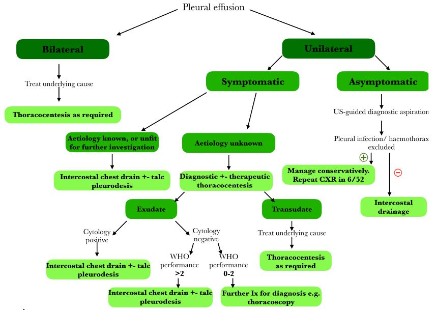
Flowchart adapted from guidance available at – Maskell N. British Thoracic Society pleural disease guidelines – 2010 update. Thorax. 2010;65(8):667–9.
Li D, Ajmal S, Tufail M, Panchal RK. Modern-day management of a unilateral pleural effusion. Clin Med J R Coll Physicians London. 2021;21(6):E561–6.
Complete Guide to Clinical Genetics ST3 Application and Recruitment (2027)
Written by Medibuddy. Last updated: October 2026. Clinical Genetics is

Complete Guide to Clinical Radiology ST3 Application and Recruitment (2027)
Recruitment paused: NHS England is not running Clinical Radiology ST3+ recru

Complete Guide to Paediatric Surgery ST3 Application and Recruitment in United Kingdom (2026)
Written by Medibuddy. Last updated: October 2026. If you’re planning


Take your subscriptions with you
Our mobile app allows you to access your interview and exam question banks wherever you are.

