


 Close
Close
 10th January 2024
Refractory Non-Malignant Pleural Effusions
Non-malignant pleural effusions that are refractory to management of the underlying cause should be considered for intercostal drainage to reduce symptomatic burden on these patients. Patients should receive optimal medical management of the underlying medical problem, ideally for 72 hours before proceeding to invasive procedures.
Management of Malignant Pleural Effusion
The presence of malignant cells in pleural fluid is diagnostic of metastatic cancer, most commonly secondary to an underlying bronchial malignancy. Management options of malignancy-associated pleural effusions are largely determined by the patient’s performance status, as per the WHO performance status categories, alongside the primary tumour, symptomatology and the predicted degree of lung re-expansion following removal of pleural fluid.
- Performance status 0. Able to carry out all normal activities without restriction.
- Performance status 1. Restricted in strenuous activity but ambulatory and able to carry out light work.
- Performance status 2. Ambulatory and capable of all self-care, but unable to carry out any work activities; up and about >50% of waking hours.
- Performance status 3. Symptomatic and chair/bed-bound>50% of the day.
- Performance status 4. Cannot carry out any self-care; confined to bed/chair.
Patients who are asymptomatic of their malignant pleural effusion should not undergo drainage routinely and should be observed in the outpatient setting. Drainage of asymptomatic pleural effusions does not prevent re-accumulation and progression of the effusion; hence, the observation method is recommended in these patients.
Patients who are symptomatic at presentation with a known malignant pleural effusion can undergo one of several options: therapeutic pleural aspiration, intercostal drainage without pleurodesis or intercostal drainage with pleurodesis. Therapeutic aspiration is not recommended for routine use in known malignant pleural effusions due to the high rate of re-accumulation but can be utilised in patients with very poor prognosis (<1 month). As such, those patients whose prognosis is >1 month should undergo intercostal drainage with the aim of pleurodesis once all pleural fluid has been removed. A chest x-ray should be completed prior to talc pleurodesis to confirm full reinflation of the lung and absence of pleural fluid.

Flowchart adapted from guidance available at – Maskell N. British Thoracic Society pleural disease guidelines – 2010 update. Thorax. 2010;65(8):667–9.
Heffner JE. Management of malignant pleural effusions [Internet]. UpToDate. 2022 [cited 2022 May 30]. Available from: https://www.uptodate.com/contents/management-of-malignant-pleural-effusions
Pleurodesis
Pleurodesis refers to a procedure that sticks the pleura to the chest wall, with the aim of preventing re-accumulation of pleural fluid. In the case of malignant pleural effusions, BTS recommends sterile talc pleurodesis once drainage and re-expansion of the lung has been confirmed. Sterile talc pleurodesis is quoted as being between “81% and 100% effective”.
Trapped Lung
Trapped lung, otherwise known as nonexpendable lung, is seen more commonly in patients with malignant pleural effusions. It refers to the inability of the lung to fully re-expand, and radiologically, the previous fluid is replaced with air in the pleural space on a chest x-ray. It can be suggested clinically by failure to improve symptomatology despite satisfactory drainage of the pleural effusion. In this situation, pleurodesis is of no benefit as visceral and parietal pleura do not come back together. In addition, recurrent thoracentesis does not improve the “trapped lung”, so repeated thoracentesis is not recommended. In patients who are significantly symptomatic with this condition, an indwelling pleural catheter (IPC) can be placed to assist with control of symptoms.
Recurrent symptomatic malignant pleural effusions
In patients who have recurrent symptomatic pleural effusions that reaccumulate despite attempts at pleurodesis, then a tunnelled pleural catheter can be considered to help alleviate symptoms associated with pleural fluid accumulation. It is particularly important for those patients with “trapped lung,” as above, but can also be utilised in patients without this phenomenon.
Management of Pleural Infection/ Empyema
Pleural infections are an ever-increasing complication of pleural fluid accumulation, particularly seen in the very young and very old populations or in those considered to be immunosuppressed. Management of pleural infections focuses on removing the source alongside intravenous antibiotics, guided by the causative agent and its sensitivities.
Pleural infections can be identified in a number of ways, and the way they are identified or suspected are important for their management strategy. The key to diagnosis is the diagnostic pleural aspiration once a pleural effusion has been identified:
- Frankly purulent pleural fluid on aspiration. Any fluid during a diagnostic aspiration which appears to be frank pus or is significantly turbid/cloudy should be considered to be a pleural infection, and immediate intercostal drainage should be completed.
- Low pH pleural fluid <7.2. In those patients who have features suggestive of possible pleural infection (high inflammatory markers, vulnerable population demographic, recurrent fevers despite antibiotics) but not a frankly purulent aspiration, pleural pH should be completed. A pH <7.2 is highly suggestive of pleural infection, and proceeding to intercostal drainage is recommended.
- Gram stain or culture-positive pleural fluid. The presence of organisms identified within pleural fluid should prompt discussion with respiratory physicians regarding the need for drainage. In some situations, these can be managed by antibiotics alone, but this is assessed on a case-by-case basis.
- Loculated pleural effusions identified on pleural USS. Locules on pleural ultrasound are suggestive of organisation of the pleural fluid and are commonly seen in pleural infection but also in haemothorax and malignant pleural effusions. It is this appearance on ultrasound that allows clinicians to refer to it as a “complicated” pleural effusion. Drainage of loculated pleural effusion is recommended due to the potential for individual locules to become infected in isolation.
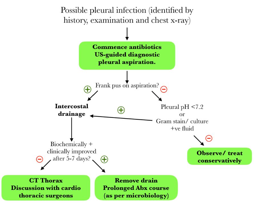
Management of the possible pleural infection (empyema).
Flowchart adapted from guidance available at – Maskell N. British Thoracic Society pleural disease guidelines – 2010 update. Thorax. 2010;65(8):667–9.
Strange C. Management and prognosis of parapneumonic pleural effusion and empyema in adults [Internet]. UpToDate. 2021 [cited 2022 May 30]. Available from: https://www.uptodate.com/contents/management-and-prognosis-of-parapneumonic-pleural-effusion-and-empyema-in-adults
In addition to drainage of the pleural infection, all patients should receive antibiotics. The antibiotics each patient gets will be dependent on several factors: the organisms identified on gram staining/ culture, the sensitivity profile of these organisms, whether the pleural infection was community or hospital-acquired and local resistance patterns.
Pleural infections have the potential to be caused by aerobic and anaerobic organisms, gram-positive and gram-negative organisms, meaning broad-spectrum cover may be needed in those patients who do not grow a specific organism from pleural sampling. Penicillin-based regimes remain the preferred choice due to both the good penetration into the pleural space and the broad-spectrum of cover they provide. Aminoglycosides, e.g. gentamicin, should be avoided due to their poor penetration into the pleural space. All antimicrobial therapy for pleural infection should be discussed with local microbiology teams to allow local resistance patterns to be accounted for. Initially, intravenous antibiotics are recommended, but these can be stepped down to the oral route once biochemical and clinical improvement is noted. Duration of treatment will be guided by local microbiology input, but it is recommended they are continued for at least 3 weeks.
| Type of Pleural Infective | Recommended First-Line Antibiotics | Penicillin-allergy |
|---|---|---|
| S. pneumoniae infection
S. milleria infection |
Penicillin-based therapy, e.g. amoxicillin.
Or third-generation cephalosporin + metronidazole. |
Carbapenem-based monotherapy, e.g. meropenem. |
| Unknown organism.
Community-acquired pleural infection |
Penicillin + beta-lactamase inhibitor e.g. co-amoxiclav or amoxicillin + metronidazole combination.
Or third-generation cephalosporin + metronidazole. |
Clindamycin
Or, Clindamycin + ciprofloxacin/ cephalosporin. |
| Unknown organism.
Hospital-acquired pleural infection. |
Penicillin + beta-lactamase inhibitor with good anaerobic cover and anti-pseudomonal cover e.g. piperacillin-tazobactam.
MRSA cover, e.g. vancomycin or teicoplanin, until results available. |
Clindamycin + ciprofloxacin/ cephalosporin.
Or antipseudomonal carbapenem e.g. meropenem. MRSA cover, e.g. vancomycin or teicoplanin, until results available. |
Table 4: Common causative organisms of empyema.
Table adapted from – Maskell N. British Thoracic Society pleural disease guidelines – 2010 update. Thorax. 2010;65(8):667–9.
Strange C. Management and prognosis of parapneumonic pleural effusion and empyema in adults [Internet]. UpToDate. 2021 [cited 2022 May 30]. Available from: https://www.uptodate.com/contents/management-and-prognosis-of-parapneumonic-pleural-effusion-and-empyema-in-adults
In patients who do not respond to drainage and antibiotic therapy after a 5-7-day treatment period, a CT thorax and referral to cardiothoracic surgeons for consideration of surgical management of pleural infection should be completed. A CT thorax allows further evaluation of pathology and planning of subsequent surgical procedures. The type of surgical procedure selected will be dependent on many factors, guided by the expertise of cardiothoracic colleagues.
Management of Haemothorax
The essential management of haemothorax is intercostal drainage, alongside appropriate resuscitation such as intravenous fluid and red blood cell replacement as required. In the case of haemothorax, large-bore chest drains should be used in order to allow the passage of clots. If there is persistent bleeding or haemodynamic stability, an urgent referral to cardiothoracic surgery for consideration of urgent thoracotomy should be completed.
Complete Guide to Clinical Genetics ST3 Application and Recruitment (2027)
Written by Medibuddy. Last updated: October 2026. Clinical Genetics is

Complete Guide to Clinical Radiology ST3 Application and Recruitment (2027)
Recruitment paused: NHS England is not running Clinical Radiology ST3+ recru

Complete Guide to Paediatric Surgery ST3 Application and Recruitment in United Kingdom (2026)
Written by Medibuddy. Last updated: October 2026. If you’re planning


Take your subscriptions with you
Our mobile app allows you to access your interview and exam question banks wherever you are.

