


 Close
Close
 22nd June 2023
Renal Physiology
Functions of the kidney
The kidney has several important functions, including:
- Filtration and excretion of waste products via the urine.
- Regulation of electrolytes, acid-base balance, serum osmolality and blood volume to ensure adequate conditions for cell function.
- Production of hormones affecting haemodynamics (renin, prostaglandins), red blood cell production (erythropoietin) and calcium homeostasis (1,25 dihydroxyvitamin D3).
Renal excretion of waste products
The kidneys eliminate metabolic waste products such as:
- Urea – from amino acids
- Creatinine – from muscle creatine
- Uric acid – from nucleic acids
Products of haemoglobin breakdown e.g. bilirubinThe kidneys also eliminate other substances which may have been ingested, such as some pharmaceutical drug metabolites, recreational drug metabolites and food additives.
Regulation of electrolytes and blood volume
The kidneys aim to maintain homeostasis so that the excretion of water and electrolytes matches the body’s intake. The kidneys can alter the excretion of water, sodium, and most other electrolytes to maintain this homeostasis. The kidneys are also an important contributor in maintaining acid-base homeostasis by excreting acids and regulating bicarbonate levels.
Hormone production
The kidneys are the main source of erythropoietin (EPO) production, a hormone which stimulates the production of red blood cells. In cases of severe kidney disease, anaemia can result from reduced EPO production.
Renin is an enzyme stored in the juxtaglomerular cells of the kidney in its inactive form and is activated and released in response to low arterial blood pressure. This triggers the renin-angiotensin-aldosterone system, a cascade of vasoactive substances and hormones which work to ultimately increase arterial blood pressure.
The kidney also has a role in the hydroxylation of inactive vitamin D to its active form, 1,25-dihydroxyvitamin D3, also known as calcitriol. Calcitriol has an important role in calcium homeostasis by increasing the deposition of calcium into the bones and increasing the reabsorption of calcium in the gastrointestinal tract.
Structure of the kidney
Anatomy
The kidneys are located retroperitoneally, either side of the spine, at the level of T12 to L3. The right kidney is typically slightly lower than the left due to the presence of the liver. The renal parenchyma is divided into the cortex (outer layer) and the medulla (inner layer), which is in turn divided into triangular-shaped renal pyramids. At the apex of each renal pyramid is the renal papilla, which is closely associated with the minor calyx, a structure which collects urine from the renal pyramids. Collections of minor calyces then form a major calyx, through which urine passes into the renal pelvis, where it drains into the ureter and subsequently the bladder.
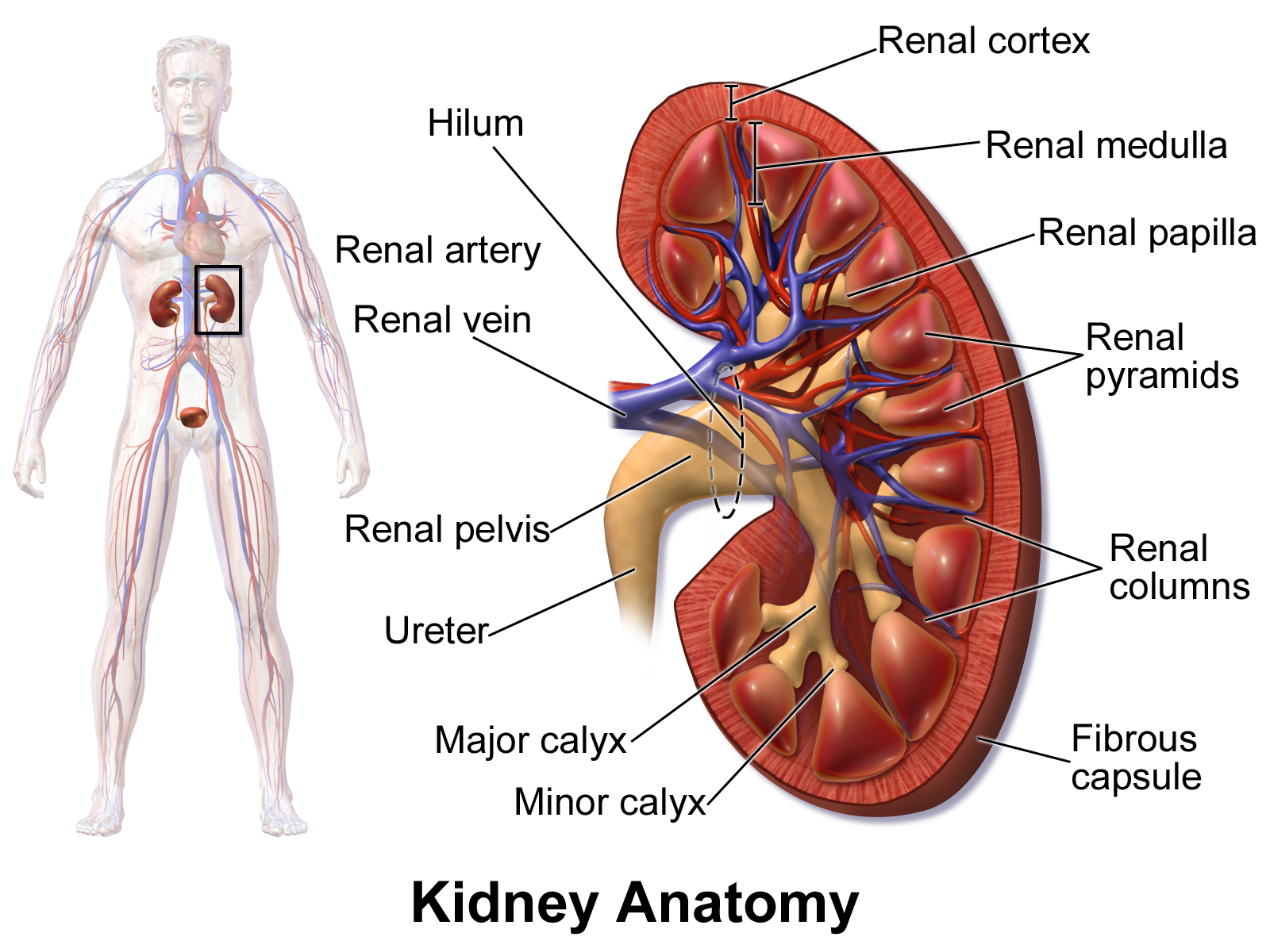
Anatomy of the kidney.
Image Source: Wikimedia Commons (2014). Blausen 0592 KidneyAnatomy 01, Licensed under a creative commons licence. Available for free at: https://commons.wikimedia.org/wiki/File:Blausen_0592_KidneyAnatomy_01.png (Accessed on: 9 February 2023).
The kidneys’ vascular supply is from the renal arteries, which arise from the abdominal aorta. The right renal artery is slightly longer than the left due to the position of the abdominal aorta just to the left of the midline. The renal artery then branches into interlobar, arcuate, interlobular arteries, and afferent arterioles, the latter of which supplies the glomerulus. The venous drainage of the kidneys is via interlobular, arcuate and interlobar veins, which run parallel to their corresponding arteries, leading to the renal veins, which drain into the inferior vena cava. Conversely to the arterial supply, the left renal vein is slightly longer than the right due to the position of the inferior vena cava just to the right of the midline.
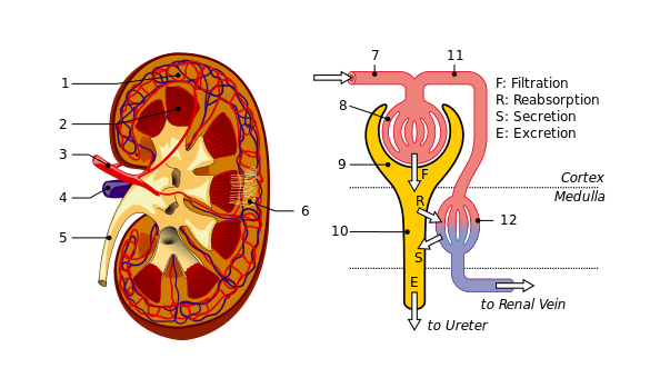

Vascular supply of the kidney and the microcirculation of a nephron.
1: Renal cortex. 2: Medulla. 3: Renal artery. 4: Renal vein. 5: Ureter. 6: Nephrons. 7: Afferent arteriole. 8: Glomerulus. 9: Bowman’s capsule. 10: Tubuli and loop of Henle. 11: Efferent arteriole. 12: Peritubular capillaries.
Image Sources:
- Wikimedia Commons (2013). KidneyAndNephron-v4 Antares42, Licensed under a creative commons licence. Available for free at: https://commons.wikimedia.org/wiki/File:KidneyAndNephron-v4_Antares42.svg (Accessed on: 9 February 2023).
- Wikimedia Commons (2012). Kidney nephron, Licensed under a creative commons licence. Available for free at: https://commons.wikimedia.org/wiki/File:Kidney_nephron.png (Accessed on: 9 February 2023).
Nephron
The nephron is the main functional unit of the kidney; there are approximately 1 million nephrons in each kidney, however, with renal disease or normal ageing, the number of nephrons decreases. Each nephron is composed of a renal corpuscle and a renal tubule.
Renal corpuscle
The renal corpuscle consists of the glomerulus and Bowman’s capsule; it is the site of filtration of the blood. Filtration of fluid from the glomerulus to the Bowman’s capsule is based on Starling forces:
- Membrane permeability
- Hydrostatic pressure gradient
- Oncotic pressure gradient
The glomerulus is a bundle of glomerular capillaries covered with a semi-permeable membrane formed of glomerular endothelial cells, the glomerular basement membrane (GBM), and podocytes; these form the filtration barrier. The glomerular capillary endothelium contains pores, or ‘fenestrae’, which are approximately 70 nm in diameter and prevent the filtration of blood cells. The GBM is composed of type IV collagen, heparan sulfate proteoglycans and laminin, and contains three layers: lamina rara externa, lamina densa and lamina rara interna. These layers limit the filtration of larger solutes, such as proteins. Surrounding the GBM is a layer of epithelial cells with foot-like processes called podocytes, separated by small ‘slit pores’ through which flows the filtrate; the slit pores prevent large molecules, e.g. proteins, from crossing. In addition to these physical barriers, the glycocalyx (surrounding the endothelium), the GBM, and the podocytes all contain, or are covered by, negatively charged glycoproteins which restrict the filtration of negatively charged anions and proteins by repelling like charges.
The glomerulus receives its blood supply from the afferent arteriole, and blood leaves the glomerulus via the efferent arteriole. These arterioles are responsible for generating pressure in the glomerulus by constriction of the efferent arteriole and dilatation of the afferent arteriole. The pressure in the glomerulus is ~50mmHg (compared to 35mmgHg in other capillaries). This high hydrostatic pressure generated within the glomerular capillaries drives the ultrafiltration of water and solutes out of the plasma.
Small molecules such as glucose can readily cross the filtration barrier, however, larger molecules such as albumin, and negatively charged anions, are not readily filtered; this creates a colloid pressure due to the presence of proteins in the blood and absence of proteins in the filtrate, forcing water and solutes into the renal tubules.
The glomerulus is encased in Bowman’s capsule, which receives filtrate from the glomerulus and passes it to the proximal tubule of the nephron.

The renal corpuscle.
Image Source: Wikimedia Commons (2019). Renal corpuscle-en, Licensed under a creative commons licence. Available for free at: https://commons.wikimedia.org/wiki/File:Renal_corpuscle-en.svg (Accessed on: 11 January 2023).
Renal tubule
The renal tubule comprises the proximal convoluted tubule, the descending and ascending limbs of the loop of Henle, the distal convoluted tubule, and the collecting duct. The function of the renal tubule is the reabsorption of certain constituents from the filtrate and secretion of molecules from the peritubular capillaries into the distal convoluted tubule.
Urinary excretion = Glomerular filtration – Tubular reabsorption + Tubular secretion
Nephrons which have glomeruli located in the cortex of the kidney are called cortical nephrons; these tend to have shorter loops of Henle and, therefore, only penetrate a small distance into the renal medulla. Nephrons with glomeruli which lie deep in the cortex, on the border of the medulla, are called juxtamedullary nephrons; these represent almost a third of nephrons and tend to have much longer loops of Henle, penetrating deeply into the medulla. The vascular supply of the juxtamedullary nephrons is via the vasa recta, specialised peritubular capillaries which originate from the efferent arterioles.
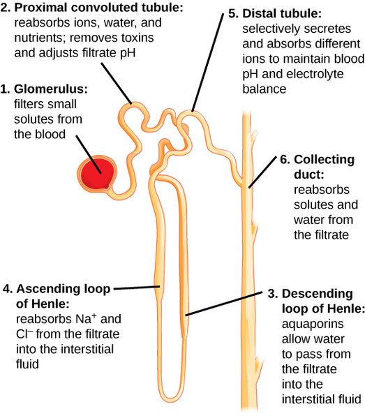
Components of the renal tubule.
Image Source: Wikimedia Commons (2016). Figure 41 03 04, Licensed under a creative commons licence. Available for free at: https://commons.wikimedia.org/wiki/File:Figure_41_03_04.jpg (Accessed on: 9 February 2023).
Glomerular Function
Filtration
Most substances contained within the plasma, with the exception of proteins and protein-bound substances, are readily filtered through the glomerulus to Bowman’s capsule. Therefore, the concentration of the filtrate in Bowman’s capsule is similar to that of the circulating plasma.
As the filtrate flows through the renal tubule, the concentration of the filtrate is modified by the reabsorption of solutes and water from the filtrate and by the secretion of other solutes into the tubular fluid from the peritubular capillaries.
End products of metabolism such as urea, creatinine and uric acid are readily filtered at the glomerulus but not reabsorbed, therefore are excreted in large quantities in the urine. Other substances requiring excretion, such as drugs, are also poorly reabsorbed but are secreted into the tubular fluid from the peritubular capillaries, resulting in excretion in the urine.
Electrolytes such as sodium, chloride, and bicarbonate ions are readily filtered and reabsorbed, resulting in mostly low levels of excretion in the urine, however, this is variable depending on the needs of the body. Amino acids and glucose are readily filtered but almost completely reabsorbed and therefore do not appear in the urine under normal circumstances.
Glomerular filtration rate
The glomerular filtration rate (GFR) is the volume of plasma passing through all the glomeruli in the kidneys in one minute; in normal functioning kidneys this is approximately 125ml/min, however normal GFR varies with sex and body size, and declines with age. The eGFR is the estimated GFR and is calculated based on the patient’s serum creatinine, age, sex and race. There are several equations used to calculate the eGFR, but in the UK, NICE recommends using the Chronic Kidney Disease Epidemiology Collaboration creatinine equation (CKD-EPI) to calculate eGFR in adults.
Tubular Function
Reabsorption and secretion
Compared to glomerular filtration, which is relatively non-selective, tubular reabsorption is highly selective, allowing for precise control of the body plasma composition. During reabsorption, solutes and water move from the renal tubule into the renal interstitium and then into the peritubular capillaries to rejoin the circulation.
Reabsorption can be passive (i.e. diffusion of solutes down a concentration gradient) or active (i.e. transport of solutes against a concentration gradient). Reabsorption can occur paracellularly (solutes are transported between cells) or transcellularly (solutes are transported through cells).
Secretion is the active movement of substances from the peritubular capillaries into the renal tubular fluid.

Reabsorption of water and solutes from tubular lumen, through the renal interstitium and to the blood.
Proximal Convoluted Tubule
The main role of the proximal convoluted tubule (PCT) is reabsorption. It is lined by cuboidal epithelial cells covered in microvilli forming a ‘brush border’, increasing surface area for reabsorption. The epithelial cells are packed with mitochondria, providing an energy supply for the active transport of solutes.
Approximately 65% of sodium and water in the glomerular filtrate is reabsorbed in the PCT.
Na⁺-K⁺-ATPase transporters on the basolateral membrane form a primary active transport mechanism in which 3 Na⁺ ions are pumped out of the cell in exchange for 2 K⁺ ions pumped into the cell. This forms an electrochemical gradient which allows diffusion of sodium from the lumen of the renal tubule into the cell.
Sodium can be co-transported with other solutes via symporters, whereby sodium moves down its concentration gradient whilst actively co-transporting other solutes, such as glucose and amino acids, against their concentration gradients. In normal physiological states, all organic solutes, such as glucose and amino acids, are reabsorbed in the PCT via these symporters.
Sodium can also be transported out of the lumen via an antiporter, which enables reabsorption of sodium with the concurrent secretion of other ions, in particular hydrogen ions, which have a role in maintaining acid/base homeostasis. H⁺ ions secreted via the Na⁺/H⁺ antiporter combine with bicarbonate ions, forming carbonic anhydrase (H₂CO₃), which subsequently splits to form CO₂ and H₂O. These are absorbed by the PCT epithelial cells, where the previous reaction reverses, releasing bicarbonate (which is absorbed into the blood) and hydrogen ions (which are recycled for further reactions). Approximately 90% of bicarbonate is reabsorbed in the PCT.

Ion absorption and secretion in the PCT.
Image Source: Wikimedia Commons (2007). Renal Diuretics, Licensed under a creative commons licence. Available for free at: https://commons.wikimedia.org/wiki/File:Renal_Diuretics.gif (Accessed on: 13 February 2023).
As the concentration of solutes within PCT decreases due to reabsorption, the solute concentration in the renal interstitium increases, and water can move out of the lumen into the interstitium by osmosis.
The PCT is also a site of secretion of organic acids and bases (e.g. bile salts, oxalate and catecholamines), and certain drug metabolites, from the peritubular capillaries into the renal tubule for rapid removal from the body.
Loop of Henle
The loop of Henle consists of the thin descending limb, the thin ascending limb, and the thick ascending limb. Despite the description, the diameter of the tubule is in fact consistent throughout the loop of Henle; the ‘thin’ limbs refer to the thin squamous epithelial cells in these segments, which lack a brush border and have low metabolic activity, whereas the thick ascending limb has cuboidal epithelium with higher metabolic activity.
The thin descending limb has a high permeability to water due to the presence of aquaporin-1 channels and lower permeability to most solutes, including sodium ions and urea.
The thin ascending limb does not contain aquaporin channels and is, therefore, impermeable to water. Passive diffusion of sodium and chloride ions can continue to occur here due to the concentration gradient between the tubular fluid and interstitium.
The thick ascending limb (TAL) is also impermeable to water. Sodium, potassium, and chloride ions are actively reabsorbed in the TAL; Na⁺-K⁺-ATPase transporters on the basolateral membrane (as also seen in the PCT) pump three sodium ions out of the cell into the interstitium, in exchange for two potassium ions. This creates a low intracellular sodium concentration and an electrochemical gradient between the lumen and the epithelial cells, allowing the movement of sodium ions out of the lumen and into the epithelial cells via Na⁺/K⁺/2Cl⁻ co-transporters (NKCC2) and Na⁺/H⁺ antiporters on the luminal membrane. The NKCC2 to-transporter is the target site of loop diuretics such as furosemide.
There is also paracellular reabsorption of magnesium, calcium, sodium, and potassium ions in the TAL down an electrical gradient due to the slight positive charge in the lumen compared to the interstitium.

Ion transport in the thick ascending limb.
Image Source: Wikimedia Commons (2007). Renal Diuretics, Licensed under a creative commons licence. Available for free at: https://commons.wikimedia.org/wiki/File:Renal_Diuretics.gif (Accessed on: 13 February 2023).
Counter-current multiplication
As the TAL is impermeable to water, but reabsorbs solutes, the interstitium surrounding the loop of Henle has a high osmolarity. This high concentration of ions outside of the lumen drives the movement of water from the thin descending limb lumen into the interstitium by osmosis. This is known as counter-current multiplication.
Distal Convoluted Tubule
The thick ascending limb of the loop of Henle flows into the distal convoluted tubule (DCT), which is subdivided into the early DCT and late DCT.
Early distal tubule
The early distal tubule forms the macula densa, a group of sensing epithelial cells which are part of the juxtaglomerular apparatus, a structure providing control of glomerular filtration rate and blood flow in the nephron.
Na⁺-K⁺-ATPase transporters on the basolateral membrane (as also seen in the PCT and TAL) generate an electrochemical gradient for the movement of sodium ions into the epithelial cells through Na⁺/Cl⁻ symporters (NCC). Chloride ion channels on the basolateral membrane then allow the diffusion of chloride into the extracellular fluid. Thiazide diuretics inhibit the NCC symporter.

Ion transport in the early distal tubule.
Late distal tubule
The late distal tubule is made up of principal cells and intercalated cells.
The principal cells again use Na⁺-K⁺-ATPase transporters on the basolateral membrane to generate an electrochemical gradient for the passage of sodium from the lumen to epithelial cells through epithelial Na⁺ channels (ENaC) and secretion of potassium ions into the lumen through K⁺ uniporter channels. Principal cells are the target of potassium-sparing diuretics (e.g. spironolactone, eplerenone, amiloride). The rate of sodium ion reabsorption and potassium ion secretion in the late DCT and cortical collecting duct is controlled largely by aldosterone (see RAAS below).
Intercalated cells secrete hydrogen ions and reabsorb bicarbonate and potassium. The hydrogen ions secreted are generated as a result of a reverse reaction to that seen in the PCT, i.e. CO₂ and H₂O absorbed into the cell combine to form carbonic acid, which subsequently splits into HCO₃- and H⁺ ions. Each hydrogen ion is secreted back into the lumen via hydrogen-ATPase and H⁺/K⁺-ATPase transporters to then combine with a bicarbonate ion to be reabsorbed, as per the reaction described in the PCT. Contrary to the PCT, the intercalated cells have the capability of secreting hydrogen ions against a large concentration gradient, allowing for H⁺ ion secretion in response to acidosis, and thus play an important role in acid-base balance.
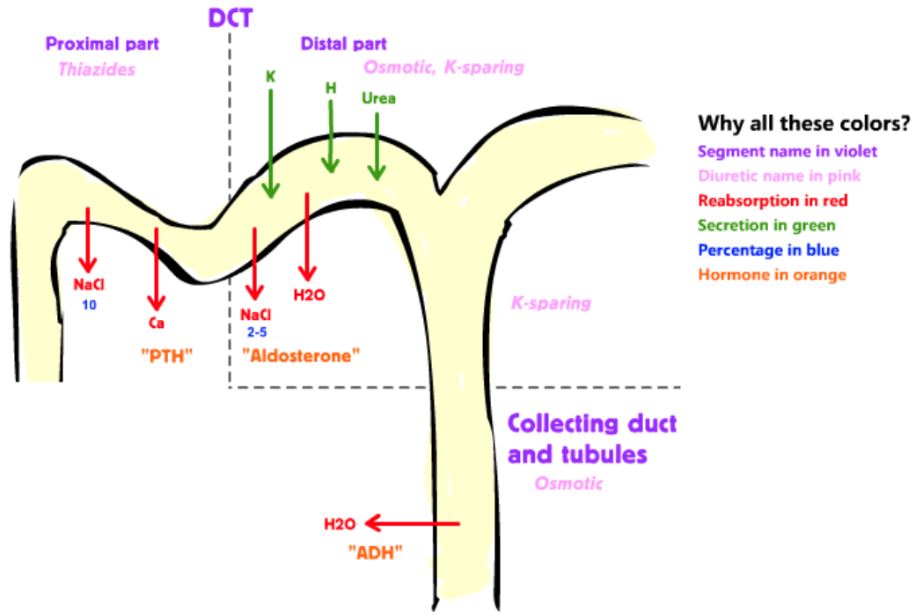
Sodium reabsorption and potassium secretion in the late distal tubule.
Image Source: Wikimedia Commons (2007). Renal Diuretics, Licensed under a creative commons licence. Available for free at: https://commons.wikimedia.org/wiki/File:Renal_Diuretics.gif (Accessed on: 13 February 2023).
Collecting Duct
The permeability of the late distal tubule, cortical and medullary collecting ducts to water is controlled by antidiuretic hormone (ADH), produced by the hypothalamus, and stored in the posterior pituitary gland. In the presence of high levels of ADH (e.g. secondary to dehydration), the number of aquaporin-2 channels in the distal convoluted tubule and collecting duct increases, increasing water reabsorption, and thereby reducing the volume and increasing the osmolality of urine. Thus, ADH has an important role in controlling the degree of concentration of the urine.
ADH is clinically relevant in conditions such as: SIADH (syndrome of inappropriate ADH secretion), resulting in excessive blood dilution leading to hyponatraemia; Diabetes insipidus (DI), an insufficient release of ADH from the posterior pituitary (central DI) or reduced response of the collecting ducts to ADH (nephrogenic DI), resulting in polyuria and subsequent polydipsia.
The late DCT and cortical collecting duct are almost completely impermeable to urea, therefore, urea in these segments is excreted in the urine. Conversely to the cortical collecting duct, the medullary collecting duct is permeable to urea. In the interstitium, urea is an effective osmole (i.e. unable to cross from extracellular fluid to intracellular fluid, raising its osmolality) which allows increased reabsorption of water from the renal tubule.
Renin-Angiotensin-Aldosterone System
The renin-angiotensin-aldosterone system (RAAS) is a hormonal system which acts to regulate fluid balance and blood pressure.
The RAAS
Renin is an enzyme synthesised and stored in an inactive form (prorenin) in juxtaglomerular cells in the juxtaglomerular apparatus (JGA), a structure adjacent to the glomerulus which regulates the function of each nephron. The JGA is made up of the macula densa (part of the DCT of the nephron), juxtaglomerular cells, and extraglomerular mesangial cells.
Prorenin is cleaved to form renin is released in response to:
- Decreased sodium delivery to the DCT (detected by the macula densa).
- Decreased perfusion pressure to the kidney (detected by baroreceptor in the afferent arteriole).
- Sympathetic stimulation of the juxtaglomerular apparatus.
Renin acts enzymatically on angiotensinogen (a precursor protein produced in the liver) to form angiotensin I. Angiotensin I is converted to angiotensin II by angiotensin converting enzyme (ACE). ACE is largely produced in the endothelium of lung vessels and thus this conversion largely occurs in the lungs. ACE is produced in much smaller quantities in the renal endothelium.
Effects of Angiotensin II
Angiotensin II has several mechanisms which act to increase blood pressure and blood volume:
- Vasoconstriction of arterioles
- Angiotensin II stimulates AT1 receptors on the arteriolar vascular endothelium, thereby inducing vasoconstriction. This increases the systemic vascular resistance and, therefore, blood pressure.
- Increased sodium reabsorption in the kidneys
- Angiotensin II increases sodium reabsorption in the PCT by increasing Na⁺/H⁺ antiporter activity and paracellular reabsorption.
- Increased noradrenaline release from the sympathetic nervous system
- Stimulation of the sympathetic nervous system results in arteriolar vasoconstriction, increased cardiac output, and further release of renin from the JGA.
- Stimulation of ADH release from the posterior pituitary
- Angiotensin II acts on the hypothalamus to increase thirst as well as stimulating the release of ADH from the posterior pituitary gland, reducing the loss of water in the urine and maintaining circulatory volume.
- Stimulation of aldosterone release from the adrenal cortex
- Aldosterone is a mineralocorticoid hormone, released from the zona glomerulosa of the adrenal cortex, and acts to increase serum sodium, and thereby subsequently increase blood volume.
- Aldosterone increases the expression of epithelial sodium channels (ENaC) on the principal cells of the late DCT and increases the activity of the basolateral membrane Na⁺-K⁺-ATPase transporters, thereby increasing sodium reabsorption from the nephron. The sodium ions in the epithelial cells are pumped into the blood by the sodium/potassium antiporter, therefore potassium is moved from the blood into the renal tubule and excreted. Therefore, increased aldosterone results in reduced serum potassium.

The Renin-Angiotensin-Aldosterone System.
Image Source: Wikimedia Commons (2018). Renin-angiotensin-aldosterone system, Licensed under a creative commons licence. Available for free at: https://commons.wikimedia.org/wiki/File:Renin-angiotensin-aldosterone_system.svg (Accessed on: 11 January 2023).
ACE-inhibitors are a class of drugs which, as the name suggests, inhibit angiotensin-converting enzymes, reducing levels of angiotensin II production, and thereby reducing the actions of angiotensin II as described above. ACE-inhibitors (e.g. ramipril, lisinopril) are commonly used in the treatment of hypertension due to their action in reducing vasoconstriction of arterioles and reducing cardiac output. ACE-inhibitors are also used in renal pathology to both reduce blood pressure and proteinuria. Angiotensin II causes preferential vasoconstriction of the efferent arteriole, increasing intraglomerular pressure and GFR. Therefore, inhibition of efferent arteriolar vasoconstriction during ACE-inhibitor use reduces the GFR and reduces urinary protein excretion.
Complete Guide to Clinical Genetics ST3 Application and Recruitment (2027)
Written by Medibuddy. Last updated: October 2026. Clinical Genetics is

Complete Guide to Clinical Radiology ST3 Application and Recruitment (2027)
Recruitment paused: NHS England is not running Clinical Radiology ST3+ recru

Complete Guide to Paediatric Surgery ST3 Application and Recruitment in United Kingdom (2026)
Written by Medibuddy. Last updated: October 2026. If you’re planning


Take your subscriptions with you
Our mobile app allows you to access your interview and exam question banks wherever you are.

