


 Close
Close
 22nd June 2023
Anatomy
The kidneys are located retroperitoneally, either side of the spine, at the level of T12 to L3. The right kidney is typically slightly lower than the left due to the presence of the liver. The renal parenchyma is divided into the cortex (outer layer) and the medulla (inner layer), which is in turn divided into triangular-shaped renal pyramids. At the apex of each renal pyramid is the renal papilla, which is closely associated with the minor calyx, a structure which collects urine from the renal pyramids. Collections of minor calyces then form a major calyx, through which urine passes into the renal pelvis, where it drains into the ureter and subsequently the bladder.
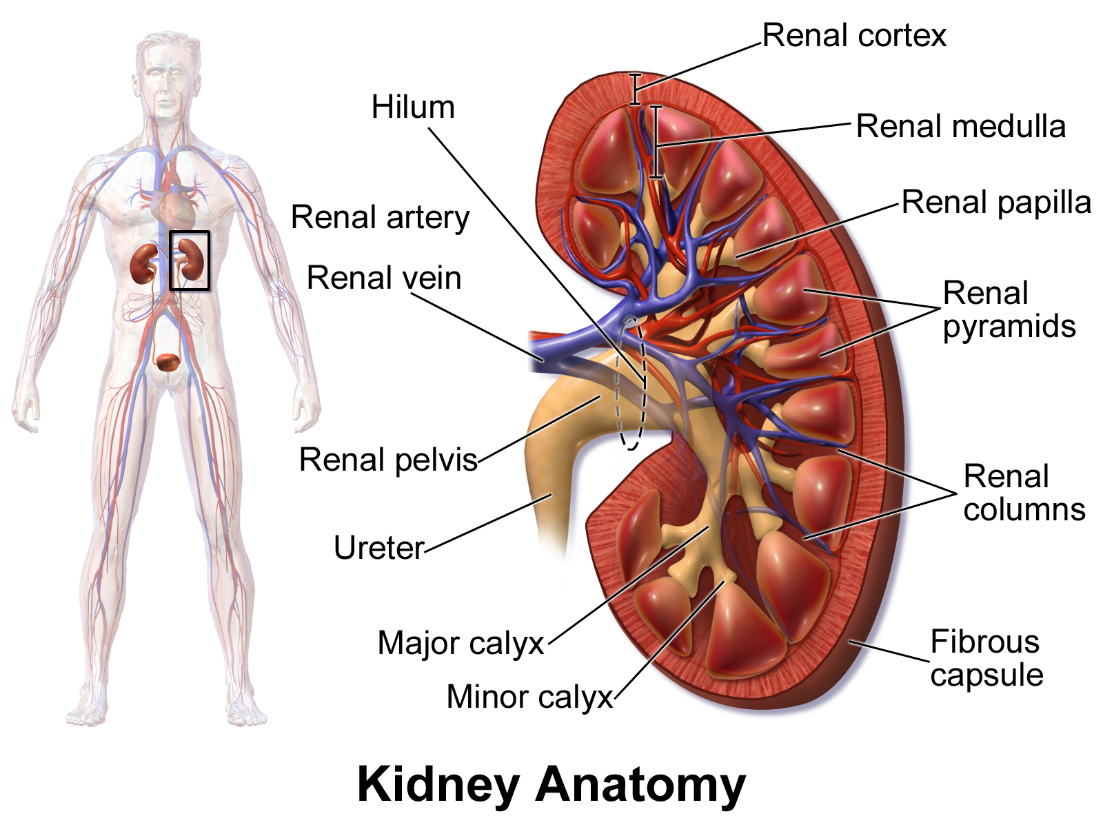
Anatomy of the kidney.
Image Source: Wikimedia Commons (2014). Blausen 0592 KidneyAnatomy 01, Licensed under a creative commons licence. Available for free at: https://commons.wikimedia.org/wiki/File:Blausen_0592_KidneyAnatomy_01.png (Accessed on: 9 February 2023).
The kidneys’ vascular supply is from the renal arteries, which arise from the abdominal aorta. The right renal artery is slightly longer than the left due to the position of the abdominal aorta just to the left of the midline. The renal artery then branches into interlobar, arcuate, interlobular arteries, and afferent arterioles, the latter of which supplies the glomerulus. The venous drainage of the kidneys is via interlobular, arcuate and interlobar veins, which run parallel to their corresponding arteries, leading to the renal veins, which drain into the inferior vena cava. Conversely to the arterial supply, the left renal vein is slightly longer than the right due to the position of the inferior vena cava just to the right of the midline.
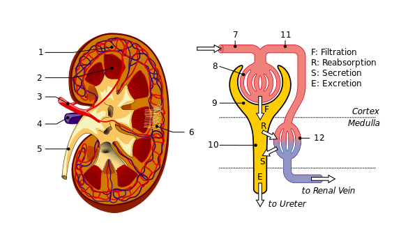

Vascular supply of the kidney and the microcirculation of a nephron.
1: Renal cortex. 2: Medulla. 3: Renal artery. 4: Renal vein. 5: Ureter. 6: Nephrons. 7: Afferent arteriole. 8: Glomerulus. 9: Bowman’s capsule. 10: Tubuli and loop of Henle. 11: Efferent arteriole. 12: Peritubular capillaries.
Image Sources:
- Wikimedia Commons (2013). KidneyAndNephron-v4 Antares42, Licensed under a creative commons licence. Available for free at: https://commons.wikimedia.org/wiki/File:KidneyAndNephron-v4_Antares42.svg (Accessed on: 9 February 2023).
- Wikimedia Commons (2012). Kidney nephron, Licensed under a creative commons licence. Available for free at: https://commons.wikimedia.org/wiki/File:Kidney_nephron.png (Accessed on: 9 February 2023).
Complete Guide to Clinical Genetics ST3 Application and Recruitment (2027)
Written by Medibuddy. Last updated: October 2026. Clinical Genetics is

Complete Guide to Clinical Radiology ST3 Application and Recruitment (2027)
Recruitment paused: NHS England is not running Clinical Radiology ST3+ recru

Complete Guide to Paediatric Surgery ST3 Application and Recruitment in United Kingdom (2026)
Written by Medibuddy. Last updated: October 2026. If you’re planning


Take your subscriptions with you
Our mobile app allows you to access your interview and exam question banks wherever you are.

