


 Close
Close
 12th January 2024
Diagnosis
Latent TB can be diagnosed by Mantoux testing (tuberculin skin testing) or interferon-ɣ release assays (IGRA). In the Mantoux test, TB antigen is injected intradermally, and the area is assessed 48-72 hours later to look for evidence of a delayed-type hypersensitivity reaction occurring, which indicates previous exposure to the antigen. This test can provide a false positive result if the patient has previously had the BCG vaccine and false negative results in individuals who are immunosuppressed (e.g. HIV-positive patients). IGRA testing involves exposing T-lymphocytes of individuals to MtB antigens and measuring interferon-ɣ to detect for previous infection. IGRA tests are not affected by prior BCG vaccination.
For suspected active pulmonary TB, a chest X-ray should be performed, with further diagnostic investigations carried out if the radiological appearance is suggestive of TB. While there are no radiographic features that are alone diagnostic of TB, a number of features are typical. Mediastinal and hilar lymphadenopathy is a common chest X-ray finding, consolidation, and cavitation, which has a predilection for the upper lobes. If appearances suggest TB, multiple respiratory samples should be sent for microscopy and culture for acid-fast bacilli (bacilli which resist acid on Ziehl-Neelsen staining). If possible, these should be three deep cough sputum samples, including an early morning sample. Bronchoscopy and lavage may be required to acquire samples. Rapid diagnostic nucleic acid amplification tests should be performed on these specimens if there is clinical suspicion of TB disease, and the person has HIV or rapid information about mycobacterial species would alter the person’s care.
In patients with suspected extra-pulmonary TB, samples should be collected again, allowing for microscopy and culture. For instance, in suspected TB meningitis cerebrospinal fluid should be taken for TB microscopy and culture; in suspected pleural TB, pleural fluid should be sent; in suspected TB lymphadenitis, a lymph node biopsy should be sent, and so on.

Image Source: Wikimedia Commons (2010). Tuberculosis image 1, Licensed under a creative commons licence. Available for free at: https://upload.wikimedia.org/wikipedia/commons/9/9a/Tuberculosis_image1.jpg (Accessed: 22 March 2021).
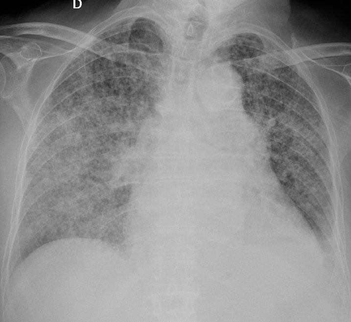
Image Source: Wikimedia Commons (2018). Chest radiograph of miliary tuberculosis. 2, Licensed under a creative commons licence. Available for free at: https://upload.wikimedia.org/wikipedia/commons/7/70/Chest_radiograph_of_miliary_tuberculosis_2.jpg (Accessed: 22 February 2022).
Complete Guide to Clinical Genetics ST3 Application and Recruitment (2027)
Written by Medibuddy. Last updated: October 2026. Clinical Genetics is

Complete Guide to Clinical Radiology ST3 Application and Recruitment (2027)
Recruitment paused: NHS England is not running Clinical Radiology ST3+ recru

Complete Guide to Paediatric Surgery ST3 Application and Recruitment in United Kingdom (2026)
Written by Medibuddy. Last updated: October 2026. If you’re planning


Take your subscriptions with you
Our mobile app allows you to access your interview and exam question banks wherever you are.

