


 Close
Close
 7th February 2024
Control of breathing
The control of breathing can be considered into separate components: receptors, controllers, and effectors.
Receptors
Receptors responsible for the control of breathing can be categorised into:
- Central chemoreceptors
- Peripheral chemoreceptors
- Other receptors
Central chemoreceptors
Central chemoreceptors are in the anterolateral surface of the medulla within the blood-brain barrier. It directly detects changes in hydrogen concentration (and, in effect, the changes in pH) in the cerebrospinal fluid (CSF). Indirectly, central chemoreceptors detect changes to blood CO2 concentration.
CO2 in the blood can pass the blood-brain barrier. After passing the blood-brain barrier into CSF, it reacts to eventually produce hydrogen ions (H+) and bicarbonate ions (HCO3-). H+ diffuses into chemoreceptor cells, directly stimulating chemoreceptor cells. Stimulation of chemoreceptor cells will then activate the respiratory centre.
H+ ions in the blood cannot pass the blood-brain barrier. Hence, respiratory acidosis will stimulate central chemoreceptors, and metabolic acidosis will not.
Compensation in hypercapnia – long-standing hypercapnia will result in compensation in the CSF and intravascular space. Long-standing hypercapnia will result in HCO3- being absorbed into CSF space to normalise pH and reabsorption of HCO3- by the kidneys.
Peripheral chemoreceptors
Peripheral chemoreceptors are in the carotid bodies and aortic bodies. Both carotid and aortic bodies respond to changes in partial pressure of carbon dioxide (PaCO2)and partial pressure of oxygen (PaO2) [not oxygen content in the blood]. However, peripheral chemoreceptors are only responsible for 20% of the body’s response to high PaCO2. Central chemoreceptors are responsible for the rest.
Carotid chemoreceptors are also stimulated by increased blood hydrogen centration (decrease in blood pH). This is the reason for hyperventilation in metabolic acidosis.
Carotid bodies send afferent signals to the respiratory centre via the glossopharyngeal nerve (CN IX)
Aortic bodies send afferent signals to the respiratory centre via the vagus nerve (CN X)
Other receptors
- Lung stretch receptors – located in bronchial smooth muscles, part of the Hering-Bruer reflex. The Hering-Bruer reflex is a reflex where overinflation of the lung (i.e. during positive pressure ventilation) causes stimulation of lung stretch receptors. Lung stretch receptors send afferent signals via the vagus nerve (CN X) to the apneustic centre, causing a reduction in the depth of breathing.
- Juxtacapillary receptors (J receptors) – These are unmyelinated C-fibres in the alveolar wall. Stimulation of these receptors leads to increased ventilation, dyspnoea, bradycardia and hypotension. These receptors are thought to be stimulated in pulmonary oedema and pulmonary embolism.
- Laryngeal epithelial irritant receptors – irritants (i.e. noxious gas) stimulate receptors, causing bronchoconstriction and increasing ventilation.
Controller
The controller of respiration is made up of a group of nuclei found throughout the brainstem. They can be considered into different groups:
- Dorsal respiratory group
- Controls diaphragmatic contraction.
- Responsible for everyday tidal volume inspiration
- Ventral respiratory group
- Controls intercostal muscles.
- Responsible for forced expiration and increased force in inspiration
- It contains the pre-Botzinger complex, thought to be the respiratory pacemaker.
- Controls intercostal muscles.
- Pneumotaxic centre
- Fine-tunes respiratory pattern
- It can increase respiratory rate.
- Apneustic centre
- Prevents over-expansion of the lung (as part of the Hering-Bruer reflex – see ‘Receptors’)
Effectors
- Diaphragm
- Responsible for 75% of volume changes in resting inspiration
- Innervated by phrenic nerve (C3,4,5)
- Intercostal muscles
- External – inspiration
- Internal – expiration
- Accessory muscles
- Include sternocleidomastoid muscles, strap muscles of the neck, and platysma.
- Used during respiratory distress, forced expiration.
Effect of PaCO2 and PaO2 on ventilation
PaO2
This can be represented by the graph below.

First, look at the line with normocapnia. Minute ventilation (MV) is unchanged when PaO2 is above 8kPa. When PaO2 falls below 8kPa, there is a steep rise in MV.
Now, compare the lines between normocapnia and hypercapnia. A rise in PaCO2 will also stimulate the chemoreceptors (both central and peripheral). Hence, the line is shifted rightwards and upwards, representing higher MV for every PaO2.
PaCO2
This can again be represented in the graph below:
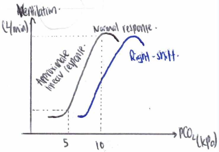
First, consider the left curve. MV will remain relatively the same below PaCO2 of 5kPa. With PaCO2 above 5kPa, there is a linear rise in MV up to approximately 11-12kPa. Above 11-12kPa, MV starts falling off due to hypercapnic respiratory depression.
A rightward shift of the line can occur in a few situations (i.e., use of opioids – blunting response to rise in PaCO2, COPD – chronic hypercapnia)
References
Levitzky, M. G. (2018). Chapter 9: Control of Breathing. In Pulmonary physiology (9th ed). McGraw-Hill education.
Complete Guide to Clinical Genetics ST3 Application and Recruitment (2027)
Written by Medibuddy. Last updated: October 2026. Clinical Genetics is

Complete Guide to Clinical Radiology ST3 Application and Recruitment (2027)
Recruitment paused: NHS England is not running Clinical Radiology ST3+ recru

Complete Guide to Paediatric Surgery ST3 Application and Recruitment in United Kingdom (2026)
Written by Medibuddy. Last updated: October 2026. If you’re planning


Take your subscriptions with you
Our mobile app allows you to access your interview and exam question banks wherever you are.

