


 Close
Close
 7th February 2024
Ventilation-perfusion relationship
Regional variations of ventilation and perfusion
Consider a person standing upright; the upper lung zones (non-dependent areas) will be expanded at rest compared to the lower lung zones (dependent areas). During inspiration, the upper lung zones will increase in size to a lesser degree than the lower lung zones. Hence, ventilation of the upper lung zone is less compared to the lower lung zone.
Perfusion of the upper lung zone will be less than the lower lung zone due to the effect of gravity. However, the increase in perfusion is more significant than the increase in ventilation as we move from upper to lower lung zones. The overall V/Q ratio of a normal lung is 0.9.
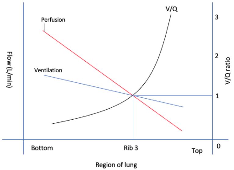
West’s zones
Dead space and shunt
When considering ventilation and perfusion of the lung, ventilation-perfusion ratio, dead space, and shunt come to everyone’s mind.
First, some definitions:
Dead space is the volume of inspired air that does not participate in gas exchange. In terms of the V/Q ratio, this translates to V of 1, Q of 0, and V/Q ratio of ∞.
A shunt is a volume of blood that enters systemic circulation without participating in gas exchange. In terms of the V/Q ratio, this translates to V of 0 and Q of 1. Hence V/Q ratio would be 0.
Dead space and shunt can be considered the opposite extremes of the V/Q ratio.
The normal V/Q ratio is around 0.8.
Dead space
Dead space is then classified further into anatomical and alveolar dead space.
Anatomical dead space = volume of air in the conducting airway.
Alveolar dead space = volume of air in the alveoli that is not perfused.
Physiological dead space = anatomical dead space + alveolar dead space.
Factors that will increase dead space include:
- Bronchodilation
- Pulmonary emboli (blood/air)
- Hypovolaemia/Hypotension
Shunt
Shunt can be classified as intrapulmonary shunt and anatomical shunt.
Intrapulmonary shunts include:
- Physiological hypoxic pulmonary vasoconstriction
- Pathological factors (e.g. consolidation in chest infection)
Anatomical shunts include:
- Physiological: Thebesian veins, bronchial vein
- Pathological: cardiac anomalies resulting in R-L shunt (e.g. ventricular septal defect)
References
Levitzky, M. G. (2018). Chapter 5: Ventilation-Perfusion Relationships. In Pulmonary physiology (9th ed). McGraw-Hill education.
Complete Guide to Clinical Genetics ST3 Application and Recruitment (2027)
Written by Medibuddy. Last updated: October 2026. Clinical Genetics is

Complete Guide to Clinical Radiology ST3 Application and Recruitment (2027)
Recruitment paused: NHS England is not running Clinical Radiology ST3+ recru

Complete Guide to Paediatric Surgery ST3 Application and Recruitment in United Kingdom (2026)
Written by Medibuddy. Last updated: October 2026. If you’re planning


Take your subscriptions with you
Our mobile app allows you to access your interview and exam question banks wherever you are.

