


 Close
Close
 7th February 2024
Ventilation
Ventilation is a process of moving air in and out of the lungs. Ventilation can be considered in two ways: minute ventilation or alveolar ventilation.
Minute ventilation is the total volume of air that is inspired or expired per minute.
Minute ventilation = Tidal volume ×Respiratory rate
Alveolar ventilation is the total volume of inspired air that participates in gas exchange. In other words, it is the minute ventilation minus the dead space. In a healthy adult, around 150mL of air is in the conducting airway, and this air volume, also referred to as dead space, does not participate in gas exchange.
Alveolar ventilation = Tidal volume-Dead space volume×Respiratory rate
Lung volumes and capacities
As clinicians, we need to be aware of several lung volumes and capacities.
Lung volumes and their definitions:
- Tidal volume (TV): volume of air that is inspired or expired during normal resting breathing.
- Inspiratory reserve volume (IRV): additional volume of air that can be maximally inspired on top of normal tidal volume inspiration.
- Expiratory reserve volume (ERV): additional air volume that can be forcefully expired after normal tidal volume expiration.
- Residual volume (RV): volume of air remaining in the lung after maximal expiration.
Lung capacities are a combination of two or more lung volumes. Lung capacities and their definitions:
- Vital capacity (VC): air volume that can be maximally expired after maximum inspiration.
- VC = IRV + TV + ERV
- Functional residual capacity (FRC): volume of air remaining in the lung at the end of resting expiration.
- FRC = ERV + RV
- Total lung capacity (TLC): volume of air in the lung after maximal inspiration
- TLC = IRV + TV + ERV + RV
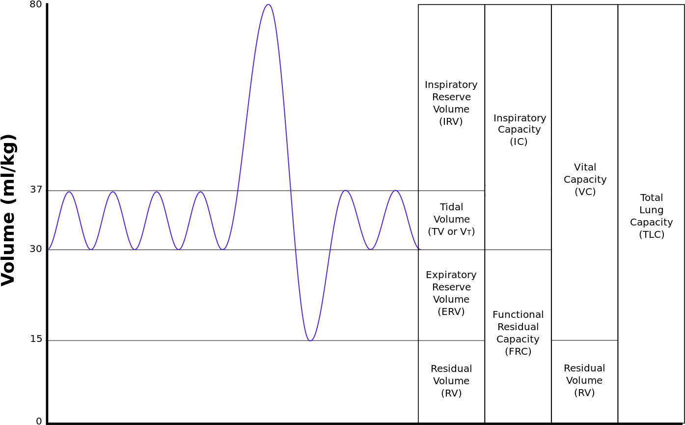
Image Source: Wikimedia Commons (2012). Lung Volumes Updated, Licensed under a creative commons licence. Available for free at: https://upload.wikimedia.org/wikipedia/commons/1/16/Lungvolumes_Updated.png (Accessed: 4 January 2024).
Other important measurements that can be obtained by the spirometry measurements include:
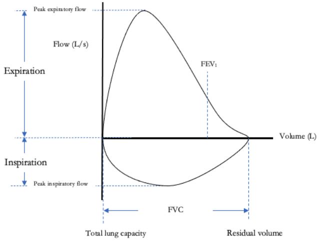
- Forced vital capacity: Volume of air that can be forcibly expired after maximal inhalation.
- FEV1: Air volume that can be forcibly expired during the first second of expiration after maximal inhalation.
This information can be summarised in a flow-volume loop. Changes in the contour of the loop can help support the diagnosis of restrictive or obstructive pulmonary conditions.
Measuring lung volumes
Methods to measure lung volumes include:
- Spirometry
- Body plethysmography
- Helium dilution technique
Spirometry cannot measure residual volume. As a result, we cannot measure functional residual volume and total lung capacity with spirometry. Residual volume can be measured using body plethysmography and helium dilution techniques.
The helium dilution technique may underestimate residual volume as it does not consider smaller airways behind an obstruction (i.e. gas trapping in COPD). Body plethysmography takes smaller airways behind obstruction into account. Hence, it is more accurate compared to the helium dilution technique.
Clinical relevance
Table of comparison of characteristics seen in pulmonary function tests in diseases
| Obstructive lung disease
(e.g. asthma, COPD, cystic fibrosis) |
Restrictive lung disease
(e.g. interstitial lung disease, neuromuscular disease, chest wall pathology) |
|
| Characteristics | ↑ airway resistance 🡪 ↓ air flow rates Air trapping Hyperinflation of lungs |
↓ lung compliance 🡪 ↓ lung volumes |
| FEV1/FVC ratio | ↓ | ↑ / normal |
| TLC | ↑ / normal | ↓ |
FEV1 can also be used in reversibility tests in the diagnosis of asthma.
Different patterns of flow-volume loops in different diseases.
Note: Flow-volume loop is produced by asking the person to fully inspire to total lung capacity from residual volume, then exhale as hard as possible back to residual volume
Normal
- The loop for the inspiratory limb is symmetric and convex.
- The expiratory limb is linear.
Restrictive disease (e.g. interstitial lung disease, neuromuscular disease, chest wall pathology)
- Narrower loop due to reduced forced vital capacity due to reduced lung compliance.
- Airflow is greater than normal during expiration at each lung volume due to increased elastic lung recoil.
Lower airway obstructive disease (e.g. asthma, emphysema)
- Airflow during expiration at all lung volumes is reduced due to increased airway resistance.
- The expiratory limb is prolonged.
Fixed upper airway obstruction (e.g. goitre, tracheal stenosis)
- Inspiratory and expiratory loops are flattened, resulting in a rectangular-looking loop. This is because fixed upper airway obstruction limits airflow during inspiration and expiration equally.
Variable extra thoracic obstruction (e.g. vocal cord paralysis, neoplasm in upper airway)
- The structures involved in pathology move according to pressure gradients across the glottis.
- Using vocal cords as an example:
- The vocal cord is drawn inwards during forced inspiration, resulting in increased airway resistance and flattening of the inspiratory limb.
- The vocal cord is pushed open during forced expiration. Hence, the expiratory limb has a normal shape.
Variable intrathoracic obstruction (e.g. tracheomalacia)
- Forced inspiration results in negative intrathoracic pressure that keeps the trachea open and patent. Hence, the normal shape of the inspiratory limb
- Forced expiration results in loss of the negative intrathoracic pressure, keeping the trachea open, narrowing the trachea and increasing airway resistance. Hence, the expiratory limb will have a flattened shape.

Pulmonary dynamics
Lung compliance
Compliance is defined as the unit change in volume per unit change in pressure. It is the inverse of elastance.
Compliance of the lung can be demonstrated using the compliance curve.

During inspiration, work is done to overcome (1) elastic recoil of the lungs and (2) resistance from airway and lung tissues sliding over each other. Work done to overcome the lung’s elastic recoil is stored as potential energy. This potential energy is released during expiration.
Hence, inspiration is active in normal, healthy patients, and expiration is passive.
This brings us to a discussion on work done during respiration.
Factors that alter compliance include (but are not limited to):
Increase compliance:
- Emphysema
- Increasing age (loss of pulmonary connective tissues)
Decrease compliance:
- Decreased lung compliance (pulmonary fibrosis, pulmonary oedema)
- Decreased chest wall compliance (obesity, burn to chest wall)
Work done during respiration.
Forgive this brief incorporation of physics. It will make sense, I promise. ( This will not be tested in the exam but will help understand the concepts below).
Work done = Pressure x Volume.
From the formula above, we can determine work done during respiration using the compliance curve.

Work done during inspiration = purple + red + green
Work done during expiration = red (to overcome airway resistance)
Purple = work done loss as heat energy
In lung diseases, the compliance curve changes, and work done changes as well.
In restrictive lung disease, lung compliance decreases, and the inspiratory limb curves downwards and rightwards. This increases work done during inspiration.
In obstructive lung disease (i.e. COPD), the expiratory limb curves leftwards and upwards. This may curve outside of the purple zone. This area represents the additional work needed for active expiration.

Airway resistance

Airway resistance is affected by:
- Airway radius
- Lung volumes
- Phase of respiration
- Autonomic activity
- Elastic recoil of the lung
- Local PaCO2
Although the airway becomes narrower towards the lung periphery, the peripheral airway contributes little airway resistance due to multiple parallel airways. The upper respiratory tract contributes 30-50% of total airway resistance in a healthy person. The large-to-medium-sized bronchi are the location along the lower respiratory tract that contributes the most resistance.
References
Levitzky, M. G. (2018). Chapter 3: Alveolar Ventilation. In Pulmonary physiology (9th ed). McGraw-Hill education.
David, S., & Edwards, C. W. (2022). Forced Expiratory Volume. In StatPearls. StatPearls Publishing. http://www.ncbi.nlm.nih.gov/books/NBK540970/
Moore, V. C. (2012). Spirometry: Step by step. Breathe, 8(3), 232–240. https://doi.org/10.1183/20734735.0021711
Ponce, M. C., & Sharma, S. (2022). Pulmonary Function Tests. In StatPearls. StatPearls Publishing. http://www.ncbi.nlm.nih.gov/books/NBK482339/
Levitzky, M. G. (2018). Chapter 2: Mechanics of Breathing. In Pulmonary physiology (9th ed). McGraw-Hill education
Complete Guide to Clinical Genetics ST3 Application and Recruitment (2027)
Written by Medibuddy. Last updated: October 2026. Clinical Genetics is

Complete Guide to Clinical Radiology ST3 Application and Recruitment (2027)
Recruitment paused: NHS England is not running Clinical Radiology ST3+ recru

Complete Guide to Paediatric Surgery ST3 Application and Recruitment in United Kingdom (2026)
Written by Medibuddy. Last updated: October 2026. If you’re planning


Take your subscriptions with you
Our mobile app allows you to access your interview and exam question banks wherever you are.

