


 Close
Close
 18th January 2024
Rheumatoid Arthritis
Background
Epidemiology
Rheumatoid Arthritis (RA) is the commonest inflammatory arthritis in the United Kingdom (UK), affecting approximately 1% of the population. The incidence of RA is greater in smokers, and it is also between 2-4 times more common in females. RA can occur at any age but peaks between the ages of 30-50 years.
Pathophysiology
RA is a chronic, systemic autoimmune disease primarily affecting the lining of synovial joints. The deformities typically associated with RA occur due to the stretching of tendons and ligaments and destruction of joints through the erosion of cartilage and bone.
A combination of genetic and environmental factors can be responsible for triggering RA. Inherited genes in the human leukocyte antigen (HLA) major histocompatibility complex (MHC) are of particular importance. The presence of HLA DR4/DR1 in RA patients is associated with increased disease severity. Smoking is the best-defined environmental stimulus, likely due to repeated activation of innate immunity, especially at mucosal surfaces.
References
- Guo Q, Wang Y, Xu D, Nossent J, Pavlos N, Xu J. Rheumatoid arthritis: pathological mechanisms and modern pharmacologic therapies. Bone Research. 2018;6(1).
- Rheumatoid arthritis | Health topics A to Z | CKS | NICE [Internet]. Cks.nice.org.uk. 2021 [cited 10 November 2021]. Available from: https://cks.nice.org.uk/topics/rheumatoid-arthritis/
- UpToDate [Internet]. Uptodate.com. 2021 [cited 10 November 2021]. Available from: https://www.uptodate.com/contents/pathogenesis-of-rheumatoid-arthritis?search=rheumatoid%20arthritis&source=search_result&selectedTitle=8~150&usage_type=default&display_rank=8#H3
Presentation
Articular manifestations
RA typically presents with symmetrically swollen, painful and stiff small joints of the hand and feet. Morning stiffness is a sign of inflammatory arthritis and typically lasts over an hour. Classically, metacarpophalangeal (MCP), proximal interphalangeal joints (PIP), and metatarsophalangeal (MTP) joints are involved (The distal interphalangeal (DIP) joints and thoracic and lumbosacral spine are spared in RA). Other synovial joints which may be involved include wrists, knees, elbows, ankles, hips, shoulders and cervical spine (C1-C2).
RA onset can vary, and initial presentation may occur in the following ways:
- Insidious onset of symmetric polyarthropathy of small joints of the wrists, hands and feet is the commonest
- Insidious onset of monoarticular or oligoarticular arthritis similar to a seronegative spondyloarthropathy. Additional small joint and symmetric involvement can occur over time.
- Sudden onset polyarticular arthritis is less common
- Palindromic RA is characterised by brief, self-limited and recurrent episodes of mono/polyarthritis of various joints
- Systemic illness with extra-articular symptoms
Signs
There is inflammation early in the disease process, but usually no joint damage. With progressive inflammation and disease progression, damage to the joints and surrounding tendons can result in the classic deformities associated with RA (now rarely seen with earlier diagnosis and initiation of disease-modifying anti-rheumatic drug (DMARD) therapy.
The following signs are some of the characteristic findings associated with RA:
- ‘Zigzag’ deformity- ulnar deviation and palmar subluxation of the MCP joints and radial deviation of the wrist
- Swan neck deformities- PIP hyperextension and DIP flexion
- Boutonniere deformities- PIP flexion and DIP hyperextension
- Trigger finger- thickening of the tendon sheaths, causing the finger to lock in flexion
- Rupture of hand extensor tendons
- Carpal tunnel syndrome- tenosynovitis in the flexor tendon sheaths can cause median nerve impingement
- Claw toe deformities of the PIP joints due to plantar subluxation of the metatarsal heads
- Valgus and pes planus deformities.

Image Source: Wikimedia Commons (2014). Swan neck deformity in a 65 year old Rheumatoid Arthritis patient- 2014-05-27 01-49, Licensed under a creative commons licence. Available for free at: https://commons.wikimedia.org/wiki/File:Swan_neck_deformity_in_a_65_year_old_Rheumatoid_Arthritis_patient-_2014-05-27_01-49.jpg (Accessed: 24 November 2021).

Image Source: Wikimedia Commons (2021). RA hand deformity. Licensed under a creative commons licence. Available for free at: https://commons.wikimedia.org/wiki/File:RA_hand_deformity.JPG (Accessed: 24 November 2021).
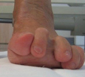
Image Source: Wikimedia Commons (2021). Luxierte Zehen bei Rheumatoidarthritis (praop), Licensed under a creative commons licence. Available for free at: https://commons.wikimedia.org/wiki/File:Luxierte_Zehen_bei_Rheumatoidarthritis_(pr%C3%A4op.)..jpg (Accessed: 24 November 2021).
Extra-articular features and complications of RA
Persistent, long-standing inflammation can lead to earlier development of cardiovascular and lung diseases and malignancy. Therefore, RA is associated with increased morbidity and premature death. Patients with high titres of Rheumatoid Factor (RF) are most likely to have extra-articular manifestations of RA.
- Skin Manifestations
- Rheumatoid nodules are the most commonly occurring skin manifestation and are associated with RF positivity. They tend to be seen on extensor surfaces which are exposed to external pressure.
- Splinter haemorrhages
- Periungual infarcts
- Leg ulcers
- Digital gangrene
- Pyoderma gangrenosum- rare
- Ocular Manifestations
- Keratoconjunctivitis sicca is the most frequent ocular manifestation and is often seen together with xerostomia in patients with secondary Sjogren’s syndrome.
- Episcleritis- inflammation of the layer superficial to the sclera, generally self-limiting and not as painful as scleritis
- Scleritis – inflammation of the sclera itself- intensely painful
- Ulcerative keratitis
- Pulmonary manifestations
- Pleural effusions- these are usually exudative in nature
- Interstitial lung disease
- Nodules can also be seen in the lung and are a feature of Caplan syndrome (Rheumatoid pneumoconiosis)- combination of seropositive RA and a characteristic pattern of fibrosis.
- Airways disease due to chronic inflammation
- Cardiac manifestations
- Increased risk of atherosclerosis with subsequent complications associated with atherosclerotic disease
- Pericarditis
- Myocarditis
- Myocardial fibrosis can lead to conduction abnormalities
- Endocarditis with formation of rheumatoid nodules in aortic or mitral valves
- Haematological manifestations
- Anaemia- multiple causes, including drug-induced, nutritional, Gastrointestinal (GI) bleed, bone marrow suppression and ineffective erythropoiesis
- Anaemia of chronic disease (normochromic and normocytic) is correlated with disease activity, especially the degree of articular inflammation
- Eosinophilia- related to disease activity or hypersensitivity to drugs
- Thrombocytosis- related to the number of active inflamed joints
- Increased risk of non-Hodgkin’s lymphoma- this risk may be further increased by immunosuppressive agents
- Felty’s syndrome- the triad of RA, splenomegaly (clinically evident or on US) and neutropenia (<1.8*10/L) on two different occasions where other causes are deemed unlikely.
- Secondary AA Amyloidosis- as with many other chronic inflammatory diseases. RA can increase the risk of developing secondary amyloidosis. The disease process is increased production of serum amyloid (AA) protein, fibrils of which get deposited in the extracellular tissues of various organs. Amyloidosis can manifest as macroglossia, cardiac failure, chronic kidney disease (usually due to nephrotic syndrome), GI bleeding or malabsorption or as hepatomegaly/ deranged liver function tests. Diagnosis is confirmed via biopsy (usually fat pad or rectal biopsy), which shows the characteristic findings of red-green birefringence when stained with Congo red. Management is through achieving control.
- GI and renal manifestations are usually iatrogenic and rare
- Neurologically- peripheral neuropathy presenting as diffuse sensorimotor neuropathy or mononeuritis multiplex.
References
- Ebel A, O’Dell J. Clinical Features, Diagnosis, and Treatment of Rheumatoid Arthritis. Physician Assistant Clinics. 2021;6(1):41-60.
- Manole Cojocaru R. Extra-articular Manifestations in Rheumatoid Arthritis [Internet]. PubMed Central (PMC). 2021 [cited 10 November 2021]. Available from: https://www.ncbi.nlm.nih.gov/pmc/articles/PMC3152850/
- Gaillard F. Caplan syndrome | Radiology Reference Article | Radiopaedia.org [Internet]. Radiopaedia.org. 2021 [cited 10 November 2021]. Available from: https://radiopaedia.org/articles/caplan-syndrome?lang=gb
- Young A, Koduri G. Extra-articular manifestations and complications of rheumatoid arthritis. Best Practice & Research Clinical Rheumatology. 2007;21(5):907-927.
- Figus F, Piga M, Azzolin I, McConnell R, Iagnocco A. Rheumatoid arthritis: Extra-articular manifestations and comorbidities. Autoimmunity Reviews. 2021;20(4):102776.
Diagnosis
RA should be suspected in any adult presenting with inflammatory polyarthritis. When taking a history in someone with such a presentation, an enquiry should be made into the presence and location of joint pain, swelling and early morning stiffness. Stiffness associated with inflammatory arthritis tends to be peripheral rather than in the lower back and lasts at least 30 minutes. The duration of symptoms is also important- if symptoms have only been present for a short time (less than 6 weeks), symptoms may be more likely due to an acute viral polyarthritis than RA. Symptoms suggestive of an alternate diagnosis, e.g. psoriasis, inflammatory bowel disease (IBD), systemic lupus erythematosus (SLE), should be excluded.
A thorough physical examination to assess for synovitis, including the presence and distribution of swollen and tender joints and limited joint motion (and extra-articular manifestations), should be completed. Whilst symmetric polyarthritis (especially of the MCPs, MTPs and PIP joints) is suggestive of RA, involvement of the DIPs is usually associated with osteoarthritis or psoriatic arthritis.
Investigations
Blood tests
Blood tests are not only useful for making the initial diagnosis of RA but are also used as baseline tests for monitoring disease activity and adverse effects of medications.
- FBC (with differential and platelet count), LFTs, U&Es, serum uric acid
- ESR and CRP are both elevated in RA- useful in initial diagnosis and for monitoring disease activity
- Immunology
- Rheumatoid Factor (RF)- present in 70-80% of patients with RA but poor specificity (also found in healthy individuals, patients with SLE, Sjogren’s syndrome, and those with mixed cryoglobulinemia, as well as in many other inflammatory conditions).
- Anti-CCP – similar sensitivity as RF but much higher specificity (95-98%)
- ANA- a negative ANA is useful in excluding SLE. It may be positive in up to ⅓ of patients with RA. If positive, anti-dsDNA and anti-Smith testing should be performed as these are highly specific for SLE.
- Seronegative RA can be diagnosed in patients lacking both RF and anti-CCP if they have findings otherwise characteristic of RA if appropriate exclusions have been met.
- Infectious screen – especially in patients with a very short history and those who are seronegative for RF and anti-CCP:
- Parvovirus B19
- HBV and HCV
- Borrelia in areas endemic for Lyme disease
- Chikungunya virus in areas where mosquito bite-transmitted viral diseases are prevalent
Imaging
Plain radiography is usually the first mode of imaging performed in patients suspected of RA, albeit not sensitive enough to pick up changes such as bony erosions in early disease. Typical radiological findings associated with RA include osteopenia, joint space narrowing, erosions and subluxation.
Ultrasound (US) may be more useful in allowing earlier diagnosis of RA, particularly in patients with seronegative disease. It can assess soft tissues and identify synovial thickening, presence of fluid in joints and abnormalities in tendons, ligaments and entheses. Doppler US assesses for soft tissue hyperaemia and is, therefore, useful for distinguishing between active and inactive inflammatory tissues.
Magnetic resonance imaging (MRI) is more sensitive still and can evaluate all structures (including soft tissue, cartilage, and bones) which can be affected by RA.
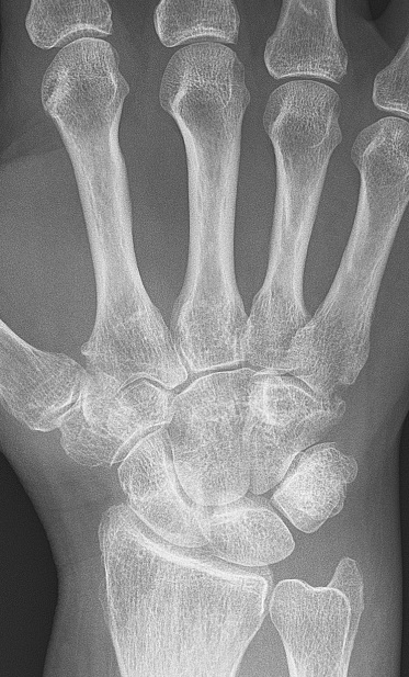
Image Source: Wikimedia Commons (2009). Rheumatoid arthritis with unaffected carpal bones 2009, Licensed under a creative commons licence. Available for free at https://en.wikipedia.org/wiki/Rheumatoid_arthritis#/media/File:Rheumatoid_arthritis_with_unaffected_carpal_bones_2009.jpg (Accessed: 24 November 2021).
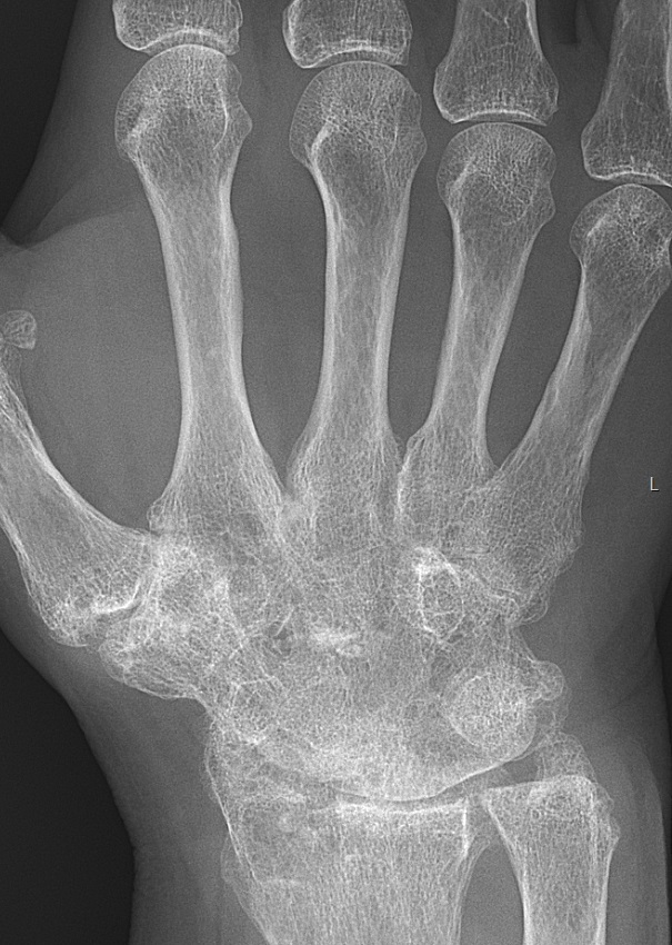
Image Source: Wikimedia Commons (2017) Rheumatoid arthritis with carpal ankylosis 2017, Licensed under a creative commons licence. Available for free at https://en.wikipedia.org/wiki/Rheumatoid_arthritis#/media/File:Rheumatoid_arthritis_with_carpal_ankylosis_2017.jpg (Accessed 24 November 2021)

Image Source: Wikimedia Commons (2006). X-ray of right fourth PIP joint with bone erosions by rheumatoid arthritis, Licensed under a creative commons licence. Available for free at: https://upload.wikimedia.org/wikipedia/commons/6/6c/X-ray_of_right_fourth_PIP_joint_with_bone_erosions_by_rheumatoid_arthritis.jpg (Accessed: 24 November 2021).
Synovial fluid analysis
- For diagnosis or exclusion of gout, pseudogout, or an infectious arthritis if there is a joint effusion
- Analysis should include:
- Cell count and differential
- Crystal search
- Gram stain and culture
Classification criteria in RA
2010 American College of Rheumatology (ACR)/ European League Against Rheumatism (EULAR) Classification Criteria
The following criteria are required to be met in order to be classified as having a definite RA diagnosis:
- Presence of synovitis in at least 1 joint
- Absence of an alternative diagnosis which better explains the synovitis
- Total score of at least 6 (out of a maximum of 10) from the individual scores in the 4 domains, which include:
- Number and site of joints involved- greater number of points for smaller joints affected.
- Serological abnormality (RF or anti-CCP positive)
- Elevated CRP or ESR
- Symptom duration at least 6 weeks
Disease Activity Score (DAS)
The DAS is used to clinically measure disease activity in patients with RA and is particularly useful for targeting and objectively measuring response to treatment. It objectively measures tenderness and swelling of the shoulder, elbow, wrist, knee, MTP and PIP joints and includes a subjective measure of the patient’s general health based on a visual analogue scale.
The DAS 28 score considers the following four variables:
- The number of swollen joints (0-28)
- The number of tender joints (0-28)
- ESR or CRP
- Visual analogue scale (VAS)disease activity (0-100mm)
The formula used to calculate the score is: (0.56*√ (tender joints) +0.28*√ (swollen joints) +0.70*Ln(ESR/CRP) +0.014*VAS)
An overall score is calculated out of 10, with a score > 5.1 indicating high disease activity and a score of <3.2 indicating low disease activity. NICE recommends using the DAS as one of the criteria for starting biological therapy.
References
- UpToDate [Internet]. Uptodate.com. 2021 [cited 10 November 2021]. Available from: https://www.uptodate.com/contents/diagnosis-and-differential-diagnosis-of-rheumatoid-arthritis/print#!#:~:text=Symmetric%20polyarthritis%2C%20particularly%20of%20the%20MCP%2C%20MTP%2C%20and%2For,suggests%20a%20diagnosis%20of%20osteoarthritis%20or%20psoriatic%20arthritis.
- Kgoebane K, Ally M, Duim-Beytell M, Suleman F. The role of imaging in rheumatoid arthritis. SA Journal of Radiology [Internet]. 2018 [cited 10 November 2021];22(1):1316. Available from: https://www.ncbi.nlm.nih.gov/pmc/articles/PMC6837821/
- Mooney J, McGee M. Early recognition of rheumatoid arthritis. Nursing and Residential Care. 2012;14(7):344-347.
Differential diagnoses
Background
When assessing patients in early disease, the ‘typical’ features associated with RA, such as joint erosions and rheumatoid nodules, are not usually present. So, joint manifestations are often difficult to distinguish from other inflammatory polyarthritis.
Septic arthritis should be excluded in any joint that is red, hot, and swollen. This should be done via means of joint aspiration as long as there is no skin infection around the site of aspiration, and there is no metalwork inside the joint (in this case, a Trauma and Orthopaedic opinion should be sought). Septic arthritis needs to be treated with antibiotics as soon as possible.
If there are episodes of joint inflammation sequentially affecting one to several joints for hours to days (with symptom-free periods lasting days to months), consider palindromic rheumatism. Some patients do, however, go on to develop a well-defined rheumatic disease, most commonly RA or SLE. Patients with anti-CCP are more likely to develop RA.
Osteoarthritis (OA)
Despite being easily distinguishable from RA in medical textbooks, OA can cause a diagnostic conundrum in middle-aged or elderly patients when small joints of the hands are involved.
In OA, DIP joints are involved with Heberden’s nodes in this area (whereas DIP joints are spared in RA). Carpometacarpal joint of the thumb is also involved. There may be joint swelling in OA as well as in RA, but the joint swelling in OA is hard and bony, as opposed to soft, warm, boggy and tender in RA.
- Stiffness is usually absent, but if present, it is worse in the evening after exercise. If present in the morning, it lasts no more than a few minutes.
- X-rays show narrowing of joint space due to cartilage loss and osteophytes due to bone remodelling, but not erosions (Remember LOSS in OA (Loss of joint space, Osteophytes, Subchondral sclerosis, Subchondral cysts) vs LESS in RA (Loss of joint space, Erosions, Soft tissue swelling, Soft bones (osteopenia)
Psoriatic arthritis
Psoriatic arthritis can be still more difficult to distinguish from RA because asymmetric polyarthritis is seen. Therefore, this diagnosis is generally made in patients who also have psoriasis and are seronegative for RF and anti-CCP. The diagnosis is made more challenging by the fact that the joint symptoms of psoriatic arthritis may precede the onset of skin disease by many years. Indeed, in some patients with a symmetric inflammatory polyarthritis, the only clue to the diagnosis of psoriatic arthritis may be a family history of psoriasis.
Although there is a clear diagnostic challenge here, the initial management of both disorders is very similar, so treatment can be initiated before necessarily distinguishing between Psoriatic arthritis and RA.
Reactive arthritis
Reactive arthritis often presents as a monoarthritis or oligoarthritis of the large joints. RA may occasionally present similarly, particularly in the knee, where the physical signs can be identical. There are, however, some findings that are more suggestive of reactive arthritis than RA. These include :
- History of recent urethritis or enteric infection
- Asymmetric pattern of joint involvement, particularly in the hands
- Involvement of not only the joint but also the tenosynovium, entheses, and surrounding tissues. This can manifest as the characteristic “sausage” swelling in the digits of the hands and feet.
- Keratoderma blennorrhagica or circinate balanitis
- Radiological evidence of sacroiliitis and/or spondylitis
- Presence of HLA B27
Other systemic rheumatic diseases
These disorders include Sjogren’s syndrome, dermatomyositis, and mixed connective tissue disease. They are characterised by presence of other systemic features (e.g. rashes, dry mouth and dry eyes, myositis, or nephritis) and autoantibodies not usually seen in RA.
In SLE, Jaccoud’s arthropathy (the joint deformities are not caused by joint destruction but by loosening and lengthening of periarticular structures and tendons may be confused with the characteristic joint deformities seen in RA. However, they can be differentiated by physical examination as the deformities are passively ‘correctable’ and on radiology as X-rays show no bony erosions. In addition, ESR and CRP are less well-correlated in other diseases than in RA. Whilst both are raised in RA, CRP is often normal or only minimally elevated in patients with active SLE, even when ESR is elevated.

Image Source: Wikimedia Commons (2021). Jaccoud arthropathy hand x-ray front, Licensed under a creative commons licence. Available for free at: https://commons.wikimedia.org/wiki/File:Jaccoud_arthropathy_hand_x-ray_front.jpg (Accessed: 24 November 2021).
Infectious arthritis
If the syndrome of polyarthritis is short-lived, lasting only from a few days to several weeks (rarely longer than 6 weeks), viral polyarthritis must be considered. Examples of organisms causing viral polyarthritis include Rubella, Parvovirus B19, hepatitis B and C (more commonly associated with arthralgias rather than polyarthritis) and Chikungunya.
Another example of an infectious arthritis is that of Lyme arthritis – a late manifestation of Lyme disease. This is characterised by intermittent or persistent inflammatory arthritis in a few large joints, most typically monoarthritis of the knee.
Miscellaneous diseases
- Enteric arthritis – Usually a large-joint oligoarthritis or spondyloarthritis with sacroiliitis. However, confusion with RA can occur in patients with peripheral polyarthritis and involvement of the MCP joints.
- Crystal arthropathy (gout or pseudogout)- Can become chronic and have a polyarticular distribution, resembling RA.
- Sarcoid arthropathy- May be oligo- or polyarticular, most often affecting the ankles, knees, hands, wrists, MCP and PIP joints.
References
- UpToDate [Internet]. Uptodate.com. 2021 [cited 10 November 2021]. Available from: https://www.uptodate.com/contents/diagnosis-and-differential-diagnosis-of-rheumatoid-arthritis/print#!#:~:text=Symmetric%20polyarthritis%2C%20particularly%20of%20the%20MCP%2C%20MTP%2C%20and%2For,suggests%20a%20diagnosis%20of%20osteoarthritis%20or%20psoriatic%20arthritis.
- Cruz Whitley J, Aronowitz P. Jaccoud’s Arthropathy. Journal of General Internal Medicine. 2018;33(9):1583-1583.
Management
The first step in the primary care management of RA is to refer any patient with suspected persistent synovitis for specialist opinion, even if the patient shows a normal acute phase response. Any investigations requested in primary care should not delay referral to secondary care services.
Management of RA is based on the ‘treat-to-target’ strategy. This means that the aim of treatment should be to achieve a target of remission or low disease activity if remission is not possible. Remission should be the target, particularly in patients at high risk of progression, including patients who are anti-CCP positive or have erosion on their X-rays at baseline assessment.
Pharmacological management
Disease-modifying anti-rheumatic drugs (DMARDs) can be classified as either conventional DMARDs or biologic DMARDs. The first-line therapy is with conventional DMARD monotherapy, either with oral methotrexate, leflunomide, or sulfasalazine, but in practice is usually Methotrexate. Treatment should be started as soon as possible and ideally within 3 months of onset of persistent symptoms. Hydroxychloroquine is an alternative for mild or palindromic disease. The dose of the initial DMARD can be escalated as tolerated. If this is not sufficient, a second DMARD can be added as combination therapy.
The beneficial effect of DMARD therapy may not be seen immediately. Therefore, short-term bridging therapy with glucocorticoids can be started when initiating therapy with a DMARD. A step-down strategy can be considered in patients who stay in remission for at least a year without requiring glucocorticoids.
Biological therapy can be considered, usually in addition to Methotrexate, if the treatment target has not been achieved despite intensive therapy with 2 or more conventional DMARDs and the DAS28 score is between 3.2 and 5.1 (moderate activity). TNF-alpha inhibitors (including Infliximab, Adalimumab and Etanercept) are usually 1st line biologic therapy. If there is a contraindication to the use of Methotrexate, Adalimumab and Etanercept can be used as monotherapy. In cases where conventional DMARDs and TNF-alpha inhibitors have failed, Rituximab (B cell depletion) can be used- again in combination with Methotrexate. Other rarer biological DMARDs include Tocilizumab (IL-6 receptor blocker) and Abatacept (disruption of T cell function). These agents may be called upon when none of conventional DMARDs, TNF alpha- inhibitors or Rituximab have managed to achieve the treatment target.
Symptom control
There is limited evidence for the use of paracetamol, opioids, and tricyclic antidepressants in symptom control. Non-steroidal anti-inflammatory drugs (NSAIDs) can be considered for pain control and stiffness. However, potential GI, liver and cardiorenal adverse effects should be considered, and a proton pump inhibitor (PPI) should be offered.
Adverse effects of DMARDs
Conventional DMARDs
Most conventional DMARDs have a similar side effect (SE) profile, including increased risk of infection and GI upset. Certain DMARDs (particularly Methotrexate and Leflunomide) are also teratogenic, so women of childbearing age should ensure they are using adequate contraception. However, there are some adverse effects which are unique to each agent.
Methotrexate, Sulfasalazine and Leflunomide share a similar adverse effect profile, including GI upset, increased incidence of common and serious infections, allergic reactions, bone marrow suppression and hepatotoxicity. Alopecia is a SE of both Methotrexate and Leflunomide. The following unique SEs are also seen:
- Methotrexate- pneumonitis and interstitial lung disease, folic acid deficiency, liver cirrhosis, oral ulcers
- Leflunomide- hypertension, peripheral neuropathy, weight loss
- Sulfasalazine- high risk of GI upset. Can cause decreased sperm count and rarely Drug Rash with Eosinophilia and Systemic Symptoms (DRESS) syndrome.
Hydroxychloroquine has perhaps the best safety profile compared to other conventional DMARDs; it does not increase the risk of serious infections or cause renal or liver dysfunction. Common SEs include rash and diarrhoea. The most serious but rare adverse effect associated with Hydroxychloroquine is retinopathy/ maculopathy. This is associated with a higher cumulative dose and greater than 5 years of therapy. Other rare adverse effects of Hydroxychloroquine include anaemia, leukopenia, myopathy and cardiomyopathy.
Biologic DMARDs
All biologic DMARDs can increase the risk of common and serious infections from bacterial, fungal and viral agents. This can also include reactivation of tuberculosis, herpes zoster and hepatitis B and C. Rarer SEs include bone marrow suppression and hepatotoxicity.
Whilst it is acceptable to use a combination of conventional DMARDs or a combination of conventional DMARD and biologic DMARD, a combination of different biologic DMARDs should not be used as the risk of severe immunosuppression reading to severe life-threatening infections is too high.
Specific SEs associated with Anti-TNF agents include:
- Worsening of severe congestive heart failure
- Drug-induced SLE
- Demyelinating central nervous system diseases
- Lymphomas and non-melanoma skin cancers
Specific SEs associated with IL-6 and JAK inhibitors include:
- Hyperlipidemia
- Deranged liver function tests
- Pancytopenia
Other miscellaneous SEs associated with particular Biologic DMARDs
- Abatacept- worsening of COPD
- IL-17 inhibitors- cause or worsen inflammatory bowel disease
- Rituximab- progressive multifocal leukoencephalopathy
Drug monitoring
In order to minimise the risk of the above adverse effects, DMARD therapy requires screening before initiation as well as monitoring during therapy.
Baseline assessment before initiating any DMARD should include height, weight, BP and blood tests (FBC, U&Es, LFTs). Any comorbidities should also be noted as this may influence DMARD choice- this should include screening for occult viral infections. Anti-Pneumococcal and Influenza vaccines are also advised.
Standard blood monitoring schedule when starting or adding a new DMARD
- Check FBC, U&Es, LFTs and albumin every 2 weeks until on stable dose for 6 weeks
- Once on a stable dose, check FBC, U&Es, LFTs and albumin every month for 3 months
- After this, check FBC, U&Es, LFTs and albumin at least every 12 weeks
In patients at higher risk of toxicity, blood monitoring should be more frequent. In addition, certain drugs require specific considerations:
- Methotrexate- all patients should be co-prescribed at least 5mg once weekly dose of folic acid supplementation.
- Azathioprine- measure a baseline thiopurine methyltransferase (TPMT)
- Hydroxychloroquine- baseline ophthalmic examination within 1 year of starting therapy
| DMARD | Blood test monitoring | Other monitoring |
|---|---|---|
| Azathioprine | Standard blood monitoring schedule | None |
| Ciclosporin | Extend standard blood monitoring schedule longer term | Blood pressure and glucose at each monitoring appointment |
| Gold | Standard blood monitoring schedule | Urinalysis (haematuria/ proteinuria) prior to each dose |
| Hydroxychloroquine | No routine blood monitoring required | Baseline ophthalmic assessment and annual assessment if continued for >5 years |
| Leflunomide | Standard blood monitoring schedule | Blood pressure and weight at each monitoring appointment |
| Methotrexate | Standard blood monitoring schedule | None |
| Sulfasalazine | Standard blood monitoring schedule for 1 year, then no routine monitoring required | None |
| Mycophenolate | Standard blood monitoring schedule | None |
Table 1: Summary of standard blood monitoring schedule for common DMARDs. Adapted from: Ledingham J, Gullick N, Irving K, Gorodkin R, Aris M, Burke J et al. BSR and BHPR guideline for the prescription and monitoring of non-biologic disease-modifying anti-rheumatic drugs. Rheumatology. 2017;56(6):865-868.BSR and BHPR guideline for the prescription and monitoring of non-biologic disease-modifying anti-rheumatic drugs | Rheumatology | Oxford Academic (oup.com)
Other considerations when prescribing and monitoring DMARDs
Prescribing DMARDs in patients with co-morbidities requires some specific considerations. Caution is required, particularly in patients with pre-existing lung disease (particularly with drugs known to cause pneumonitis) and deranged liver function tests. In patients who have chronic viral hepatitis, anti-viral therapy should be initiated before commencing immunosuppressive DMARDs. In patients with chronic kidney disease, dose reduction and increased frequency of blood monitoring may be required.
In the perioperative period, DMARD therapy does not usually need to be stopped, but each case should be assessed on an individual basis. In the case of serious infections, many of the DMARDs, including methotrexate, leflunomide, and sulfasalazine, should be paused while the patient recovers from the infection.
References
- Ledingham J, Gullick N, Irving K, Gorodkin R, Aris M, Burke J et al. BSR and BHPR guideline for the prescription and monitoring of non-biologic disease-modifying anti-rheumatic drugs. Rheumatology. 2017;56(6):865-868.
- Rheumatoid arthritis – NICE Pathways [Internet]. Pathways.nice.org.uk. 2021 [cited 10 November 2021]. Available from: https://pathways.nice.org.uk/pathways/rheumatoid-arthritis
- Allen A, Carville S, McKenna F. Diagnosis and management of rheumatoid arthritis in adults: summary of updated NICE guidance. BMJ. 2018;:k3015.
- Benjamin, O., Bansal, P., Goyal, A. and Lappin, S., 2021. Disease Modifying Anti-rheumatic Drugs (DMARD). [online] Ncbi.nlm.nih.gov. Available at: <https://www.ncbi.nlm.nih.gov/books/NBK507863/> [Accessed 3 November 2021].
Complete Guide to Clinical Genetics ST3 Application and Recruitment (2027)
Written by Medibuddy. Last updated: October 2026. Clinical Genetics is

Complete Guide to Clinical Radiology ST3 Application and Recruitment (2027)
Recruitment paused: NHS England is not running Clinical Radiology ST3+ recru

Complete Guide to Paediatric Surgery ST3 Application and Recruitment in United Kingdom (2026)
Written by Medibuddy. Last updated: October 2026. If you’re planning


Take your subscriptions with you
Our mobile app allows you to access your interview and exam question banks wherever you are.

