


 Close
Close
 19th January 2024
Spondyloarthritides
Background
The spondyloarthritides are a group of inflammatory rheumatological conditions predominantly affecting the spine or peripheral joints. They are characterised by inflammatory back pain, stiffness and swelling of the joints, and there is a strong association with HLA-B27 antigens. The synovitis is similar to that seen in rheumatoid arthritis; however, rheumatoid factor and anti-CCP are typically negative.
The spondyloarthritides can be subdivided into:
- Axial – affecting the spine or sacroiliac joints, e.g., ankylosing spondylitis
- Peripheral – affecting peripheral joints, enthesitis or dactylitis, e.g. psoriatic arthritis, reactive arthritis, enteric arthritis
Ankylosing spondylitis
Background
Definition
Axial spondylitis is classified as a chronic seronegative spondyloarthropathy, primarily involving the axial skeleton. The hallmark clinical features are inflammatory back pain and spinal stiffness, which tend to respond well to non-steroidal anti-inflammatory drugs (NSAIDS). Ankylosing spondylitis (AS) is the name given to the radiological changes in the spine and sacroiliac joints, which can result from the progressive inflammatory changes. Further progression of the disease can result in spinal fusion and subsequent severe disability. HLA-B27 is found in 90% of patients with AS. It occurs most commonly between the ages of 20 and 30 years, and is more common in men than women.
Causes and pathophysiology
The aetiology of AS is unknown, but is thought to be a result of interaction between genetic and environmental factors. The heritability is estimated at 97%. The pathophysiological processes underlying AS include inflammation, erosion, and subsequent repair of cartilage. An increased number of osteoclasts are found in the axial skeleton of patients with AS.
Presentation
AS usually presents with an insidious onset before the age of 30 years. Symptoms may be mild in early stages, with inflammatory back pain being a hallmark feature of the disease. Early morning stiffness is also characteristic. Systemic features and extra-articular manifestations are common.
Symptoms
- Inflammatory back pain:
- Worse first thing in the morning and can awaken patient from sleep
- Often improves with physical activity but not rest
- Originates in sacroiliac joints
- Can be felt as diffuse, non-specific buttock pain
- Improves with NSAIDs
- Can cause loss of spinal mobility
- Back stiffness
- Usually occurs in the early morning
- Improves with exercise
- Peripheral enthesitis:
- Inflammation at points where tendons and ligaments attach to bones
- Commonly affected sites: Achilles tendonitis, plantar fasciitis, tibial tuberosity
- Painful lesions, especially in the morning
- May be associated with swelling, stiffness, and reduced mobility
- Peripheral arthritis:
- Usually monoarticular or oligoarticular
- Usually asymmetrical and involves hips, shoulder joints, chest wall joints and pubic symphysis
- Temporomandibular joints can also be affected occasionally
- More common in females
- Extra-articular manifestations:
- Uveitis
- Psoriasis
- Inflammatory bowel disease
- Osteoporosis
- Cardiac complications: aortic regurgitation, mitral regurgitation and non-aortic valvular heart disease, heart failure, arrhythmias, cardiovascular disease, AV node block
- Lung complications: restrictive lung disease, apical fibrosis
- Neurological complications: vertebral fractures, cauda equina
- Renal complications: amyloidosis, IgA nephropathy
- Systemic features: fatigue, sleep disturbance
Signs
- Loss of lumbar lordosis and increased kyphosis (in chronic cases) 🡪 ‘question mark posture’
- Reduced spinal flexion
- Schober test: mark is made at 5th lumbar spinous process and 10cm above with patient in erect position. Distance should increase to 15cm on bending forwards in individuals without ankylosing spondylitis
- Pelvic and chest wall tenderness
- Sacroiliac joint tenderness
- Reduced chest expansion:
- Signifies costovertebral joint involvement
- Less than 2.5cm on deep inspiration measured at 4th intercostal space
- Increased tragus-to-wall distance
- Measure of kyphosis and cervical mobility
- Measured as the distance between tragus of the ear and the wall with patient standing with heels and back against the wall and head in the neutral position
Diagnosis
The diagnosis of AS can be challenging, as symptoms may be diverse, and patients may present with either musculoskeletal or extra-articular complaints or both. According to NICE guidelines, AS should be suspected in patients below the age of 45 years who present with lower inflammatory back pain and stiffness lasting more than 3 months. It should also be suspected in the presence of buttock, thoracic or cervical pain, asymmetric and peripheral arthritis, enthesitis, anterior uveitis, or in those with psoriasis, inflammatory bowel disease or genitourinary infections. Symptoms typically wake patients up during the night and respond to NSAIDs. There may be a family history of AS or other spondyloarthritides. As the diagnosis can be difficult, referral to a rheumatologist is recommended.
Classifications
Modified New York criteria for ankylosing spondylitis:
The ‘Modified New York Criteria’ or the ‘Assessment of Spondyloarthritis International Society (ASAS) Classification Criteria’ can be used by rheumatologists as an aid for the diagnosis of AS.
A diagnosis of AS can be made in the presence of criterion 4 plus any one of the other criteria:
- Low back pain of at least 3 months’ duration that is improved by exercise and not relieved by rest.
- Limited lumbar spinal motion in sagittal and frontal planes.
- Chest expansion decreased relative to normal values for sex and age.
- Bilateral sacroiliitis grade 2 to 4, or unilateral sacroiliitis grade 3 or 4.
ASAS classification criteria of axial spondyloarthritis
The Assessment of Spondyloarthritis (ASAS) International Society has developed the following criteria for the classification of AS for patients under the age of 45 years, with back pain lasting more than 3 months:
Either
- Sacroiliitis on MRI or x-ray imaging
- Plus at least 1 spondyloarthritis feature from:
- Inflammatory back pain
- Arthritis
- Heel enthesitis
- Uveitis
- Dactylitis
- Psoriasis
- Crohn’s/colitis
- Good response to NSAIDs
- Family history of spondyloarthritis
- HLA-B27
- Elevated C-reactive protein
or
- HLA-B27-positive
- Plus at least 2 spondyloarthritis features
Investigations
The diagnosis of AS cannot be made reliably based on a single test, and often, a number of investigations are carried out, in addition to suggestive clinical features, in order to confirm the diagnosis.
Blood tests:
- Useful for ruling out other diagnoses mimicking AS (see differential diagnoses section below).
- FBC: could reveal normocytic normochromic anaemia of chronic disease
- ESR and CRP should be checked, and may be raised in AS, although a negative result does not rule out the disease
- HLA B-27 testing can be performed in patients presenting with highly suggestive features of AS prior to referral to rheumatology
- Rheumatoid factor and antinuclear antibodies are not necessary to make a diagnosis

Image Source: Wikimedia Commons (2020). Bamboo spine ankylosing spondylitis, Licensed under a creative commons licence. Available for free at: https://commons.wikimedia.org/wiki/File:Bamboo_spine_ankylosing_spondylitis.jpg (Accessed: 30 June 2021).
If X-rays are negative for sacroiliac joint involvement, but there remains a high index of suspicion for AS, an MRI should be requested. MRI may be useful in early disease to identify sacroiliitis, which would present as bone marrow oedema.
Spirometry may be useful for diagnosing restrictive lung disease in patients with apical fibrosis, kyphosis and involvement of the costovertebral joints.
Differential diagnoses
- Osteoarthritis – presents with mechanical pain, usually worse at the end of the day and post-activity. Radiographs may show osteophytes or evidence of degenerative disease in OA.
- Diffuse idiopathic skeletal hyperostosis (DISH) – can be asymptomatic or present with back pain and stiffness worse in the morning, in patients between the ages of 50 and 75 years. Radiographs may demonstrate flowing osteophytes along anterior margin of vertebra with normal vertebral bodies and discs.
- Psoriatic arthritis – usually presents in patients between the ages of 35 and 45 years, with no preponderance for male sex. Can get unilateral sacroiliitis, and there may be a history of psoriatic skin disease. Dactylitis is more common in psoriatic arthritis. Radiographs may show erosive disease in psoriatic arthritis.
- Reactive arthritis – symptoms may be preceded by infection. Skin manifestations and dactylitis are more common in reactive arthritis. Patients usually present with urethritis and conjunctivitis in addition to arthritis. Radiographs may reveal unilateral sacroiliitis in reactive arthritis.
- Enteric arthritis – associated with inflammatory bowel disease. Peripheral joints are commonly affected, and patients may also present with extra-articular features such as erythema nodosum or pyoderma gangrenosum. Radiographs may show unilateral sacroiliitis in enteric arthritis.
- Discitis – may be accompanied with features of systemic upset, including fever, fatigue, and reduced appetite alongside back pain. MRI or X-rays can show focal spinal abnormalities, and inflammatory markers may be raised in spinal infections.
- Vertebral fractures – can be seen in patients with ankylosing spondylitis or occur in patients without the disease. Present with spinal pain and can be identified on radiographs
- Primary bone diseases or bony metastases – patients may present with signs and symptoms of the primary malignancy or systemic features such as weight loss and night sweats. Radiographs can reveal vertebral collapse or metastatic deposits in malignancy.
- Spinal stenosis – presents with bilateral pain in the buttocks or legs, which is exacerbated by standing, walking or extension of the lumbar region and relieved by bending forwards or sitting. Can also present with neurological symptoms such as numbness and tingling in the lower limbs. Lumbar spine MRI is the investigation of choice of spinal stenosis and may show disc space narrowing.
Management
All patients with suspected AS should be referred to a rheumatologist for confirmation of diagnosis and initiation of management. The main outcomes of treatment are prevention of disease progression and control of symptoms to maintain a good quality of life for patients. The management plans should be tailored to each individual patient and take into consideration the patient’s disease activity level and degree of functional impairment. A combination of non-pharmacological and pharmacological therapies is recommended.
Non-pharmacological management
All patients should be provided with specific information regarding their disease, prognosis, likely symptoms, potential extra-articular features, and management options. Physiotherapy is essential for patients to optimise their posture, flexibility and motility and can include stretching and strengthening, spinal extension and deep-breathing exercises. Hydrotherapy can also be beneficial in pain management. Patients with AS are more prone to develop cardiovascular disease; therefore, should be routinely assessed for modifiable risk factors. Smoking has been shown to be a key predictor of spinal radiological disease progression; therefore, smoking cessation advice must be offered to patients.
Pharmacological management
- Non-steroidal anti-inflammatory drugs (NSAIDS): NSAIDs are first-line treatment for patients with pain and stiffness in active AS. The lowest effective dose should be used in conjunction with a proton pump inhibitor to minimise the risk of side effects. If NSAIDs are contraindicated, paracetamol and/or codeine can be trialled. If NSAIDs are taken at the maximum tolerated dose for 2-4 weeks and fail to provide adequate control of symptoms, NICE guidelines recommend switching to another NSAID or seeking specialist advice.
- Corticosteroid injections: Steroid injections may be offered to patients with localised inflammation, e.g. peripheral enthesitis.
- Conventional disease-modifying anti-rheumatic drugs (DMARDs): DMARDS such as Sulfasalazine and Methotrexate can be used in the presence of peripheral joint involvement but have not been shown to be effective in treating axial disease
- Biological disease-modifying anti-rheumatic drugs (DMARDs): The European League against Rheumatism (EULAR) recommends the use of anti-tumour necrosis factor (Anti-TNF) therapy for patients with persistently high disease activity, despite the optimal use of conventional therapy such as physiotherapy and NSAIDs. It can also be considered in patients with severe disease who cannot tolerate NSAIDs. Patients do not need to have received a trial of DMARDs before progressing to anti-TNF therapy. Examples of anti-TNF drugs which can be used are Adalimumab, Certolizumab pegol, Etanercept, Golimumab and Infliximab. NSAIDs should be taken in conjunction with anti-TNF inhibitors upon initiation of treatment. Treatment should be assessed after 12 weeks of initiation and only be continued if there is clear evidence of response. In patients who do not receive an adequate response to an anti-TNF medication, NICE recommends switching to another anti-TNF drug. Potential complications of anti-TNF therapy include increased risk of serious infections, malignancy such as lymphoma, worsening of heart failure and increased incidence of demyelinating disease. A baseline chest x-ray and hepatitis B and tuberculosis status should be checked before commencing treatment.
Complications
Patients with AS are at an increased risk of developing spinal fractures, cauda equina syndrome and cardiovascular disease. Patients can also suffer from adverse effects from treatment, e.g., infection, immunosuppression and malignancy resulting from long-term use of biological DMARDs.
References
- Ballinger A. Essentials of Kumar and Clark’s Clinical Medicine E-Book. Elsevier Health Sciences; 2011 Sep 29.
- Braun J, Sieper J. Ankylosing spondylitis. The Lancet. 2007 Apr 21;369(9570):1379-90.
- Braun JV, Van Den Berg R, Baraliakos X, Boehm H, Burgos-Vargas R, Collantes-Estevez E, Dagfinrud H, Dijkmans B, Dougados M, Emery P, Geher P. 2010 update of the ASAS/EULAR recommendations for the management of ankylosing spondylitis. Annals of the rheumatic diseases. 2011 Jun 1;70(6):896-904.
- El Maghraoui A. Extra-articular manifestations of ankylosing spondylitis: prevalence, characteristics and therapeutic implications. European Journal of Internal Medicine. 2011 Dec 1;22(6):554-60.
- Elyan M, Khan MA. Diagnosing ankylosing spondylitis. The Journal of Rheumatology Supplement. 2006 Sep 1;78:12-23
- Linden SV, Valkenburg HA, Cats A. Evaluation of diagnostic criteria for ankylosing spondylitis. Arthritis & Rheumatism. 1984 Apr;27(4):361-8.
- McAllister K, Goodson N, Warburton L, Rogers G. Spondyloarthritis: diagnosis and management: summary of NICE guidance. Bmj. 2017 Mar 1;356.
- McVeigh CM, Cairns AP. Diagnosis and management of ankylosing spondylitis. Bmj. 2006 Sep 14;333(7568):581-5.
- Poddubnyy D, Haibel H, Listing J, Märker‐Hermann E, Zeidler H, Braun J, Sieper J, Rudwaleit M. Baseline radiographic damage, elevated acute‐phase reactant levels, and cigarette smoking status predict spinal radiographic progression in early axial spondyloarthritis. Arthritis & Rheumatism. 2012 May;64(5):1388-98.
- Rudwaleit M, Van Der Heijde D, Landewé R, Listing J, Akkoc N, Brandt J, Braun J, Chou CT, Collantes-Estevez E, Dougados M, Huang F. The development of Assessment of SpondyloArthritis international Society classification criteria for axial spondyloarthritis (part II): validation and final selection. Annals of the rheumatic diseases. 2009 Jun 1;68(6):777-83
- Vaishya R, Vijay V, Nwagbara IC, Agarwal AK. Diffuse idiopathic skeletal hyperostosis (DISH)–A common but less known cause of back pain. Journal of clinical orthopaedics and trauma. 2017 Apr 1;8(2):191-6
- Passmedicine [Internet]. [place unknown: publisher unknown]; 2021; cited 2022 August]. Available from: https://passmedicine.com/menu/menu.php
Psoriatic arthritis
Background
Definition
Psoriatic arthritis is a chronic seronegative inflammatory arthritis which affects joints and connective tissue and is associated with psoriasis of the skin or nails. Psoriatic arthritis can also affect tendons, leading to dactylitis (swelling of digits) or enthesitis. It is a progressive disorder, and the presentation can range from mild synovitis to the development of severe erosive arthropathy in around 40-60% of patients.
Epidemiology
Psoriatic arthritis has been shown to affect around 20-30% of patients with psoriasis and is more common in the middle-aged Caucasian population. It correlates poorly with skin psoriasis and can often precede the development of skin symptoms. Due to the clinical heterogeneity of symptoms and the absence of skin signs in some patients, the prevalence and incidence of psoriatic arthritis may be underreported. Males and females are equally affected; however, males tend to experience an earlier onset of disease.
Causes and risk factors
The aetiology of psoriatic arthritis is unknown; however, there appears to be an increased incidence of the disease in first-degree relatives. Risk factors include family history of psoriatic arthritis, presence of HLA-B27 genotype, obesity, smoking and psoriatic nail disease. Those patients who have sustained trauma to joints or tendons, or those with a history of infections, particularly HIV, are also at increased risk for the development of psoriatic arthropathy.
Presentation
The clinical presentation of psoriatic arthritis varies greatly, with a diverse pattern of joint involvement and dermatological features. Patients often have a large variation in disease course and outcomes and may or may not have pre-existing psoriatic skin manifestations. The disease presents most commonly in young or middle-aged adults who either have a personal or family history of psoriasis. Symptoms can vary from mild to severe, and the arthritis tends to run a relapsing and remitting course. All patients with any type of psoriasis should be reviewed annually for the development of arthritis, particularly within the first 10 years of the diagnosis of skin disease.
The hallmark feature of psoriatic arthritis is inflammatory pain, stiffness and swelling in the joints; however, other features may be present. The most commonly affected areas are the knees, ankles, hands and feet, with axial disease also occurring occasionally. The most common pattern of joint disease is monoarticular or oligoarticular with asymmetric involvement; however, polyarticular disease can occur.
Symptoms and signs
- Joint symptoms
- Pain, tenderness, swelling in peripheral joints or axial skeleton
- Prolonged morning stiffness longer than 30 minutes
- Symptoms improve with activity and worsen with prolonged rest
- Dactylitis – swelling and tenderness of whole digit
- Psoriatic skin disease- red or silvery white, inflamed well-circumscribed scaly papules and plaques mostly found on extensor surfaces and the scalp
- Psoriatic nail disease
- Pitting – shallow dents on nail
- Oncolysis – painless separation of nail from the nail bed
- Enthesitis – mostly affecting Achilles tendon and plantar fascia
- Ocular features:
- Conjunctivitis – most common
- Anterior uveitis
- Mostly seen in patients with sacroiliitis and HLA-B27 positivity
- Features of metabolic disease, e.g. hypercholesterolaemia, high blood pressure, etc
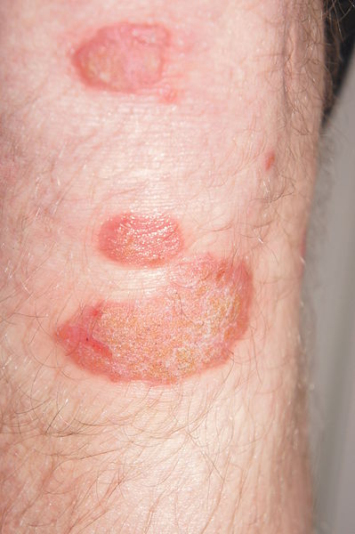
Image Source: Wikimedia Commons (2013). Psoriasis am Unterschenkel, Licensed under a creative commons licence. Available for free at: https://commons.wikimedia.org/wiki/File:Psoriasis_am_Unterschenkel.JPG (Accessed: 30 June 2021).
Patterns of presentation
The Moll and Wright diagnostic criterion is the simplest and most often used classification system to describe the 5 main patterns of presentations in psoriatic arthritis:
- Symmetric polyarthritis (30-40%, most common type)- symmetric polyarthritis most commonly occurs in women and can mimic the pattern seen in rheumatoid arthritis. Wrists, hands, feet, and ankles are usually affected. Factors which can help in differentiating psoriatic arthritis from rheumatoid arthritis are distal interphalangeal joint involvement rather than metacarpophalangeal joints, absence of skin nodules and absence of rheumatoid factor or anti-CCP in blood results.
- Asymmetric oligoarthritis (20-30%)- most commonly affected areas are hands and feet, initially with enthesitis causing dactylitis (“sausage fingers”). Usually, less than 5 joints are involved.
- Distal interphalangeal joint (DIP) arthritis (10%)- sole DIP joint involvement usually occurs in men. The nail and surrounding tissues can also be involved.
- Arthritis mutilans (5%)- arthritis mutilans is a rare type of severe and deformative DIP disease, which gives the appearance of “telescoping fingers” due to resorption of the terminal phalanx. The classic “pencil in cup” appearance on radiographs can be seen. This is the most severe and debilitating form of psoriatic arthritis.
- Psoriatic spondylitis with sacroiliac and spinal involvement- spondylitis most commonly occurs in men and presents with morning stiffness and limited range of back movement. Patients may be asymptomatic, and there may not be any obvious radiological findings. This can mimic ankylosing spondylitis; however, tends to affect vertebrae asymmetrically rather than symmetrically.

Image Source: Wikimedia Commons (2010). Psoriatic arthritis2010, Licensed under a creative commons licence. Available for free at: https://commons.wikimedia.org/wiki/File:Psoriatic_arthritis2010.JPG (Accessed: 30 June 2021).
Diagnosis
The diagnosis of psoriatic arthritis is clinical and is based largely on history and examination. Blood tests and imaging are performed either to establish severity of symptoms or exclude other diagnoses, particularly rheumatoid arthritis. Inflammatory joint pain and stiffness lasting more than 30 minutes, and joint or digit swelling are highly suggestive features of psoriatic arthritis in patients with pre-existing skin disease.
Investigations
Blood tests:
- ESR and CRP – can be normal or raised in polyarticular and progressive arthritis
- Rheumatoid factor – a negative result can be used to distinguish between rheumatoid arthritis and psoriatic arthritis
- Anti-CCP – useful for excluding rheumatoid arthritis (although note that anti-CCP is not always positive in RA)
- Lipid profile, fasting blood glucose and uric acid level – metabolic screen due to the increased risk of cardiovascular disease in patients with psoriatic arthritis
Imaging:
Patients who present with evidence of joint inflammation should receive plain x-rays of the hands and feet. Early disease may not produce characteristic radiological changes, and x-rays may, therefore, only reveal soft tissue swelling. The following radiological features can be demonstrated in patients with psoriatic arthritis:
- DIP joint erosions
- Joint subluxation
- Periarticular new-bone formation
- Periostitis
- Pencil-in-cup deformity: due to periarticular erosions and bone resorption in advanced disease
- Arthritis mutilans: osteolysis and articular collapse can cause “telescoping fingers” in advanced disease
MRI or CT scanning may be a more specific and sensitive method of investigating subtle psoriatic arthritis signs, especially in the hands and feet. MRI imaging of the sacroiliac joints can detect early pre-radiographic sacroiliitis, which is characteristically asymmetric and can be asymptomatic. Spinal changes tend to mostly affect the cervical spine, and subchondral bone oedema is a characteristic finding.
Other investigations:
Synovial fluid aspiration can be performed if there is a high suspicion of gout. The aspirate should not contain crystals; however, the white cell count is often high. Ultrasound of the joints can also be used to demonstrate involvement of entheses in psoriatic arthritis.
Differential diagnoses
It can often be difficult to differentiate between psoriatic arthritis and other types of inflammatory joint disease. However, distinguishing features can include pattern of joint involvement, dactylitis, enthesitis and the absence of rheumatoid factor or anti-CCP antibodies.
- Rheumatoid arthritis (RA) – RA is an important differential diagnosis. It tends to present as a symmetric polyarthritis affecting small joints of the hands and feet. This pattern of disease can be observed in patients with psoriatic arthritis; however, these patients often have distal interphalangeal joint involvement. Dactylitis and sacroiliitis are not seen in RA, and rheumatoid factor and anti-CCP are usually positive. Hand x-rays of patients with RA would reveal osteopenia, joint space narrowing, erosions, and subluxation.
- Gout – gout usually presents with acutely swollen, warm and tender joints, and most commonly has either a monoarticular or oligoarticular pattern; however, can also affect several joints in the hands and feet, especially in the elderly. Synovial fluid examination would reveal negatively birefringent, needle-shaped crystals.
- Erosive osteoarthritis (OA) – OA often occurs in middle-aged women and affects distal and proximal interphalangeal joints of the fingers. X-rays of the hand will show characteristic changes such as osteophytes and joint space narrowing.
- Ankylosing spondylitis (AS) – some patients with psoriasis may also present with inflammatory spinal disease, which can be difficult to distinguish from AS.
- Enteric arthritis – patients with enteric arthritis would have a co-existing diagnosis of inflammatory bowel disease.
- Septic arthritis – patients with septic arthritis usually present with an acutely swollen, warm, and tender joint with systemic features such as fever. Synovial fluid culture would reveal the presence of underlying organisms.
- Reactive arthritis – reactive arthritis often presents with oligoarthritis, usually in weight-bearing joints and can also include dactylitis and enthesitis. However, there may often be a history of preceding gastrointestinal or genitourinary infection, and features such as conjunctivitis and urethritis may also be present. X-rays may show soft tissue swelling.
- Mycobacterial tenosynovitis – patients with mycobacterium tuberculosis and atypical mycobacteria may also present with dactylitis; however, synovial biopsy and culture would reveal the underlying organisms in these cases
- Sarcoid dactylitis – chronic sarcoidosis can also present with dactylitis, but patients will often have other features suggestive of underlying sarcoid, e.g., lymphadenopathy, respiratory symptoms. Chest x-ray may reveal hilar lymphadenopathy, and serum ACE levels may be raised.
Management
Non-pharmacological management
All patients with suspected psoriatic arthritis should be referred to a rheumatologist for assessment and management. Non-pharmacological therapies include physiotherapy, exercise, and optimization of metabolic risk factors such as smoking cessation and weight loss.
Pharmacological management
In patients with a combination of psoriatic arthritis and psoriasis, monotherapy, which can adequately treat skin and joint disease, is preferred. The European League Against Rheumatism (EULAR) guidelines recommend progression of therapies to achieve adequate disease control and individualised treatment plans.
- Non-steroidal anti-inflammatories (NSAIDs): NSAIDs can be used for symptomatic relief and may even be sufficient for patients with limited disease (i.e., monoarthritis, oligoarthritis and absence of joint erosions). An adequate trial consists of 6-12 weeks of at least 2 NSAIDs. If this does not produce a sufficient improvement in symptoms, other therapies are considered.
- Corticosteroid injections: Intra-articular steroid injections can be used as adjunctive therapy if response to NSAIDs is inadequate. Systemic steroids should be avoided due to the risk of rebound worsening of skin disease once withdrawn.
- Conventional disease-modifying anti-rheumatic drugs (DMARDs): Conventional DMARDs such as Methotrexate, Sulfasalazine or Leflunomide should be considered at an early stage in patients with multiple affected joints, structural damage, raised inflammatory markers and extra-articular manifestations. Methotrexate is often given first line due to ease of administration and rapid onset of action. Hydroxychloroquine should be avoided due to risk of worsening psoriasis. Conventional DMARDs, however, are only used for peripheral arthritis, as they have not been shown to have any benefit in treating axial disease.
- Biological disease-modifying anti-rheumatic drugs (DMARDs): Anti-TNF drugs such as Etanercept, Infliximab, Adalimumab or Golimumab can be offered in patients presenting with arthritis and three or more tender or swollen joints, when at least two other DMARDs have not produced a sufficient response. They are also considered for patients who present with progressive axial disease despite receiving an adequate course of treatment with at least 2 NSAIDs. If there is no response after 12 weeks of receiving a biological agent, treatment should be stopped. Other drugs which can be used if anti-TNF medications are contraindicated include Ustekinumab (human monoclonal antibody which inhibits receptor binding of interleukin 12 and interleukin 23), Secukinumab (monoclonal antibody to interleukin 17) and Apremilast (oral phosphodiesterase-4 inhibitor).

Summary of the management of psoriatic arthritis, adapted from BMJ Best Practice: Psoriatic Arthritis [Internet] Bestpractice.BMJ.com [cited June 2022]. Available from: https://bestpractice.bmj.com/topics/en-gb/524/management-approach
References
- Ballinger A. Essentials of Kumar and Clark’s Clinical Medicine E-Book. Elsevier Health Sciences; 2011 Sep 29.
- BMJ Best Practice [Internet] Bestpractice.BMJ.com [cited April 2022]. Available from: https://bestpractice.bmj.com/topics/en-gb/524
- Gossec L, Baraliakos X, Kerschbaumer A, de Wit M, McInnes I, Dougados M, Primdahl J, McGonagle DG, Aletaha D, Balanescu A, Balint PV. EULAR recommendations for the management of psoriatic arthritis with pharmacological therapies: 2019 update. Annals of the rheumatic diseases. 2020 Jun 1;79(6):700-12.
- Helliwell PS, Taylor W. Classification and diagnostic criteria for psoriatic arthritis. Annals of the rheumatic diseases. 2005 Mar 1;64(suppl 2):ii3-8.
- Jacobson JA, Girish G, Jiang Y, Resnick D. Radiographic evaluation of arthritis: inflammatory conditions. Radiology. 2008 Aug;248(2):378-89.
- Liu JT, Yeh HM, Liu SY, Chen KT. Psoriatic arthritis: epidemiology, diagnosis, and treatment. World journal of orthopedics. 2014 Sep 18;5(4):537.
- Menter A. Psoriasis and psoriatic arthritis treatment. Am J Manag Care. 2016 Jun 1;22(8 Suppl):s225-37.
- Moll JM, Wright V. Psoriatic arthritis. InSeminars in arthritis and rheumatism 1973 Jan 1 (Vol. 3, No. 1, pp. 55-78). WB Saunders.
- National Institute for Health and Clinical Excellence. CG153. Psoriasis: the assessment and management of psoriasis. NICE. London. 2012
- Ory PA, Gladman DD, Mease PJ. Psoriatic arthritis and imaging. Annals of the Rheumatic Diseases. 2005 Mar 1;64(suppl 2):ii55-7.
- Samarasekera E, Sawyer L, Parnham J, Smith CH. Assessment and management of psoriasis: summary of NICE guidance. Bmj. 2012 Oct 24;345.
- Samarasekera E, Sawyer L, Parnham J, Smith CH. Assessment and management of psoriasis: summary of NICE guidance. Bmj. 2012 Oct 24;345.
Reactive arthritis
Background
Definition
Reactive arthritis is an inflammatory arthritis which occurs typically following a gastrointestinal or genitourinary infection. The infection may precede symptom onset by around 1 to 4 weeks. The classical triad of arthritis, urethritis and conjunctivitis, also known as Reiter’s syndrome, is frequently described but only occurs in a minority of patients.
Causes and pathophysiology
Reactive arthritis commonly affects young Caucasian adults. The syndrome is often subdivided into two groups depending on the underlying cause:
- Post-enteric – associated with gram-negative bacteria such as Campylobacter (most common), Salmonella, Shigella, and Yersinia.
- Post-venereal – associated with Chlamydia infection, Neisseria gonorrhoeae or human immunodeficiency virus (HIV).
Reactive arthritis associated with sexually transmitted infections is most commonly seen in men, whereas post-enteric arthritis has an equal sex incidence. The most frequent causative organism of reactive arthritis is Chlamydia trachomatis, with Chlamydia pneumoniae also being a known trigger. Bacterial DNA of causative organisms have been identified in the synovial tissue of patients presenting with reactive arthritis. A strong association with HLA B27 has been noted.
Presentation
Reactive arthritis generally develops around 2-4 weeks after an infection; however, in 10% of patients, no symptomatic infection is identified. The onset is usually acute, and systemic features such as fever, weight loss and fatigue may be present.
- Classic triad: urethritis, conjunctivitis, arthritis = Reiter’s syndrome.
- Peripheral arthritis:
- Usually asymmetrical
- Predominantly affects large joints in the lower limbs
- Usually oligoarticular – affects less than 6 joints
- Tends to present as tender, swollen, stiff joints, usually in the morning
- Axial arthritis:
- Sacroiliitis and inflammation in the lumbosacral spine occurs commonly
- May present as lower back pain and stiffness, particularly in the morning and after periods of inactivity – symptoms are generally alleviated with exercise
- Enthesitis
- Achilles tendonitis, which presents as heel pain and plantar fasciitis can occur
- Dactylitis
- Skin manifestations:
- Erythema nodosum (palpable, tender, erythematous subcutaneous nodules)
- Circinate balanitis (painless ulcers and plaque-like lesions on the shaft or glans of the penis)
- Keratoderma blennorrhagicum (vesicles which can develop into plaque-like or pustular lesions, most commonly on the palms and soles of the feet)
- Dystrophic nail changes
- Mucous membrane involvement:
- Mouth ulcers – usually superficial and painless
- Genital ulcers
- Cystitis
- Prostatitis
- Ocular manifestations:
- Conjunctivitis – can present with eye erythema, tearing and purulent discharge
- Anterior uveitis (iritis) – associated with HLA B27 positivity. Can present with pain, erythema and photophobia
- Episcleritis
- Keratitis
- Corneal ulcerations
- Gastrointestinal manifestations:
- Some patients may have intermittent symptoms of abdominal pain and diarrhoea
- Cardiovascular manifestations:
- Aortitis – can occur with or without aortic regurgitation and may precipitate heart failure
- Cardiac conduction defects leading to arrhythmias

Image Source: Wikimedia Commons (2022). Swollen eye with conjunctivitis, Licensed under a creative commons licence. Available for free at: https://commons.wikimedia.org/wiki/File:Swollen_eye_with_conjunctivitis.jpg (Accessed: 30 June 2021).

Image Source: Wikimedia Commons (2022). Keratoderma blennorrhagic, Licensed under a creative commons licence. Available for free at: https://commons.wikimedia.org/wiki/File:Keratoderma_blennorrhagica.jpg (Accessed: 30 June 2021).
Diagnosis
Investigations
There are no confirmatory tests for reactive arthritis, and diagnosis is based on clinical suspicion in a patient with inflammatory arthritis and a history of a preceding genitourinary or gastrointestinal infection. Investigations are often performed to rule out other diagnoses.
Blood tests:
- FBC: may reveal normocytic normochromic anaemia, mild leucocytosis, and thrombocytosis in the acute stage
- ESR and CRP are usually very high in the acute stage
- HLA B27 is positive in most affected patients; however, is not specific for reactive arthritis
- Rheumatoid factors and antinuclear antibodies are negative
Identifying causative organism:
- Urogenital and stool cultures: positive at the time of infection, but are usually negative post onset of arthritis
- Men: Urine nucleic acid amplification test (NAAT) for C. trachomatis and N. gonorrhoeae and urethral culture for N. gonorrhoeae
- Women: Vulvovaginal NAAT for C. trachomatis and N. gonorrhoeae and endocervical culture for N. gonorrhoeae
- Both sexes: HIV, syphilis, hepatitis B and hepatitis C screening depending on clinical history
- Serology for Yersinia, Campylobacter, Salmonella and Shigella if indicated
Joint aspiration
- Joint aspiration may be needed to rule out septic arthritis or crystal arthropathy.
- Synovial fluid in patients with reactive arthritis would demonstrate a high white cell count (2000-50,000 cells/mm3. ) with a neutrophil count above 50% and may reveal triggering organisms, e.g. gonorrhoea.
Imaging
- X-rays: may be normal in early disease or show non-specific changes such as soft tissue swelling. In advanced disease, joint space narrowing and erosions in the joints of the hands and feet may be seen. X-rays may also reveal bilateral but asymmetrical sacroiliitis or enthesitis as fluffy periosteal new bone formation. Non-marginal syndesmophytes are typically seen.
- MRI: may be more sensitive in detecting sacroiliitis than x-rays in patients with lower back pain
- Ultrasound: may detect synovial hypertrophy and increased vascularity using doppler imaging
Differential diagnoses
- Ankylosing spondylitis – can present similarly to reactive arthritis; however, spinal involvement tends to be symmetrical, and x-rays can reveal the presence of syndesmophytes. AS also tends to also affect peripheral joints less frequently than reactive arthritis.
- Psoriatic arthritis – tends to affect mostly distal interphalangeal joints, which can be identified on x-rays
- Rheumatoid arthritis – usually presents as a symmetrical polyarthritis and does not affect lumbar spine or sacral joints. Rheumatoid factors and anti-CCP are frequently positive.
- Rheumatic fever – associated with an upper respiratory tract infection. Rheumatic fever tends to present as a migratory arthritis in the upper and lower limbs and does not involve the spine. Antistreptolysin-O titre may be positive.
- Adult-onset Still’s disease – may also present with fever and joint pain. Joint pain tends to occur in two spikes throughout the day and is associated with a salmon-coloured rash. Serum LDH and ferritin may be elevated.
- Enteric arthritis – patients will have a history of inflammatory bowel disease and tend to present with bilateral and symmetrical sacroiliitis. X-rays reveal marginal syndesmophytes in contrast to reactive arthritis, where non-marginal syndesmophytes are usually seen.
- Gout – patients usually present with an acutely swollen and tender joint, and distal interphalangeal joint involvement is common. Gout can be differentiated from reactive arthritis by synovial fluid examination, which would reveal crystals in gout.
- Septic arthritis – usually affects a single joint, and presents as a swollen, erythematous and warm joint in the presence of a fever. Synovial fluid analysis and culture would reveal a very high white cell count (>50,000mm ³) and neutrophil count >75%, with identification of the underlying organism. Blood cultures may also be positive in septic arthritis.
- Post-viral arthritis – parvovirus infection can present with a flu-like illness followed by arthralgia. This tends to mostly demonstrate a symmetrical small joint polyarticular pattern, and patients may often have a maculopapular rash. Serology would reveal the presence of parvovirus B19
Management
The main aim of treatment is directed towards symptomatic relief and prevention or slowing down of progressive joint damage. In the acute phase, rest is recommended, but physiotherapy can be used in later stages for symptomatic management.
Pharmacological management
- NSAIDS: NSAIDs are often used first line to treat symptomatic axial and peripheral arthritis, and can improve functional status if used early in the disease process.
- Corticosteroids: In patients unresponsive to NSAIDs, corticosteroids can be trialled. Steroids have been shown to be beneficial in improving symptoms in peripheral joints, but their efficacy in the management of axial disease is limited. Patients with monoarticular or oligoarticular joint involvement may be offered intra-articular steroid injections in acute flares, but if multiple joints are affected or if there are cardiac or ocular manifestations, systemic corticosteroids may be required. Topical corticosteroids can be used to treat skin disease associated with reactive arthritis, i.e. circinate balanitis and keratoderma blennorrhagicum.
- Conventional DMARDs: DMARDs such as Sulfasalazine can be used in patients who do not respond sufficiently to NSAIDs and demonstrate progressive joint damage and functional impairment
- Antibiotics: NICE guidelines recommend treating the initial infection if a causative organism is found, but to not offer long-term treatment to manage the arthritic symptoms following the infection.
References
- Barth WF, Segal K. Reactive arthritis (Reiter’s syndrome). American family physician. 1999 Aug 1;60(2):499.
- BMJ Best Practice [Internet] Bestpractice.BMJ.com [cited April 2022]. Available from: https://bestpractice.bmj.com/topics/en-gb/597/aetiology
- Carlin E, Marzo-Ortega H, Flew S. British Association of Sexual Health and HIV national guideline on the management of sexually acquired reactive arthritis 2021. International journal of STD & AIDS. 2021 Oct;32(11):986-97.
- Carlin E, Marzo-Ortega H, Flew S. British Association of Sexual Health and HIV national guideline on the management of sexually acquired reactive arthritis 2021. International journal of STD & AIDS. 2021 Oct;32(11):986-97.
- Cheeti A, Chakraborty RK, Ramphul K. Reactive Arthritis. 2018
- Martel W, Braunstein EM, Borlaza G, Good AE, Griffin Jr PE. Radiologic features of Reiter disease. Radiology. 1979 Jul;132(1):1-0.BMJ Best Practice [Internet] Bestpractice.BMJ.com [cited April 2022]. Available from: https://bestpractice.bmj.com/topics/en-gb/597/aetiology
- McAllister K, Goodson N, Warburton L, Rogers G. Spondyloarthritis: diagnosis and management: summary of NICE guidance. Bmj. 2017 Mar 1;356.
Enteric arthritis
Background
Definition
Enteric or enteropathic arthritis is a term used to describe patterns of inflammatory arthritis associated with gastrointestinal diseases such as inflammatory bowel disease (IBD – Crohn’s disease and ulcerative colitis), coeliac disease, Whipple’s disease and post-intestinal bypass surgery. It is a part of the spondyloarthritides, therefore, primarily affects entheses, small and large joints and the axial skeleton.
Epidemiology
IBD-associated joint disease can either affect the axial skeleton, which is more common in men, or peripheral joints primarily in the lower limbs, which is more common in women. Peripheral arthritis is the most frequent extra-intestinal manifestation of IBD, with a prevalence of between 17% and 20%, and occurs more commonly in Crohn’s disease than in ulcerative colitis.
Causes and risk factors
HLA B27 positivity is a known association of enteric arthritis affecting the axial skeleton, but not the peripheral joints. Risk factors for the development of arthritis in patients with IBD include smoking, active bowel disease, family history, and the presence of other extra-intestinal manifestations, particularly skin disease.
Presentation
Joint symptoms
Peripheral arthritis can occur before, simultaneously, or after the diagnosis of IBD is established. The most common pattern of peripheral disease is that of an asymmetric mono- or oligoarthritis, affecting mostly the large joints of the lower limbs. Patients present with typical inflammatory joint pain, prolonged morning stiffness and improvement of symptoms with activity. It tends to run an episodic and recurrent course, with occasional spontaneous improvement in symptoms within 6 months; however, a minority of patients can progress to chronic disease. Exacerbations of arthritic symptoms tend to correlate closely with extent and severity of intestinal involvement in IBD, and to the presence of extra-intestinal manifestations. Enthesitis can occur, particularly in the Achilles tendon or plantar fascia.
Extra-articular manifestations of IBD
- Intestinal: abdominal pain, diarrhoea, bloody stools
- Dermatological: erythema nodosum and pyoderma gangrenosum
- Mucous membranes: painful recurrent aphthous ulcers
- Ocular: anterior uveitis
- Systemic features: fever, malaise, fatigue, weight loss
Arthritis in other gastrointestinal disorders
- Intestinal bypass surgery tends to present as polyarthritis with an associated dermatitis due to bacterial overgrowth and pathological immune complex deposition in the bypassed section of bowel.
- Whipple’s disease: mostly occurs in middle-aged men and presents as a migratory polyarthritis
- Coeliac disease: arthritis is a rare manifestation of coeliac disease but can present as a symmetrical arthritis of the lumbar spine, hips, knees, and shoulders. There is a resolution in symptoms following a gluten-free diet.
Diagnosis
There is no gold standard criterion for the diagnosis of enteric arthritis; however, patients with a history of IBD presenting with inflammatory joint pain or synovitis are considered to have spondyloarthropathy unless proven otherwise. Investigations may be carried out to rule out other similarly presenting diagnoses.
Investigations
Blood tests
- FBC – to detect anaemia due to chronic disease or deficiency of iron, B12 or folate, or leucocytosis seen in patients with IBD
- ESR and CRP – usually elevated
- Rheumatoid factor and anti-CCP – to rule out rheumatoid arthritis
Imaging
- X-ray of affected joints – similar radiological findings to other spondyloarthropathies. Sacroiliitis tends to be asymmetrical.
Other
- Stool microscopy and culture – to investigate aetiology of undetermined bowel disease
- Sigmoidoscopy/colonoscopy/upper GI endoscopy/small bowel imaging – dependent on suspected underlying gastrointestinal disorder
- Synovial fluid aspiration – to rule out septic joint or crystal arthritis
Differential diagnoses
- Any cause of peripheral polyarthritis, e.g., rheumatoid arthritis, gout, sarcoidosis, Bechet’s disease
- Any cause of acutely swollen joint, e.g., septic arthritis, gout
- Acute cause of spondyloarthritis, e.g., ankylosing spondylitis, reactive arthritis, psoriatic arthritis
Management
Any patient with a known history of inflammatory bowel disease who develops new-onset inflammatory arthritis should be referred to a rheumatologist for further assessment and management. The treatment for enteric arthritis is similar to that for other spondyloarthropathies, i.e., physiotherapy, symptomatic control, and use of DMARDs in resistant cases. Peripheral arthritis generally improves with treatment of the bowel disease, but this is usually not seen in axial disease. NSAIDs can be used for symptomatic management; however, should be used cautiously due to the risk of worsening gastrointestinal symptoms.
References
- Ballinger A. Essentials of Kumar and Clark’s Clinical Medicine E-Book. Elsevier Health Sciences; 2011 Sep 29.
- McAllister K, Goodson N, Warburton L, Rogers G. Spondyloarthritis: diagnosis and management: summary of NICE guidance. Bmj. 2017 Mar 1;356.
- Peluso, R., Di Minno, M.N.D., Iervolino, S., Manguso, F., Tramontano, G., Ambrosino, P., Esposito, C., Scalera, A., Castiglione, F. and Scarpa, R., 2013. Enteropathic spondyloarthritis: from diagnosis to treatment. Clinical and Developmental Immunology, 2013.
- Weerakkody, Y., Feger, J. Enteropathic arthritis. Reference article, Radiopaedia.org. (accessed on 25 Jun 2022) https://doi.org/10.53347/rID-34060
Complete Guide to Clinical Genetics ST3 Application and Recruitment (2027)
Written by Medibuddy. Last updated: October 2026. Clinical Genetics is

Complete Guide to Clinical Radiology ST3 Application and Recruitment (2027)
Recruitment paused: NHS England is not running Clinical Radiology ST3+ recru

Complete Guide to Paediatric Surgery ST3 Application and Recruitment in United Kingdom (2026)
Written by Medibuddy. Last updated: October 2026. If you’re planning


Take your subscriptions with you
Our mobile app allows you to access your interview and exam question banks wherever you are.

