


 Close
Close
 8th December 2023
Infectious Red Eye
Infectious Conjunctivitis
Conjunctivitis is inflammation of the conjunctiva as a response to either exogenous or endogenous processes. In infectious conjunctivitis, there is an inflamed conjunctiva in response to infection by a pathogen. It is a common cause of red eye in primary and secondary care. It is usually benign and self-limiting. It can be classified as acute or chronic.
Viral conjunctivitis
This is the commonest cause of infectious conjunctivitis, and adenoviruses account for up to 90% of cases. Viral conjunctivitis may also be caused by viruses of the herpes family or enteroviruses. It is transmitted by direct contact with ocular or respiratory secretions, or via fomites such as towels. Common symptoms include redness, burning sensation, foreign body sensation, mild photophobia, watery discharge and transient blurred vision. It may be bilateral or unilateral. There may be prodromal symptoms such as malaise or recent history of a viral upper respiratory tract infection. This is especially true in children, where pharyngoconjunctival fever presents fever, pharyngitis, periauricular lymphadenopathy, and acute follicular conjunctivitis. This condition is self-limiting and resolves within weeks. The conjunctiva is usually diffusely red, and there may be follicles present. There may be haemorrhage, especially in severe infection and pseudo-membranes might be present. Pre-auricular lymphadenopathy is common in viral conjunctivitis.
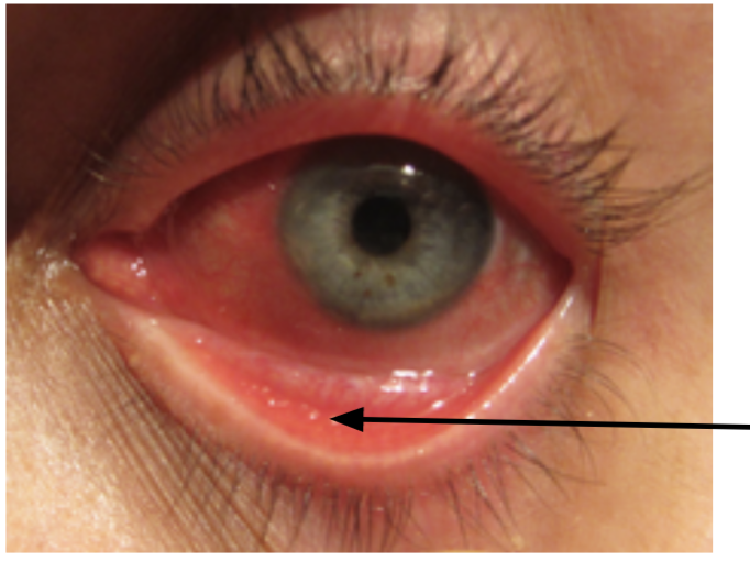
Image Source: Wikimedia Commons (2015). Conjuntivitis (RPS 03-06-2015), Licensed under a creative commons licence. Available for free at: https://commons.wikimedia.org/wiki/File:Conjuntivitis_(RPS_03-06-2015).png (Accessed on: 5 May 2022).
Diagnosis is clinical, but investigations may be useful in specific cases where there is uncertainty and viruses such as the herpes family are suspected. As it is a self-limiting condition and viral, topical antibiotics are not usually required. Cold compresses and artificial tears will provide symptomatic relief. Adenovirus conjunctivitis is very contagious, and patients should be advised on infection control procedures, such as not sharing towels at home. Herpes simplex keratitis should be treated with antivirals.
Bacterial conjunctivitis
Bacterial conjunctivitis is typically a self-limiting condition that occurs on direct inoculation with bacteria. Common pathogens include Staphyloccoccus Aureus, Streptococcus Pneumoniae, Haemophilus influenza and Moraxella Catarrhis. Other less common but more serious causes include Chlamydia and Gonorrhoea. It is usually more common in children.
Symptoms include mucopurulent discharge, particularly in the morning, redness, burning sensation, foreign body sensation, transient blurred vision and mild photophobia. Eyelid oedema may be present in severe cases. Gonococcal infection presents hyperacutely with copious mucopurulent discharge and rapid progression of symptoms. It is important to take a sexual history in these patients, particularly those with persistent bacterial conjunctivitis not responding to topical treatment such as chloramphenicol.
Examination shows conjunctival hyperaemia, mucopurulent discharge, which may be present only in fornices if the eye has been recently cleaned; papillae and follicles may be present in chlamydia conjunctivitis. Pre-auricular lymphadenopathy can occur in gonococcal conjunctivitis. Less commonly, there may be formation of pseudo-membranes or membranes.
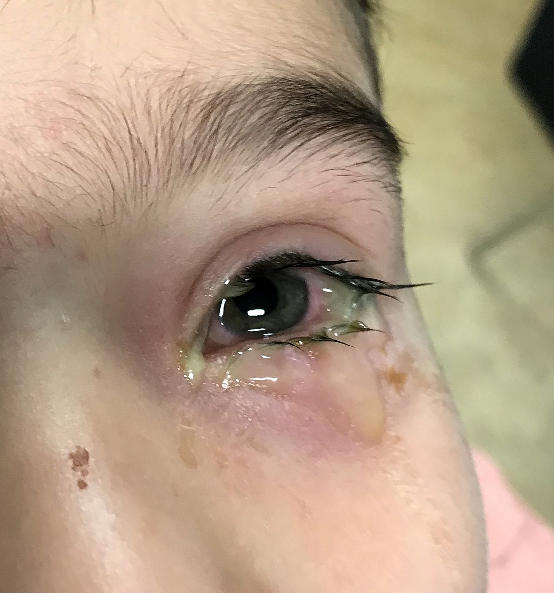
Image Source: Wikimedia Commons (2019). Bacterial conjunctivitis, Licensed under a creative commons licence. Available for free at: https://commons.wikimedia.org/wiki/File:Bacterial_conjunctivitis.jpg (Accessed on: 5 May 2022).

Image Source: Wikimedia Commons (2019). Swollen eye with conjunctivitis, Licensed under a creative commons licence. Available for free at: https://commons.wikimedia.org/wiki/File:Swollen_eye_with_conjunctivitis.jpg (Accessed on: 5 May 2022).
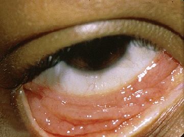
Image Source: Wikimedia Commons (2011). Chlamydial-conjunctivitis, Licensed under a creative commons licence. Available for free at: https://commons.wikimedia.org/wiki/File:Chlamydial-conjunctivitis.jpg (Accessed on: 5 May 2022).

Image Source: Wikimedia Commons (2016). Membranous conjunctivitis, Licensed under a creative commons licence. Available for free at: https://commons.wikimedia.org/wiki/File:Membranous_conjunctivitis.jpg (Accessed on: 5 May 2022).
Bacterial conjunctivitis is a clinical diagnosis, but investigations may be appropriate if gonococcal, chlamydial or meningococcal infection is suspected. Investigations are also useful in cases where there is failure of treatment. Although bacterial conjunctivitis is self-limiting, topical treatment can be given to hasten resolution. This includes anti-bacterials such as chloramphenicol and fluoroquinolones. Gonococcal conjunctivitis is treated with one dose of intramuscular Ceftriaxone, and systemic Azithromycin is given for Chlamydial infection. Advice should be given regarding handwashing and not sharing towels.
Neonatal conjunctivitis
Inflammation of the conjunctiva within the first 28 days of life is termed neonatal conjunctivitis. It can be chemical or infectious in nature. It is the most common infection in neonates. Chlamydia is a leading cause9. Other organisms include staphylococci, streptococci, and chlamydia. Route is typically via vertical transmission during delivery or from caregivers in the post-natal period. History taking should include parental symptoms of sexually transmitted infection, prophylactic eyedrops instilled at birth and systemic illness in the neonate. Symptoms include sticky eyes, mucopurulent or watery discharge, red eyes and eyelid oedema. Examination should include corneal examination to look for ulceration and the tarsal and bulbar conjunctiva.

Image Source: Wikimedia Commons (2016). OSC Microbio 21 02 neonatorum, Licensed under a creative commons licence. Available for free at: https://commons.wikimedia.org/wiki/File:OSC_Microbio_21_02_neonatorum.jpg (Accessed on: 5 May 2022).
Investigation is unnecessary in mild cases, but conjunctival swabs and/or scrapes can be taken and sent for analysis in moderate to severe cases. Untreated, there is a risk of blindness, disability, and significant morbidity. Neonates with suspected infective conjunctivitis should be referred urgently for same-day Ophthalmology assessment9. Mild conjunctivitis can be treated with broad-spectrum topical antibiotics such as Chloramphenicol. Moderate to severe cases Gonococcal conjunctivitis is treated with Ceftriaxone according to weight and chlamydial infections with Azithromycin according to weight.
Keratitis
Inflammation of the cornea can result from either exogenous insults or endogenous processes. For the purposes of this, keratitis will be discussed in terms of infective processes. Various pathogens such as bacterial, viral, fungi or protozoa can cause corneal infections.
Bacterial keratitis
Bacterial keratitis (bacterial infection of the cornea) is synonymously referred to as corneal ulcer and is the most common infectious keratitis. Although commonly presenting with an epithelial defect (ulcer), there might be a focus of infection (infiltrate) without an ulcer. A healthy cornea with an intact epithelium is a good defensive barrier against pathogens. Predisposing factors such as: contact lens wear (commonest), ocular surface disease, trauma, previous ocular surgery, immunosuppression, and systemic diseases are associated with an increased risk of corneal ulcers. Gram-positive bacteria such as staphylococci and gram-negative species such as pseudomonas are pathogens common in isolates. There are four pathophysiological states: infiltration of the cornea by the pathogen, active ulceration, regression and healing. Disease severity depends on the pathogenicity of the invading organism.
Common presenting complaints include redness, watering, photophobia, foreign body sensation and blurred vision. Patients should be asked about contact lens use/misuse (sleeping, swimming, sleeping, showering, or washing lenses in tap water).
Examination of the cornea should include, if possible, the use of fluorescein. Signs include staining epithelial defect (usually circular), whitish infiltrate or focal haziness of the cornea, mucopurulent discharge, conjunctival hyperaemia, anterior chamber reaction such as cells or hypopyon and eyelid oedema.

Image Source: Wikimedia Commons (2016). Contact Lens Related Small Corneal Ulcer with Sodium Fluorescein Staining 7 copy, Licensed under a creative commons licence. Available for free at: https://commons.wikimedia.org/wiki/File:Contact_Lens_Related_Small_Corneal_Ulcer_with_Sodium_Fluorescein_Staining_7_copy.png (Accessed on: 5 May 2022).

Image Source: Wikimedia Commons (2019). Corneal Ulcer, Licensed under a creative commons licence. Available for free at: https://commons.wikimedia.org/wiki/File:Corneal_Ulcer.png (Accessed on: 5 May 2022).

Diagnosis is primarily clinical. Corneal scrapes for microscopy, culture and sensitivities are usually sent for ulcer >2mm, deeper or not responding to treatment. Contact lenses should be discontinued. Treatment typically involves topical broad-spectrum antibiotics, which can be changed according to sensitivities. In patient with anterior chamber reaction will benefit from cycloplegia and/or initiation of steroids once antibiotic therapy has been established. Patients with suspected corneal ulcers need urgent ophthalmology assessment. Sequelae of bacteria keratitis include corneal abscess, perforation and corneal scarring.
Herpes Simplex Keratitis (HSK)
Most people would have been exposed to herpes simplex, and the prevalence of herpes simplex is high in developing countries. Herpes simplex can cause a primary or secondary infection. Primary infection occurs on direct contact of the eye with the virus and is usually subclinical. The virus then stays dormant in the trigeminal nerve with reactivation, causing a secondary infection. Risk factors for reactivation are shown below. Recurrent activation has sight-threatening complications. Herpes can cause inflammation in every ocular tissue, but for the purpose of this, we shall only discuss herpes simplex keratitis. HSK is mainly caused by HSV-1.
Based on the Herpetic Eye Disease Study Group’s ‘Predictors of Recurrent Herpes Simplex Virus Keratitis’, reactivation can be triggered by a variety of stimuli:
- Psychological stress
- UV light
- Fever
- Immunosuppression
- Trauma such as injury or surgery
- Intercurrent illness
- Previous episodes of disease recurrence
Herpes Simplex Keratitis can present clinically as two entities: Herpes Simplex Epithelitis and stromal keratitis.
Herpes Simplex Epithelitis occurs due to reactivation and anterograde migration of the virus through the trigeminal nerves to the corneal nerves. Active replication of the virus leads to loss of epithelial cells in a geographic, dendritic/branching pattern. Risk factors for activation include sunlight, stress, trauma and immunosuppression. Patient presents with unilateral (rarely bilateral) redness, pain, photophobia, eye-watering and blurry vision. Examination of the Fluorescein- stained cornea will reveal a characteristic branching defect with terminal bulbs. Diagnosis is usually clinical. In cases of diagnostic uncertainty, a corneal swab or scrap can be sent for PCR; PCR is highly sensitive in epithelial disease. Treatment consists of topical antiviral agents such as Ganciclovir. Antiviral agents are only beneficial in cases of active viral replication such as this.
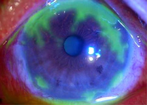
Image Source: Wikimedia Commons (2012). Herpes simplex geographic corneal ulcer, Licensed under a creative commons licence. Available for free at: https://commons.wikimedia.org/wiki/File:Herpes_simplex_geographic_corneal_ulcer.jpeg (Accessed on: 5 May 2022).

Image Source: Wikimedia Commons (2017). Dendritic corneal ulcer, Licensed under a creative commons licence. Available for free at: https://commons.wikimedia.org/wiki/File:Dendritic_corneal_ulcer.jpg (Accessed on: 5 May 2022).
Stromal keratitis occurs as a result of both the viral-mediated destruction and the host’s immunological response to the virus. Inflammation in the stroma leads to a stromal opacity, which is often accompanied by neovascularisation. Stromal keratitis can be further classified based on the integrity of the epithelium. In the necrotising form, there is an overlying epithelial defect and a high risk of cornea perforation. In the non-necrotising form, the epithelium is intact. Presenting symptoms can include redness, pain, photophobia, decrease in visual acuity and noticeable opacification of the cornea. Clinical signs include evidence of active inflammation such as redness, vascularisation, evidence of anterior chamber activity and raised intraocular pressure. There is often corneal stromal opacity, which can present in a classic disciform shape. Diagnosis is clinical. PCR and viral culture may have a positive yield, but this is greatly reduced in the absence of epithelial defects. The principles of treatment are to inhibit viral replication and reduce damage caused by inflammation. First line treatment often includes topical steroids and antivirals. In cases of epithelial defects and raised intraocular pressure, antibacterial and intraocular pressure-lowering drugs are indicated respectively. The Herpetic Eye Disease study showed that in patients with recurrent disease, systemic antiviral can reduce recurrence by up to 50%. Complications from recurrent or severe infections include corneal scarring, corneal vascularisation, lipid keratopathy and increased risk of transplant rejection. There are also associated complications from prolonged steroid therapy.
Herpes Zoster Ophthalmicus (Shingles)
Primary infection with Varizella Zoster typically occurs in childhood, with the virus then lying dormant with a sensory nerve ganglion. 10-20% of Herpes Zoster infections present with Herpes Zoster Ophthalmicus. Herpes Zoster Ophthalmicus (HZO)occurs when a reactivation of latent Varicella Zoster Virus (VZV) hidden in the sensory ganglia affects the ophthalmic division (V1) of the trigeminal nerve. V1 is a sensory nerve supplying:
- Related areas of the meninges and tentorium cerebelli
- The forehead and scalp
- Sinuses – Frontal, ethmoidal and sphenoidal
- Lacrimal gland
- Upper eyelid and conjunctiva
- Cornea
- Dorsum of the nose
Herpes Zoster Ophthalmicus does not in itself imply ocular involvement, but there is ocular infection in about 50% of cases. The reactivated virus travels to the sensory axons in the skin, forming a vesicular rash which blisters. The local immune response results in blisters and perinueritis, leading to significant neuropathic pain in the nerve distribution. Ocular involvement results in inflammatory conditions such as conjunctivitis, keratitis and uveitis.
Risk factors for reactivation are:
- Increasing Age
- Immunosuppression
- Stress
- Chronic illness such as diabetes
- Organ or bone marrow transplantation
- Malignancy
- Chemotherapy
An episode presents as a blistering rash in V1 (usually unilateral) associated with subcutaneous oedema. The rash starts off as a maculopapular rash, which then becomes vesicular and pustular before blistering and scabbing over. There is usually a prodromal phase of malaise, fever, paraesthesia or neuropathic pain (typically burning or shooting) in the affected dermatome and headache. In cases of ocular involvement, patients can present with redness, watering, pain, photophobia and blurry vision. General examination should assess for the presence of Hutchinson’s sign. A positive Hutchinson’s sign is when the rash involves the nasociliary branch of the trigeminal nerve (any area from the root to the tip of the nose) and is a strong predictor of ocular involvement. The eye should be examined for conjunctival redness and cornea stained with fluorescein to look for punctate keratitis or pseudo-dendrites. Decrease in corneal sensation may be present. Eyelid swelling and ptosis might occur due to gravitational oedema from the forehead.
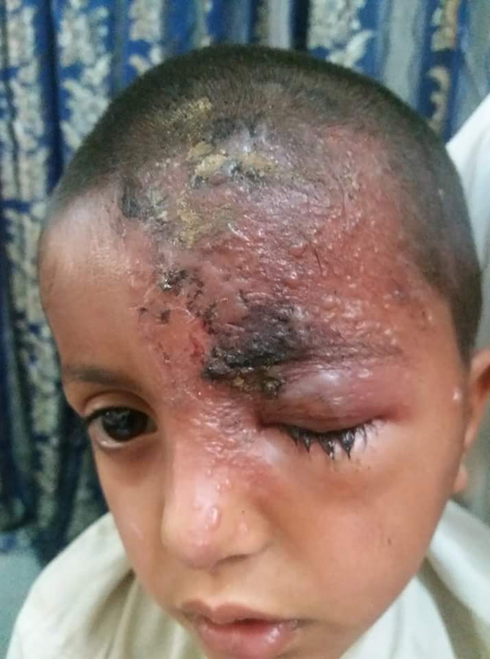
Image Source: Wikimedia Commons (2018). Herps zoster ophthalmicus, Licensed under a creative commons licence. Available for free at: https://commons.wikimedia.org/wiki/File:Herps_zoster_ophthalmicus.jpg (Accessed on: 5 May 2022).

Image Source: Wikimedia Commons (2015). Day07 shingles or Herpes Zoster Virus attacking forehead and eye, Licensed under a creative commons licence. Available for free at: https://commons.wikimedia.org/wiki/File:Day07_shingles_or_Herpes_Zoster_Virus_attacking_forehead_and_eye.jpg (Accessed on: 5 May 2022).
Diagnosis is clinical, but on occasion of diagnostic uncertainty, a viral swab can be sent for PCR. Antiviral treatment should be started – ideally within 72 hours of onset. PO Aciclovir 800mg 5x/day for 7-10 days is the mainstay of oral treatment. Immunocompromised patients might require intravenous antiviral agents. In patients with suspected ocular involvement, ophthalmology input should be sought within 24 hours. Surface ocular disease can be treated with topical Ganciclovir, but substantial inflammation will require steroids. Bacterial superinfection should also be treated. Vesicles blister and typically heal by 6 weeks. Patients commonly develop post-herpetic neuralgia, which can be debilitating and will require treatment with neuropathic pain medication. Inflammation around the eyelids can cause scarring, which expose the cornea, leading to exposure keratopathy and its sequelae – this is a sight-threatening condition. Direct corneal inflammation, if prolonged, also risks sight loss. Shingles vaccination has been demonstrated to decrease disease recurrence and is currently available on the NHS to people in their 70s.
Acanthamoeba keratitis
Acanthamoeba is a free-living protozoan which can rarely cause a severe opportunistic corneal infection. It exists in two forms: trophozoites (active organism) and cysts (encased and resistant to a variable of external factors). Risk factors include contact lens wear inoculation through contaminated water or via trauma. Patients typically present with pain disproportionate to clinical findings. They also complain of non-specific symptoms of keratitis. Early clinical findings include an irregular epithelium, infiltrates and pseudodendrites. Late signs can include characteristic ring-shaped or disciform stromal infiltrates, as shown in the image below, corneal perforation, anterior uveitis and scleritis. Diagnosis is often through corneal scrapes grown on specific agar or confocal microscopy.
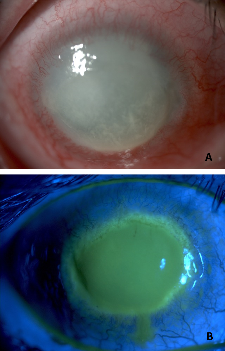
Image Source: Wikimedia Commons (2015). Parasite140120-fig1 Acanthamoeba keratitis, Licensed under a creative commons licence. Available for free at: https://commons.wikimedia.org/wiki/File:Parasite140120-fig1_Acanthamoeba_keratitis.png (Accessed on: 5 May 2022).
Urgent ophthalmology assessment is needed with even a marginal suspicion of acanthamoeba keratitis. Current treatment options include combinations of antibiotics, antivirals, antifungals and anti-parasitic agents, some of which are toxic and require lengthy treatment.
Orbital cellulitis
Orbital cellulitis is an ophthalmic emergency. It is a sight-threatening condition which occurs when there is a bacterial infection of soft tissues posterior to the orbital septum. This differs from pre-septal cellulitis, where there is infection of the soft tissue anterior to the orbital septum without involvement of the globe or orbit. The infection frequently originates from the paranasal sinuses, pre-septal cellulitis, dental infection, dacryocystitis and trauma. Causative organisms include S.Pneumonaie and S.Aureus. It is more common in children and in immunocompromised states. Patients present with a painful swollen eye, change in vision, fever, malaise and less common features of intracranial spread such as meningism or neurological impairment. Typically, there is a history regarding the source of infection, such as recent dental work or upper respiratory tract infection. Examination shows reduced vision and/or signs of optic nerve dysfunction, eyelid swelling and erythema, conjunctival oedema and injection, proptosis, decreased eye movements associated with pain, and the patient is usually febrile. Patients should also be examined for any signs of intracranial spread, such as meningism or focal neurological signs, and if available, fundoscopy should be carried out.

Image Source: Wikimedia Commons (2011). Orbital cellulitis, Licensed under a creative commons licence. Available for free at: https://commons.wikimedia.org/wiki/File:Orbital_cellulitis.jpg (Accessed on: 5 May 2022).

Image Source: Wikimedia Commons (2011). RtmaxobitinfectteethCT, Licensed under a creative commons licence. Available for free at: https://commons.wikimedia.org/wiki/File:RtmaxobitinfectteethCT.png (Accessed on: 5 May 2022).
Investigations include blood tests to assess inflammatory markers, blood cultures if febrile and/or culture of the nasopharynx and conjunctiva -although the yield is low, and imaging (contrast enhanced CT scan of orbit, sinuses and brain). Patients presenting with pre-septal cellulitis will need thorough examination to rule out orbital cellulitis. The table below shows differentiating features between orbital cellulitis and pre-septal cellulitis.
| Pre-septal cellulitis | Orbital cellulitis |
|---|---|
| White-eye | Conjuctival injection and oedema |
| Full painless extraocular movements | Painful and/or restricted extraocular movements |
| No proptosis | Proptosis |
| Normal vision | Impaired vision and/or signs of optic nerve dysfunction |
| Systemically well | Fever and/or malaise |
Adapted from Kim HJ, Kersten R. Peri-orbital and orbital cellulitis – Symptoms, diagnosis and treatment | BMJ Best Practice, https://bestpractice.bmj.com/topics/en-gb/734 (accessed 31 May 2022).
Patients suspected of having orbital cellulitis need urgent hospital admission and initiation of empirical intravenous antibiotics, which can be tailored to any positive cultures. Antibiotics should cover for anaerobic infections and also have good penetrance of the CSF to cover for intracranial spread. Prompt imaging should be arranged to assess for any evidence of sinusitis, confirm diagnosis and also assess for intracranial spread or presence of abscesses. Children will be managed under paediatrics, and all patients will require ophthalmology input. If necessary, dental, max-fax or ENT input may also be sought. Complications can be localised to the orbit (optic neuropathy, endophthalmitis, central retinal artery occlusion, subperiosteal abscess, ocular empyema) or intracranial (meningitis, abscess and thrombosis).
References
- Ryder EC, Benson S. Conjunctivitis. StatPearls, https://www.ncbi.nlm.nih.gov/books/NBK541034/ (2022, accessed 15 June 2022).
- Azari AA, Arabi A. Conjunctivitis: A Systematic Review. J Ophthalmic Vis Res 2020; 15: 372.
- Manasseh GSL, Amarakoon S, Photiou V, et al. Approach to conjunctivitis in newborns. BMJ; 376. Epub ahead of print 23 March 2022. DOI: 10.1136/BMJ-2021-068023.
- Bourcier T, Thomas F, Borderie V, et al. Bacterial keratitis: Predisposing factors, clinical and microbiological review of 300 cases. Br J Ophthalmol 2003; 87: 834–838.
- Lin A, Rhee MK, Akpek EK, et al. Bacterial Keratitis Preferred Practice Pattern®. Ophthalmology 2019; 126: P1–P55.
- Sibley D, Larkin DFP. Update on Herpes simplex keratitis management. Eye 2020 3412 2020; 34: 2219–2226.
- Herpetic Eye Disease Study Group. Predictors of Recurrent Herpes Simplex Virus Keratitis. Cornea: March 2001 – Volume 20 – Issue 2 – p 123-128
- Ting DSJ, Ghosh N, Ghosh S. Herpes zoster ophthalmicus. BMJ; 364. Epub ahead of print 2019. DOI: 10.1136/BMJ.K5234.
- Meduri A, Grenga PL, Kaufman SC. Herpes Zoster Ophthalmicus. Expert Rev Ophthalmol 2021; 4: 537–545.
- Lorenzo-Morales J, Khan NA, Walochnik J. An update on Acanthamoeba keratitis: diagnosis, pathogenesis and treatment. Parasite; 22. Epub ahead of print 2015. DOI: 10.1051/PARASITE/2015010.
- Adamson J, Waterfield T. Fifteen-minute consultation: Preseptal and orbital cellulitis. Arch Dis Child – Educ Pract 2019; 104: 79–83.
- Kim HJ, Kersten R. Peri-orbital and orbital cellulitis – Symptoms, diagnosis and treatment | BMJ Best Practice, https://bestpractice.bmj.com/topics/en-gb/734 (accessed 31 May 2022).
Complete Guide to Clinical Genetics ST3 Application and Recruitment (2027)
Written by Medibuddy. Last updated: October 2026. Clinical Genetics is

Complete Guide to Clinical Radiology ST3 Application and Recruitment (2027)
Recruitment paused: NHS England is not running Clinical Radiology ST3+ recru

Complete Guide to Paediatric Surgery ST3 Application and Recruitment in United Kingdom (2026)
Written by Medibuddy. Last updated: October 2026. If you’re planning


Take your subscriptions with you
Our mobile app allows you to access your interview and exam question banks wherever you are.

