


 Close
Close
 8th December 2023
Allergic eye disease
Allergic eye disease covers a spectrum of disease. This section covers the forms of allergic conjunctivitis. Allergic conjunctivitis is an umbrella term for a spectrum of disorders where conjunctivitis results typically from Type I hypersensitivity reaction to an allergen. It is estimated to affect up to 40% of the population and is usually seasonal. Allergic conjunctivitis can be classified as shown in the image below. Allergic conjunctivitis is particularly common during the spring season, but only a proportion of individuals will experience symptoms severe enough to seek medical treatment.

Image source: Adapted from La Rosa M, Lionetti E, Reibaldi M, et al. Allergic conjunctivitis: a comprehensive review of the literature. Ital J Pediatr 2013; 39: 18.
Allergic conjunctivitis without corneal involvement occurs on secondary contact with an allergen. It is due to a type I hypersensitivity reaction with subsequent mast cell degranulation. Allergic conjunctivitis with corneal involvement is a lesser-understood process with both type I and type IV hypersensitivity reaction and changes in T cell signalling. The accompanying changes in T cell behaviour confer chronicity.
Allergic conjunctivitis without corneal involvement (simple allergic conjunctivitis) tends to occur in response to allergens present on the ocular surface. In this form, only the conjunctiva is involved. Incidence decreases with increasing age.
Seasonal
This occurs in response to allergens which are more abundant in certain seasons, such as pollen or grass. Sufferers tend to have a history of hay fever.
Perennial
This is a form of simple allergic conjunctivitis which occurs all year round and is triggered by environmental allergens such as dust mites or animal dander.
Acute
A term loosely used to describe an acute onset of simple allergic conjunctivitis as opposed to an episode persisting throughout the spring and summer or all year round.
Simple allergic conjunctivitis typically presents with bilateral watering eyes, itching, conjunctival swelling, and eyelid swelling. Pain is rarely a feature.

Image Source: Wikimedia Commons (2013). Allergicconjunctivitis, Licensed under a creative commons licence. Available for free at: https://commons.wikimedia.org/wiki/File:Allergicconjunctivitis.jpg (Accessed on: 5 May 2022).

Image Source: Wikimedia Commons (2017). Pterygium (conjunctiva), Licensed under a creative commons licence. Available for free at: https://commons.wikimedia.org/wiki/File:Pterygium_(conjunctiva).jpg (Accessed on: 5 May 2022).
Atopic Keratoconjunctivitis (AKC)
This tends to occur in patients with a history of atopy, and incidence decreases with age. It arises from allergen exposure in an individual with a genetic predisposition to atopy. Patients typically have a history of dermatitis, usually around the eyelids. Symptoms are usually present all year around and will include corneal symptoms. It is a chronic disease and persists for several years. Chronic unchecked inflammation can lead to corneal scarring and vascularisation with resultant decrease in vision.
Vernal Keratoconjunctivitis (VKC)
VKC occurs due to an immune-mediated response to both allergens and other factors in the environment, such as climate. It is more common in hot, arid environments. There is a male preponderance, and patients typically have a history of atopy and asthma. It usually resolves after adolescence. Symptoms are typically worse in springtime. Patients present with itching, watering (thick discharge secondary to goblet cell hyperplasia), pain and corneal symptoms. Foreign body sensation is a common symptom. Distinguishing features for VKC include giant papillae in the tarsal conjunctiva, which can present in a cobblestone pattern and perilimbal Horner-Trantas dots, which are aggregates of epithelial debris and eosinophils.
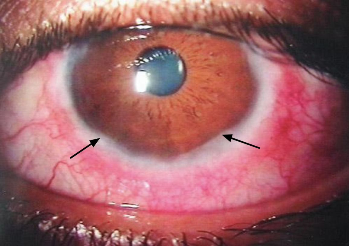
Image Source: Wikimedia Commons (2006). Vernal, Licensed under a creative commons licence. Available for free at: https://commons.wikimedia.org/wiki/File:Vernal.jpg (Accessed on: 5 May 2022).
Giant Papillary Conjunctivitis (GPC)
GPC results from an immune hyperactivity reaction to chronic exposure to a foreign object. Chronic micro-trauma from the foreign object also facilitates entry of the allergen. This is typically seen in cases with contact lens use, prosthesis, or sutures. Patients typically experience itching, thick discharge, pain and foreign body sensation. Examination reveals features such as conjunctival hyperaemia and chemosis, as well as papillae in the tarsal conjunctiva.
Itching is a hallmark feature of allergic eye disease, and symptoms tend to be bilateral, although they may be asymmetrical. Patients also present with redness, watering, conjunctival chemosis, eyelid swelling and skin changes (if there is periorbital dermatitis). In patients with corneal involvement, accompanying symptoms will include pain, photophobia, blurred vision and foreign body sensation. Patients have a history of recurrent episodes of symptoms which may be seasonal or worsen in certain situations. Examination shows evidence of allergic conjunctivitis such as conjunctival hyperaemia, chemosis, papillae, corneal epithelial defects or punctate staining in patients with corneal involvement – these patients can also have ulcers with infiltrate in cases of infection. Diagnosis is clinical. Red flag features in the history or examination (according to NICE guidelines) should prompt referral for urgent ophthalmology assessment. Refractory cases with no red flag features can be referred routinely.
Adapted from NICE Conjunctivitis guidance, the following are red flag features in allergic conjunctivitis necessitating ophthalmology referral:
- Reduced visual acuity
- Marked eye pain, headache or photophobia
- Inability to open the eye or keep it open
- Red sticky eye in a neonate (within 30 days of birth)
- History of trauma (mechanical, chemical or ultraviolet) or possible foreign body
- Copious rapidly progressive discharge
- Possible infection with a herpes virus:
- Herpes simplex — may present as unilateral red eye with vesicular lesions on the eyelid
- Herpes zoster
- Soft contact lens use with corneal symptoms
- Recent intraocular surgery
- Systemic disease or immunosuppressed
- Suspected periorbital or orbital cellulitis
Treatment focuses on symptomatic relief, reducing inflammation and immunomodulation if necessary. Treatment Box 1 shows the ladder of treatment in patients with allergic eye disease.
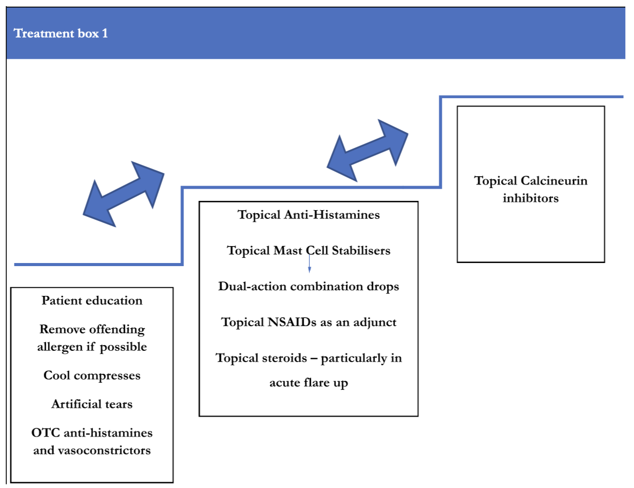
Image source: Adapted from La Rosa M, Lionetti E, Reibaldi M, et al. Allergic conjunctivitis: a comprehensive review of the literature. Ital J Pediatr 2013; 39: 18.
Initial treatment for all forms of allergic conjunctivitis are similar, and further therapies are targeted to complications or response to treatment, as shown in treatment box 1. Corneal involvement requires more aggressive treatment than patients in whom the conjunctiva only is affected, and these patients require ophthalmology referral.
Simple therapies include:
- avoiding the allergen if possible
- cool compresses (these induce local vasoconstriction and provide symptomatic relief)
- lubricating eye drops and short courses of over-the-counter topical antihistamines
Topical mast cell stabilisers and antihistamines are effective in refractory cases, and a short course of steroids can break the cycle of inflammation. Topical calcineurin inhibitors are used in severe or refractory disease, usually with corneal involvement and are initiated by specialists. Superimposed bacterial infection can be treated with anti-bacterials.
Management in primary care should include:
- patient education (discourage patient from eye rubbing as this increases the risk of corneal ectasias)
- topical antihistamines and/or mast cell stabilisers (combination preparations are available)
- monitoring with prompt referral if necessary
- referring patients with multi-system disease to an allergy specialist
VKC and AKC will include treatment for simple allergic conjunctivitis as well as topical mast cell stabilisers. Acute flares might require treatment with topical corticosteroids, and this should be initiated by an ophthalmologist. Cases refractory to steroid therapy or who are difficult to wean off steroids will require treatment with steroid-sparing agents such as topical ciclosporin or tacrolimus. Patients with periocular dermatitis will require control of this and, if severe, will need dermatological input.
In GPC, first-line treatment will include removal of the offending foreign object. Chronic inflammation affecting the cornea can lead to corneal scarring and vascularisation with subsequent visual loss. This is particularly important in young children who can develop amblyopia deprived of visual stimuli.
Complete Guide to Clinical Genetics ST3 Application and Recruitment (2027)
Written by Medibuddy. Last updated: October 2026. Clinical Genetics is

Complete Guide to Clinical Radiology ST3 Application and Recruitment (2027)
Recruitment paused: NHS England is not running Clinical Radiology ST3+ recru

Complete Guide to Paediatric Surgery ST3 Application and Recruitment in United Kingdom (2026)
Written by Medibuddy. Last updated: October 2026. If you’re planning


Take your subscriptions with you
Our mobile app allows you to access your interview and exam question banks wherever you are.

