


 Close
Close
 8th December 2023
Scleritis
Scleritis is an uncommon, potentially blinding condition characterised by inflammation of the scleral tissue. Scleritis may be categorised by anatomical location, pathogenesis, or presence of necrosis. The image below shows the common classification of scleritis.
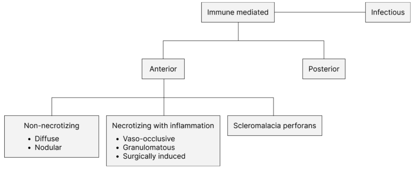
Image source: Adapted from Sims J. Scleritis: presentations, disease associations and management. Postgrad Med J 2012; 88: 713–718.
The most common form of scleritis is anterior scleritis, where there is inflammation anterior to the recti muscles. This presents as a painful red eye with or without associated loss of vision. There are different forms of anterior scleritis. Pathophysiologically, there is oedema and cellular infiltration of the entire thickness of the sclera. Commonly, inflammation begins in a focal area and then spreads over the anterior segment. Inflammation can be granulomatous (histologically epitheloid cells and multinucleate giant cells) or non-granulomatous (histologically plasma cells, macrophages and lymphocytes).
Non-necrotising
This can be diffuse or nodular. In diffuse non-necrotising scleritis, there is diffuse inflammation of the affected portion of the sclera. In nodular, there is a distinct nodule(s) of inflamed sclera. The nodular form is more common in patients who have a previous history of Herpes Zoster Ophthalmicus. The nodules, in this case, are immobile compared to nodular episcleritis. In the non-necrotising form, the sclera becomes translucent due to rearrangement of scleral collagen following inflammation.
Necrotising with inflammation
This is the most severe form with tissue necrosis. It is characterised by severe pain and scleral tenderness and is often bilateral. Necrosis of the sclera leads to exposure of the underlying choroid. The vaso-occlusive form is more common in rheumatoid arthritis with patches of scleral oedema, which eventually coalesce and necrose. Granulomatous scleritis can occur in medium vessel vasculitis. Surgically induced scleritis can occur after repeated ocular surgery and commonly presents within 1-month post-procedure. It is more common in surgical procedures involving the ocular surface and with administration of mitomycin C. The inflammation is usually localised to the surgical wound, although it might spread. Here, the sclera thins out and exposes the underlying uvea.
Scleromalacia perforans
This a form of necrotising scleritis where there is progressive, painless thinning of the sclera in the absence of inflammation. It is more common in older patients with a long history of rheumatoid arthritis. Scleral perforation rarely occurs due to scarring.
There is an association with systemic diseases such as rheumatoid arthritis, vasculitis and connective tissue disorders, particularly in necrotising disease. There is a female preponderance, and it typically presents between the fifth and sixth decades. The presentation of scleritis is varied depending on disease course. It can present as an acute episode, a relapsing-remitting illness or as a chronic process. Patients can present with gradual onset, severe boring pain, which wakes them up at night. It is worse in the morning and improves during the course of the day, and may radiate to the temples. The eye may be diffusely red and tender or localised to an area, and there may be associated visual loss.

Image Source: Wikimedia Commons (2012). Lederhaut MUC0367xxk, Licensed under a creative commons licence. Available for free at: https://commons.wikimedia.org/wiki/File:Lederhaut_MUC0367xxk.jpg (Accessed on: 5 May 2022).

Image Source: Wikimedia Commons (2017). Recurrent scleritis, Licensed under a creative commons licence. Available for free at: https://commons.wikimedia.org/wiki/File:Recurrent_scleritis.jpg (Accessed on: 5 May 2022).
On examination, there is an area of diffusely swollen sclera with injection of the scleral vessels. It typically has a violaceous hue, and the inflamed vessels are deep and cannot be moved with a cotton bud. The globe is tender to touch, and application of 2.5% Phenylephrine distinguishes scleritis (non-blanching) from episcleritis (blanching). They may be associated keratitis or uveitis. Areas of necrosis appear thinned out with visibility of the underlying choroid. Patients can also be examined for features of associated systemic disease. Diagnosis is clinical, but investigations can be carried out to confirm diagnosis (in cases of uncertainty) or to test for underlying or associated diseases. Ocular imaging can include B-Scan ultrasonography and MRI to detect cases of posterior scleritis with more generalised imaging to look for conditions such as sarcoidosis or tuberculosis. Laboratory tests should screen for associated conditions. The principle of treatment is to control inflammation and, therefore, decrease damage to ocular structures. Treatment summary for scleritis is shown in treatment box 2.

Image source: Adapted from Sims J. Scleritis: presentations, disease associations and management. Postgrad Med J 2012; 88: 713–718.
Patients suspected of having scleritis need urgent same-day ophthalmology assessment. In patients with associated diseases, multi-disciplinary management is required. Complications of scleritis include keratitis, uveitis, glaucoma, cataract formation, rarely scleral perforation and loss of vision.
Complete Guide to Clinical Genetics ST3 Application and Recruitment (2027)
Written by Medibuddy. Last updated: October 2026. Clinical Genetics is

Complete Guide to Clinical Radiology ST3 Application and Recruitment (2027)
Recruitment paused: NHS England is not running Clinical Radiology ST3+ recru

Complete Guide to Paediatric Surgery ST3 Application and Recruitment in United Kingdom (2026)
Written by Medibuddy. Last updated: October 2026. If you’re planning


Take your subscriptions with you
Our mobile app allows you to access your interview and exam question banks wherever you are.

