


 Close
Close
 8th December 2023
Uveitis
Uveitis is an umbrella term for a heterogenous group of disorders causing inflammation of the uvea (iris, ciliary body and choroid). Classification of uveitis has been standardised by the International Uveitis Study Group. It is primarily classified anatomically, as shown in the illustrative table below.
| Classification | Primary site | Disorders included | Aetiology |
|---|---|---|---|
| Anterior | Anterior chamber | Iritis, iridocyclitis, anterior cyclitis | Systemic disorders such as spondyloarthropathies, non-systemic such as Fuch’s heterochromic Iridocyclitis and infectious causes such as HSV |
| Intermediate | Vitreous | Pars planitis, Posterior cyclitis, Hyalitis | Systemic disorders such as sarcoidosis, non-systemic such as undifferentiated intermediate uveitis and infections such as syphilis |
| Posterior | Retina and choroid | Focal or diffuse choroiditis, chorioretinitis, Retinitis, retinochoroiditis, Neuroretinitis | Systemic disorders such as sarcoidosis, non-systemic such as birdshot chorioretinopathy and infections such as toxoplasma retinitis |
| Panuveitis | Anterior chamber, vitreous, retina and/or choroid | Systemic disorders such as Behcet disease, non-systemic such as sympathetic ophthalmia and infections such as syphilis |
Source: Adapted from Deschenes J, Murray PI, Rao NA, et al. International Uveitis Study Group (IUSG) Clinical Classification of Uveitis. http://dx.doi.org/101080/09273940801899822 2009; 16: 1–2. (accessed 31 May 2022)
Uveitis can also be characterised by laterality, onset of symptoms, duration (limited or persistent), and course (acute -usually resolves within 3 months, recurrent, repeated episodes separated by periods of inactivity; and chronic – persisting for longer than 3 months). The most common presentation of uveitis is acute anterior uveitis (AAU), accounting for up to 90% of cases in primary care settings22 and will be the focus of this section. In anterior uveitis, the anterior uvea – iris and ciliary body – are affected by inflammation. The peak age of presentation is between 30 and 40 years.
Aetiology
AAU may occur as a response to systemic disease, non-systemic disease, or infection. Disorders associated with anterior uveitis ae shown below. In most cases of uveitis, there is no known cause.
Adapted from ‘Uveitis for the non-ophthalmologist’, the following are common conditions associated with uveitis:
Systemic
- Spondyloarthropathies – associated with the HLA B27 genotype
- Inflammatory bowel disease
- Sarcoidosis
- Behcet disease
- Vasculitides
- Interstitial nephritis
- Systemic Lupus Erythematosus
Non-systemic
- Fuch’s heterochromic Iridocyclitis
Infection
- Herpes Simplex
- Varicella Zoster
- Tuberculosis
- Syphillis
- Lyme disease
Symptoms and signs
AAU typically presents with sudden onset (1-2 days) pain, redness, photophobia, watering and change in vision. It can affect all age groups. Some causes of anterior uveitis may present sub-acutely (1-2 weeks). Evidence of inflammation can be seen in the anterior chamber and sometimes just behind the lens in the anterior vitreous (usually the case in iridocyclitis). The hallmark features of anterior uveitis which may be present on examination with a slit lamp are listed below.
Adapted from ‘Uveitis Straight to the point of care’, the following are examination findings which may be present in anterior uveitis:
- Cells in the aqueous (visible in the anterior chamber)
- Flare (due to proteinaceous exudate)
- Fibrin in the anterior chamber
- Keratic precipitate (aggregate of inflammatory cells on corneal endothelium, can be granulomatous)
- Hypopyon
- Posterior synechiae (sticking of the iris to the lens capsule)
- Iris nodules (uncommon)
- Iris atrophy (associated with HSV and VZV)
- Cells in the anterior vitreous
Patients have a visibly red eye and with cases of posterior synechiae an irregular pupil. The pattern of redness is typically ciliary – a ring-like pattern of dilated vessels around the cornea due to involvement of the branches of the anterior ciliary arteries supplying the cornea, iris and ciliary body.
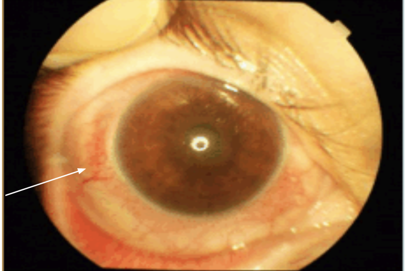
Image Source: Wikimedia Commons (2019). Fig-1-Photograph-showed-non-granulomatous-anterior-uveitis-with-nuclear-cataract-and-ciliary-injection-in-the-right-eye, Licensed under a creative commons licence. Available for free at: https://commons.wikimedia.org/wiki/File:Fig-1-Photograph-showed-non-granulomatous-anterior-uveitis-with-nuclear-cataract-and-ciliary-injection-in-the-right-eye.gif (Accessed on: 5 May 2022).
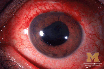
Image Source: Wikimedia Commons (2013). Anterior-uveitis, Licensed under a creative commons licence. Available for free at: https://commons.wikimedia.org/wiki/File:Anterior-uveitis.jpg (Accessed on: 5 May 2022).
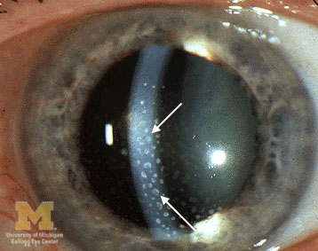
Image Source: Wikimedia Commons (2013). Keratic-precipitates, Licensed under a creative commons licence. Available for free at: https://commons.wikimedia.org/wiki/File:Keratic-precipitates.jpg (Accessed on: 5 May 2022).

Image Source: Wikimedia Commons (no date). Manual_of_ophthalmology_(1917)_(14592285118), Licensed under a creative commons licence. Available for free at: https://commons.wikimedia.org/wiki/File:Manual_of_ophthalmology_(1917)_(14592285118).jpg (Accessed on: 5 May 2022).

Image Source: Wikimedia Commons (2006). Hypopyon, Licensed under a creative commons licence. Available for free at: https://commons.wikimedia.org/wiki/File:Hypopyon.jpg (Accessed on: 5 May 2022).
Diagnosis
Patients suspected of having uveitis should be referred for urgent ophthalmology assessment, usually within 24 hours. Once a provisional diagnosis of AAU is made clinically, a uveitis screen should be carried out to determine the aetiology (if possible, although this is typically done in recurrent cases). Blood tests should include serum ACE, T-spot test, syphilis, ANA, ANCA and rheumatoid factor. CXR to assess for evidence of tuberculosis or bilateral hilar lymphadenopathy to suggest sarcoidosis. The ophthalmologist might carry out further investigations, such as OCT scanning.
Management
Principles of treatment are to treat inflammation and prevent complications, and in patients with chronic disease, to prevent relapses. First-line treatment is topical corticosteroids, and intensity of drops is tailored to the degree of inflammation according to the standardised grading system. Cycloplegics can also be given to relieve pain from ciliary spasm and to break or prevent formation of posterior synechiae by dilating the iris. Episodes refractory to topical treatment or those which are severe enough may require subconjunctival/intravitreal steroids or oral therapy. Raised intraocular pressure is treated with anti-glaucoma medication. Infectious uveitis will require treatment of the infection itself. Treatment should be initiated urgently in infectious cases as it tends to progress rapidly, is more aggressive and can lead to blindness. Long-term use of steroids increases the risk of cataracts and glaucoma. Steroid-sparing agents may be used in select patients with chronic uveitis, and biologics are an emerging successful therapy in refractory cases. Uveitis tends to recur. Patients with chronic disease have no overt signs such as redness or pain, even with ongoing inflammation. Typically, inflammation recurs when treatment is stopped or tapered. Early and prompt treatment can reduce morbidity and prevent blindness. Complications include posterior synechiae, which becomes permanent due to scarring, secondary glaucoma, band keratopathy, chronic macular oedema and cataracts.
Complete Guide to Clinical Genetics ST3 Application and Recruitment (2027)
Written by Medibuddy. Last updated: October 2026. Clinical Genetics is

Complete Guide to Clinical Radiology ST3 Application and Recruitment (2027)
Recruitment paused: NHS England is not running Clinical Radiology ST3+ recru

Complete Guide to Paediatric Surgery ST3 Application and Recruitment in United Kingdom (2026)
Written by Medibuddy. Last updated: October 2026. If you’re planning


Take your subscriptions with you
Our mobile app allows you to access your interview and exam question banks wherever you are.

