


 Close
Close
 12th July 2023
Upper GI Pathology
Diseases of the Mouth
Primary Oral Disease
Recurrent Aphthous Stomatitis (RAS)
RAS, also known as canker sores, is a common condition resulting in recurrent episodes of painful oral ulcers. It is important to consider other differentials which may be causing oral ulcers (e.g., Behçet’s disease, IBD, HIV, coeliac disease) before coming to a diagnosis of RAS.
Oral Squamous Cell Carcinoma (SCC)
SSC is responsible for the majority of tumours found in the mouth. Certain pre-cancerous lesions can predispose to SSC, such as leukoplakia, erythroplakia and lichen planus. Risk factors are tobacco (including oral tobacco products), excess alcohol, HPV (type 16), obesity, age, male sex, and poor nutrition.
Persistent unexplained oral ulcers or lumps should be referred for biopsy to rule out SCC.
Other Primary Oral Diseases
Geographic tongue ― recurrent idiopathic inflammatory disorder, which results in glossitis and patches of depapillation.
Black hairy tongue ― a benign condition which can be seen in smokers, those with poor oral hygiene and some antibiotics.
Leukoplakia ― white patches on the oral mucosa, which is very common. In some patients this may progress to SCC. This is not to be confused with oral hairy leukoplakia (caused by EBV), which is almost entirely seen in patients with HIV.
Erythroplakia ― presents as red patches in the oral mucosa. Erythroplakia is also a premalignant lesion with a high risk of progression to SCC. Surgical excision is a common treatment.
Secondary Oral Disease
Oral manifestations secondary to systemic diseases are often tested in the MRCP examinations. Some of the more common oral manifestations that are tested are summarised below (table 1).
| Speciality | Condition | Oral manifestations |
|---|---|---|
| Dermatology | Stevens-Johnson syndrome | Painful haemorrhagic erosions with a grey membrane |
| Autoimmune bullous disorders | Pemphigus vulgaris – oral mucosal ulceration is common | |
| Lichen planus | Wickham striae – fine white lines in the oral mucosa | |
| Angioedema | Hereditary angioedema caused by C1 esterase inhibitor deficiency. ACE inhibitors are a common trigger | |
| Haematology | Amyloidosis | Infiltration in the mouth – most commonly resulting in macroglossia |
| Leukaemia | Gingival hypertrophy (particularly in AML and APML) | |
| Endocrine | Addison’s disease | Hyperpigmentation – buccal mucosa, vermillion border, gums |
| Diabetes Mellitus | Gingivitis, periodontal disease | |
| Gastroenterology | Inflammatory Bowel Disease | Aphthous ulcers
Pyoderma vegetans – rare, pustules on the oral mucosa (more commonly seen in UC) |
| Coeliac disease | Enamel defects, mouth ulcers, atrophic glossitis | |
| Peutz-Jeghers syndrome | Hyperpigmented macules on the oral mucosa and lips | |
| Osler-Weber-Rendu syndrome (Hereditary Haemorrhagic Telangiectasia) | Telangiectasia can be seen around the mouth and face | |
| GORD | Acid erosion of the teeth | |
| Nutritional deficiencies | Angular cheilitis can be seen with iron/B12 deficiency | |
| Infectious Diseases | Oral candidiasis | White patches on the oral mucosa which can be scraped off leaving a raw area underneath. Caused by candida species |
| Oral hairy leukoplakia | Painless white plaques (typically on the lateral aspects of the tongue) which cannot be scraped off. Caused by EBV. | |
| Herpes Simplex Virus (HSV) | Cold sores
Herpetic gingivostomatitis can be seen in children who have not been exposed to HSV/immunosuppressed adults |
|
| Measles | Koplik’s spots are pathognomonic of measles infection (may precede rash) | |
| HIV seroconversion | Aphthous ulcers | |
| Kaposi’s sarcoma (KS) | Oral KS can be seen in patients with advanced HIV. It is associated with Human Herpesvirus-8 (HHV-8) | |
| Rheumatology | Behçet’s disease | Aphthous ulcers (genital ulcers also seen) |
| Systemic Lupus Erythematosus (SLE) | Aphthous ulcers, honeycomb plaques, non-specific erythema | |
| Genetic | Marfan’s syndrome | High-arched palate
Autosomal dominant mutation of FBN1 gene coding for fibrillin |
| Neurofibromatosis type 1 | Oral neurofibromas
Autosomal dominant mutation of NF-1 gene |
|
| Down’s syndrome | Macroglossia, crowded teeth
Trisomy 21 |
|
| Other | Radiotherapy | Oral mucositis |
Table 1: Secondary oral manifestations of systemic conditions
Gingival Hypertrophy and Macroglossia
The causes of gingival hypertrophy and macroglossia are frequently tested in the MRCP examinations. Causes of each are summarised below.
Gingival hypertrophy:
- Inflammatory enlargement
- Periodontal disease and poor dentition
- Drugs:
- Anticonvulsants e.g. phenytoin, lamotrigine
- Calcium-channel blockers (nifedipine in particular)
- Ciclosporin
- Neoplastic
- Leukaemia – particularly Acute Myeloblastic Leukaemia (AML)
- Infiltrative
- Granulomatosis with polyangiitis, sarcoidosis, amyloidosis
- Pregnancy
- Scurvy
Macroglossia
- Inherited
- Down syndrome
- Neurofibromatosis 1
- Mucopolysaccharidosis
- Glycogen storage disorders
- Acquired
- Endocrine
- Acromegaly
- Hypothyroidism
- Haematological
- Amyloidosis
- Lymphomas
- Multiple Myeloma
- Angioedema
- Carcinoma
- Endocrine
Oral Flora
The healthy oral cavity is polymicrobial and home to aerobic and anaerobic bacteria. As such, human bites resulting in skin and soft tissue infection are often polymicrobial in origin. This requires treatment with a broad-spectrum antibiotic such as co-amoxiclav. Transmission of blood-borne viruses such as HIV, hepatitis B and C can also occur if there has been blood exposure from a human bite.
Cat and dog bites, on the other hand, often isolate Pasteurella species. Infected cats may result in infection with Bartonella henselae, which causes ‘cat-scratch disease’ – a disease characterised by localised, painful lymphadenopathy.
Disorders of the Salivary Glands and Swallowing
Anatomy and Physiology
- Parotid glands
- Largest salivary gland, located behind the angle of the jaw
- 2 lobes with the facial nerve passing between the lobes
- Submandibular glands
- Located below the jaw
- Most common site for salivary gland stones (sialolithiasis)
- Sublingual glands
- Located under the floor of the mouth
- Minor salivary glands
The main digestive enzymes produced by the salivary glands are amylase (digestion of sugars) and lingual lipase (digestion of fats).
Salivary Gland Swelling
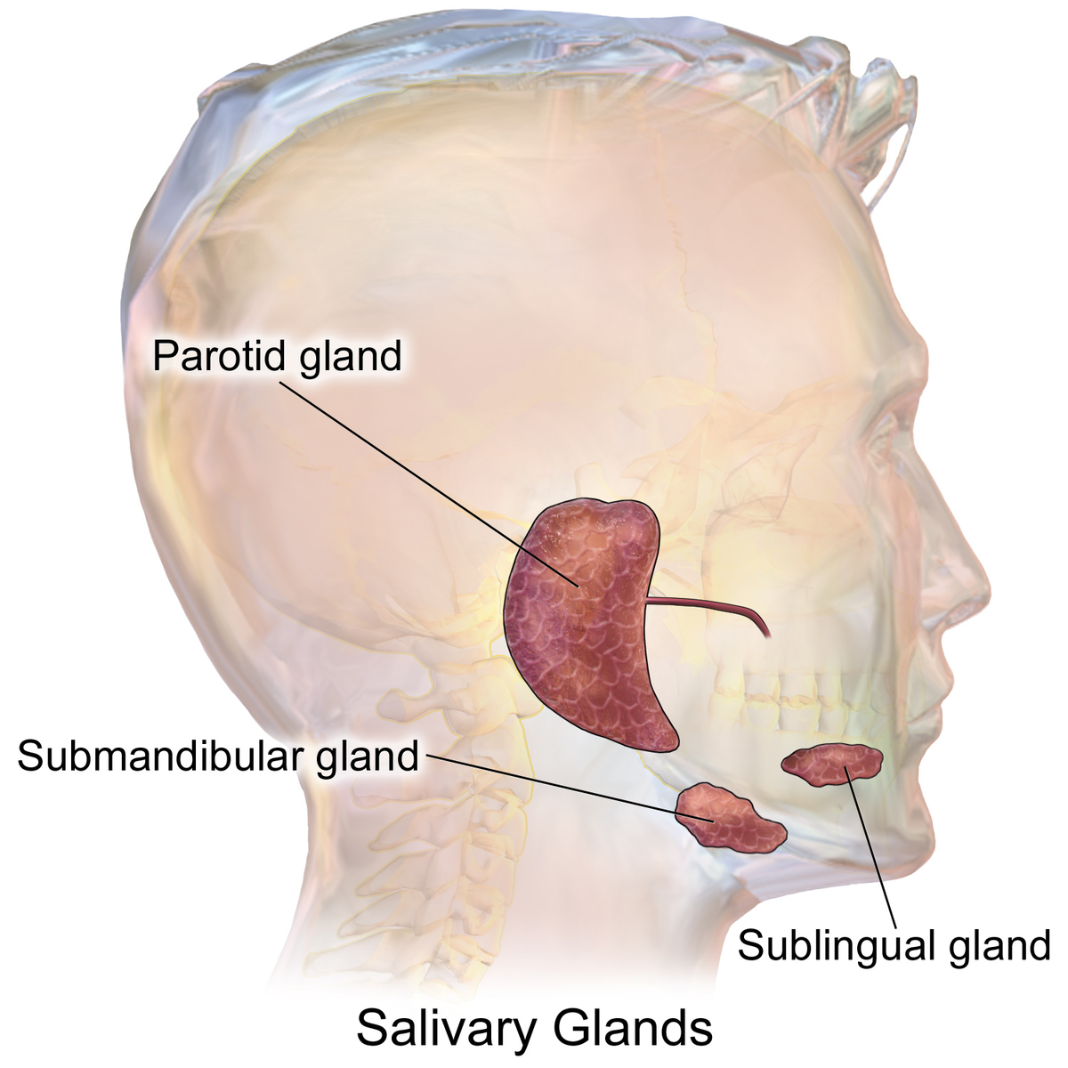
Salivary gland anatomy
Image Source: Wikimedia Commons (2022). Blausen 0780 SalivaryGlands, Licensed under a creative commons licence. Available for free at: https://commons.wikimedia.org/wiki/File:Blausen_0780_SalivaryGlands.png (Accessed on: 9 June 2022).

| Acute | Chronic | |
|---|---|---|
| Unilateral | Bacterial infection (sialadenitis)
Obstruction (sialithiasis or stricture) Radiation |
Chronic sialadenitis
Neoplasm:
Radiation |
| Bilateral | Viral infections:
Radio-iodine treatment |
Blood-borne viruses (HIV, hepatitis B and C)
Sjögren’s (primary and secondary) Endocrine:
Sarcoidosis:
Alcohol Tuberculosis Amyloidosis Rheumatological:
Anorexia/bulimia |
Table 2: Common causes of salivary gland swelling
Neoplasm
Approximately 85% of salivary gland neoplasms affect the parotid glands, for which the majority (about 75%) are benign. Tumours in the other salivary glands (sublingual, submandibular and minor salivary glands), however, are more likely to be malignant.
- The most common benign tumour of the salivary glands is a pleomorphic adenoma (≈50% of benign neoplasms). Pleomorphic adenoma, though a benign lesion, has a high malignant transformation rate. In the absence of contraindications, parotidectomy should be performed in patients.
- The most common malignant tumour of the salivary glands is the mucoepidermoid carcinoma (≈30% of malignant neoplasms).
Tumours in the salivary glands may also be secondary to metastases from lymphoma, squamous cell carcinomas of the head and neck and melanomas. It is worth noting that patients with Sjögren’s syndrome are at a marked increased risk of lymphoid malignancy (40-60-fold), which frequently occur in the salivary glands.
Xerostomia
A reduction in salivary gland output into the oral cavity can result in xerostomia, which is defined as the sensation of oral dryness. There are several temporary and permanent causes of xerostomia (see table 3).
| Temporary | Permanent |
|---|---|
| Physiological
Psychological (anxiety and depression) Drugs (many causes! Just a few listed below)
Sialolithiasis Infections |
Sjögren’s syndrome
Diabetes mellitus Sarcoidosis Cystic fibrosis Parkinson’s disease Bone marrow transplant (60`% of recipients) Trauma (accidental or postoperative) Radiation |
Table 3: Causes of xerostomia
After physiological xerostomia, the next most common cause is drug-induced xerostomia.
Complications of xerostomia include altered taste, dysphagia, dental caries and an increased risk of oral candidiasis and bacterial infections.
Oropharyngeal Dysphagia
Dysphagia can be broadly split into:
- Oropharyngeal dysphagia – difficulty initiating a swallow (choking, coughing, nasopharyngeal regurgitation, aspiration, sensation of residual food in the pharynx)
- Oesophageal dysphagia – food ‘sticking’ after swallowing. This will be discussed in more depth in the diseases of the oesophagus section.
In clinical practice, it is important to try to differentiate oropharyngeal and oesophageal dysphagia in a history as they have different aetiologies and, therefore, require different investigations. Sometimes, however, patients can present with a mixed picture as some causes listed can also give an oesophageal dysphagia – particularly neurological and neuromuscular pathology.
| Anatomical | Neurological | Muscular/neuromuscular | Other |
|---|---|---|---|
| Pharyngeal (Zenker’s) diverticulum
Inflammatory:
Tumours Postoperative Radiation Extrinsic compression:
Corrosive injury |
Cerebrovascular accident
Parkinson’s disease Alzheimer’s disease Multiple sclerosis Motor neuron disease Brain tumour |
Polymyositis
Myasthenia gravis Myotonic dystrophy Muscular dystrophies Metabolic myopathy (e.g. thyroid myopathy) Radiation Botulism |
Psychogenic
Drugs:
|
Table 4: Causes of oropharyngeal dysphagia
Swallow assessment
- Videofluoroscopy ― anteroposterior and lateral images are taken while a patient swallows a barium solution to assess muscle coordination and detect aspiration.
- Fibreoptic Endoscopic Evaluation of Swallowing (FEES) ― allows for direct visualisation of structures as well as a functional assessment of swallowing.
- Imaging – if there is concern of extraluminal compression, e.g. MRI scan for osteophytes or soft tissue masses in the throat, or USS to assess the thyroid
Diseases of the Oesophagus
Oesophageal Dysphagia
Oesophageal dysphagia is characterised by the sensation of food being obstructed in the oesophagus after eating (patients often describe that ‘food sticks’). The dysphagia occurs several seconds after initiating a swallow. We can broadly divide causes of oesophageal dysphagia into 1) mechanical obstruction and 2) motility/neuromuscular disorders. As mentioned above, some causes listed here may also give an oropharyngeal picture.
| Mechanical Obstruction | Motility/neuromuscular disorders |
|---|---|
Intrinsic:
Extrinsic:
|
Primary:
Secondary:
|
Table 5: Causes of oesophageal dysphagia
Investigations
- Fluoroscopy (i.e. barium swallow)
- Gastroscopy – good at visualising mechanical obstructions and is important to exclude cancers
- Oesophageal manometry – performed in patients with non-diagnostic gastroscopies and/or clinical suspicion of oesophageal motility disorders
- Achalasia will show increased pressure at the lower oesophageal sphincter, whereas CREST syndrome will show lower pressure
Patients with ongoing symptoms but with no evidence of structural, endoscopic, histopathological or manometric abnormality may have functional dysphagia.
Achalasia
Achalasia is characterised by the failure of the Lower Oesophageal Sphincter (LES) to relax as a result of the degeneration of ganglion nerve cells in the myenteric plexus. Patients typically describe dysphagia to both solids and liquids as well as retrosternal discomfort following meals. Secondary causes of achalasia include malignancy, Chagas disease and amyloidosis.
Investigations: Barium swallow studies may reveal a typical bird’s beak appearance at the lower oesophagus (see image below). However, oesophageal manometry is the gold standard diagnostic test in which the LES will fail to relax.
Treatment: this is guided by the patient’s preference and surgical risk. More definitive treatments include surgical myotomy or endoscopic pneumatic dilatation. If this is not suitable, alternative treatments include botulinum injections at the LES and/or medications such as calcium channel blockers and nitrates (medical management is often the least effective).

Barium swallow showing bird’s beak appearance that is seen in achalasia
Image Source: Wikimedia Commons (2007). Acha, Licensed under a creative commons licence. Available for free at: https://commons.wikimedia.org/wiki/File:Acha.JPG (Accessed on: 9 June 2022).

Gastro-Oesophageal Reflux Disease (GORD)
GORD is a common condition, which can be defined as the reflux of gastric acid into the oesophagus, causing mucosal injury (erosive oesophagitis) or symptoms. Mild cases may be diagnosed clinically and managed in primary care without the need for further investigation. However, the gold standard diagnostic test is ambulatory 24-hour pH monitoring ― this will help differentiate GORD from functional dyspepsia. Endoscopy may be required if patients have ALARM symptoms or have refractory disease.
Management
- Lifestyle measures ― weight management, smoking cessation, modify caffeine, alcohol, and spicy foods
- Medication review (common culprit medications – bisphosphonates, NSAIDs, steroids, calcium channel blockers)
- Medical management ― H2 antagonists, proton-pump inhibitors, antacids, surface agents (sucralfate)
- Invasive procedures ― endoscopic v surgical (Nissen’s fundoplication)
Complications
GORD can result in a number of oesophageal and extra-oesophageal complications.
Oesophageal:
- Erosive oesophagitis
- Barrett’s oesophagus ― normal oesophageal squamous epithelium undergoes metaplasia and becomes columnar epithelium in response to acid exposure. This increases the risk of oesophageal adenocarcinoma. Therefore, patients are risk stratified to determine the frequency of endoscopic surveillance. Some patients may require interventions such as radio-frequency ablation (RFA)
- Oesophageal strictures
Extra-oesophageal:
- Dental issues
- Asthma
- Chronic laryngitis
Oesophageal Cancers
There are two main forms of oesophageal malignancy:
- Squamous Cell Carcinoma (SCC) – most commonly located in the upper ⅔ of the oesophagus
- Adenocarcinoma – most commonly located in the lower ⅓ of the oesophagus
Traditionally, SSC has been more prevalent in Western nations, however, in the last century, adenocarcinoma has become the leading cause of oesophageal cancer. Key symptoms include dysphagia and weight loss. Unfortunately, many patients with oesophageal cancer present late with unresectable, locally advanced disease. There are various risk factors (Table 6).
| Specific Risk Factors | Shared Risk Factors | |
|---|---|---|
| Squamous Cell Carcinoma (SCC) | Achalasia (10-fold risk)
Plummer-Vinson syndrome Coeliac disease |
Age
Male sex Smoking Alcohol Radiation |
| Adenocarcinoma | Barrett’s oesophagus
GORD Obesity |
Table 6: Risk factors for squamous cell carcinoma and adenocarcinoma of the oesophagus
Other Oesophageal Disorders
- Mallory-Weiss Tear ― an oesophageal mucosal tear, often caused by severe vomiting or coughing. This can result in an upper GI bleed.
- Boerhaave’s perforation ― a sudden rupture of the oesophagus due to raised intra-oesophageal pressure and reduced intrathoracic pressure (e.g. severe vomiting). Patients may have severe retrosternal chest pain (intrathoracic perforation), abdominal pain with radiation to the shoulders (intra-abdominal perforation) and subcutaneous emphysema.
- Eosinophilic oesophagitis ― a history may mention a young patient with a history of recurrent food bolus obstruction (perhaps with certain trigger foods causing an allergic reaction). Patients will show no improvement to PPI trials. Endoscopy may be normal but oesophageal rings may be present. Diagnostic biopsies show ≥15 eosinophils per high power field. Treatments can be difficult and include eliminating trigger foods, topical steroids and dilatation where oesophageal strictures are present.
- Plummer-Vinson syndrome ― a triad of iron deficiency anaemia, dysphagia (secondary to an oesophageal web) and glossitis/angular cheilitis. Oesophageal webs can result in dysphagia.
Diseases of the Stomach
Anatomy and Physiology
Vasculature
The arterial supply of the stomach comes from branches of the coeliac trunk.
- Cardia – left gastric artery
- Fundus – short gastric arteries (arising from splenic artery)
- Lesser curvature – right gastric artery (inferiorly) and left gastric artery (superiorly)
- Greater curvature – left gastroepiploic artery (superiorly) and right gastroepiploic artery (inferiorly)

Blood supply to the stomach
Image Source: Wikimedia Commons (2017). Stomach blood supply, Licensed under a creative commons licence. Available for free at: https://commons.wikimedia.org/wiki/File:Stomach_blood_supply.svg (Accessed on: 9 June 2022).

Stomach acid
The normal pH of the stomach ranges from 1.5 to 2.5. Gastric acid secretion increases dramatically during a meal and can be split into three phases:
- Cephalic phase (20%) – activated by sight, smell and act of chewing food. Vagally mediated.
- Gastric phase (50-60%) – activated by the presence of digestive products in the stomach and distention of the stomach by food. Mediated by the hormone gastrin.
- Intestinal phase (5-10%) – activated by the presence of acidic chyme in the duodenum.
Gastrointestinal hormones
There are a large number of hormones in the normal functioning of the gastrointestinal tract. Some of these are tested in the MRCP examinations.
| Hormone | Source | Function |
|---|---|---|
| Gastrin | G-cells of stomach | Increases gastric acid secretion |
| Cholecystokinin (CCK) | I-cells in duodenum | Increases pancreatic enzyme secretion
Increases gallbladder contraction |
| Somatostatin | Pancreatic delta cells | Decreases secretion of many other hormones (e.g. gastrin and digestive enzymes)
Prolongs gastric emptying time |
| Vasoactive Intestinal Peptide (VIP) | Parasympathetic ganglia | Increases enzyme secretion
Reduces gastric acid secretion |
Table 7: Function of gastrointestinal hormones
Satiety is also mediated by gastrointestinal hormones:
- Appetite is increased by ghrelin
- Appetite is decreased by cholecystokinin (CCK), pancreatic polypeptide (PP), glucagon-like peptide-1 (GLP-1) and leptin
Gastric transit
In a normal functioning stomach, food transits in 4-6 hours. This process is controlled by the autonomic nervous system, smooth muscle, and pacemaker cells (interstitial cells of Cajal) within the stomach. Symptomatic slow gastric transit, in the absence of mechanical obstruction, is termed gastroparesis. Symptoms may include: nausea, vomiting, early satiety, bloating and abdominal pain.
Common aetiologies of gastroparesis include:
- Idiopathic – this is the most common form of gastroparesis (50% of cases)
- Neurogenic – seen postoperatively (especially after vagotomy), diabetic neuropathy, neurological problems such as stroke and MS, following viral infection (VZV and EBV) and Chagas disease.
- Myogenic – rheumatological (scleroderma, polymyositis, SLE), muscular dystrophies, amyloidosis.
- Drug-induced – listed below
- Other aetiologies – Zollinger-Ellison syndrome, peptic ulcer disease, anorexia, endocrine (hypothyroidism), radiation
| Drugs that speed up gastric emptying | Drugs that slow gastric emptying |
|---|---|
| Anticholinesterases – e.g. neostigmine
Dopamine antagonists – e.g. domperidone, metoclopramide Macrolide antibiotics – e.g. erythromycin |
Opioids
Ondansetron Anticholinergics – including tricyclic antidepressants Dopamine agonists – e.g. anti-Parkinsonian medication Proton pump inhibitors Alcohol Calcium channel blockers |
Table 8: Drugs that impact gastric emptying time
Investigation of gastroparesis involves assessing gastric motility using gastric emptying scintigraphy, as well as excluding mechanical obstruction. The latter is often done by direct visualisation endoscopically but may also involve cross-sectional imaging or fluoroscopic assessment.
Management hinges on dietary modification, maintaining adequate nutrition and managing any precipitating factors (such as diabetes). Patients with ongoing symptoms despite this may be treated with oral prokinetics (e.g. dopamine antagonists). With patients presenting with acute dysmotility, it is important to manage their nutrition. In general, enteral feeding (of some form) is always the preferred option.
Helicobacter Pylori Infection
H.pylori is a gram-negative bacterium that has been implicated in a number of gastrointestinal pathologies.
| Pathology | Details |
|---|---|
| Chronic gastritis | H. pylori can predispose patients to other conditions, such as neoplasm |
| Peptic ulcer disease | H. pylori has been implicated in:
|
| Gastric adenocarcinoma | H. pylori associated with a 1-2% lifetime risk of gastric adenocarcinoma |
| B cell lymphoma of MALT tissue | 75% of MALT lymphomas are successfully treated with H. pylori eradication therapy |
Table 9: Sequelae of chronic Helicobacter pylori infection.
Investigations (BSG guidelines)
- 1st line: Urea breath test/stool antigen testing
- Requires a 2-week PPI washout period and 4 weeks for antibiotics
- Urea breath test is the preferred investigation to test for eradication post treatment
- 2nd line: Serology (does not differentiate between active and past infection, so cannot be used for retesting)
If a patient is undergoing OGD, the endoscopist can take biopsies to perform a rapid urease test (also known as the CLO test – Campylobacter-Like Organism) to test for H. pylori.
Treatment
7 days high-dose PPI + amoxicillin + metronidazole/clarithromycin
- If the patient is penicillin allergic, use high-dose PPI + metronidazole + clarithromycin (7 days).
Antiulcer medication
Reduction/neutralisation of stomach acid:
- Proton pump inhibitors (antisecretory)
- Histamine-2 receptor antagonists (antisecretory)
- Antacids (neutralising)
Other mechanisms:
- Sucralfate (forms a protective barrier over the ulcer)
Bleeding Lesions in the Stomach
Bleeding gastric lesions, which are commonly tested in the MRCP examination, are listed in table 10.
| Lesion | Details |
|---|---|
| Peptic ulcer | Breaks in the mucosal layer can penetrate down the muscularis mucosa of the stomach and duodenum.
They are associated with burning epigastric pain.
|
| Gastric varices | Can occur secondary to portal hypertension. Causes:
|
| Erosive gastritis | Multiple erosions seen in the gastric mucosa.
Causes: H. pylori, medication (steroids, NSAIDs), alcohol. |
| Gastric neoplasms | Gastric adenocarcinoma – most often found in the cardia
Gastrointestinal stromal tumours (GISTs) Neuroendocrine tumours Gastric lymphomas |
| Dieulafoy’s lesion | Vascular malformations where aberrant tortuous arterioles can erode through the submucosal layer and cause bleeding. They account for 5% of UGIB. |
| Gastric Antral Vascular Ectasia (GAVE) | An uncommon cause of chronic gastric bleeding caused by fragile antral vessels.
Question clue: the classic ‘watermelon stomach’ appearance described on endoscopy. |
| Zollinger-Ellison Syndrome (ZES) | Gastrin producing tumours (gastrinomas) secrete excess gastrin in the duodenum and pancreas. Suspicion for ZES should be raised in cases of multiple peptic ulcers, ulcers in the jejunum, ulcers that are non-responsive to PPI therapy. 20% of patients have MEN-1.
Investigation:
Question clue: diarrhoea + multiple peptic ulcers + not responding to PPIs |
| Angiodysplasia | Small vascular malformations which can be found throughout the GI tract. |
Table 10: Bleeding lesions in the stomach
Other Stomach Pathology
Ménétrier’s Disease: a rare disorder characterised by giant gastric folds. Patients have decreased acid secretion and may develop a protein-losing gastropathy. This results in hypoalbuminaemia, which may manifest with oedema.
Autoimmune Gastritis: autoimmune destruction of the stomach results in atrophic gastritis evidenced by flattened rugal folds and visible subcutaneous vessels on endoscopy. Patients may have antibodies to parietal cells and/or intrinsic factor. Intrinsic factor is required for B12 absorption in the terminal ileum, so a lack of this can result in pernicious anaemia.
Cyclical vomiting syndrome (CVS): characterised by bouts of severe vomiting that may last several days, with intervening periods of good health. A similar condition, cannabis hyperemesis syndrome (CHS), is seen in people with heavy, often daily use of cannabis – it has a similar presentation to CVS. Symptoms often improve with abstinence from cannabis. Many patients with CVS and CHS find that their symptoms are relieved by hot showers or baths.
Bariatric Surgery
Bariatric procedures can be considered for the management of severe obesity and its complications. Surgery is reserved for those with:
- A BMI of 40 kg/m2 or more, or
- Between 35 kg/m2 and 40 kg/m2 with evidence of other significant disease (e.g. type 2 diabetes mellitus or obstructive sleep apnoea) which will improve with weight loss
In these patients, surgery is only considered when all appropriate non-surgical measures have been tried. There are two main surgeries to be aware of for the purposes of the MRCP:
- Roux-en-Y gastric bypass – a small gastric pouch is created, which is anastomosed to the jejunum (restrictive and malabsorptive)
- Sleeve gastrectomy – a partial gastrectomy reducing the volume of the stomach (restrictive)
Complications:
There are many complications for which the following could be tested in the MRCP examination:
- Nutritional deficiencies – malabsorptive procedures are at increased risk of deficiencies. Risk of anaemia and metabolic bone disease.
- Anatomical – Stricture formation, marginal ulcers, small capacity (early satiety)
- Gallstones – because of distorted anatomy, ERCP can be difficult in these patients!
- Short-bowel syndrome
- Dumping syndrome
- Change in bowel habits
Complete Guide to Clinical Genetics ST3 Application and Recruitment (2027)
Written by Medibuddy. Last updated: October 2026. Clinical Genetics is

Complete Guide to Clinical Radiology ST3 Application and Recruitment (2027)
Recruitment paused: NHS England is not running Clinical Radiology ST3+ recru

Complete Guide to Paediatric Surgery ST3 Application and Recruitment in United Kingdom (2026)
Written by Medibuddy. Last updated: October 2026. If you’re planning


Take your subscriptions with you
Our mobile app allows you to access your interview and exam question banks wherever you are.

