


 Close
Close
 15th July 2023
Disorders of the Salivary Glands and Swallowing
Anatomy and Physiology
- Parotid glands
- Largest salivary gland, located behind the angle of the jaw
- 2 lobes with the facial nerve passing between the lobes
- Submandibular glands
- Located below the jaw
- Most common site for salivary gland stones (sialolithiasis)
- Sublingual glands
- Located under the floor of the mouth
- Minor salivary glands
The main digestive enzymes produced by the salivary glands are amylase (digestion of sugars) and lingual lipase (digestion of fats).
Salivary Gland Swelling
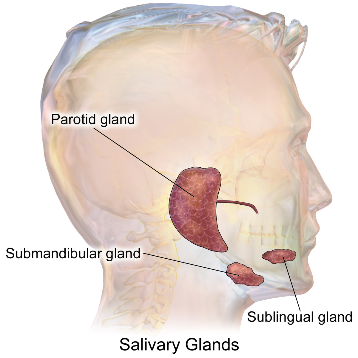
Salivary gland anatomy
Image Source: Wikimedia Commons (2022). Blausen 0780 SalivaryGlands, Licensed under a creative commons licence. Available for free at: https://commons.wikimedia.org/wiki/File:Blausen_0780_SalivaryGlands.png (Accessed on: 9 June 2022).

| Acute | Chronic | |
|---|---|---|
| Unilateral | Bacterial infection (sialadenitis)
Obstruction (sialithiasis or stricture) Radiation |
Chronic sialadenitis
Neoplasm:
Radiation |
| Bilateral | Viral infections:
Radio-iodine treatment |
Blood-borne viruses (HIV, hepatitis B and C)
Sjögren’s (primary and secondary) Endocrine:
Sarcoidosis:
Alcohol Tuberculosis Amyloidosis Rheumatological:
Anorexia/bulimia |
Table 2: Common causes of salivary gland swelling
Neoplasm
Approximately 85% of salivary gland neoplasms affect the parotid glands, for which the majority (about 75%) are benign. Tumours in the other salivary glands (sublingual, submandibular and minor salivary glands), however, are more likely to be malignant.
- The most common benign tumour of the salivary glands is a pleomorphic adenoma (≈50% of benign neoplasms). Pleomorphic adenoma, though a benign lesion, has a high malignant transformation rate. In the absence of contraindications, parotidectomy should be performed in patients.
- The most common malignant tumour of the salivary glands is the mucoepidermoid carcinoma (≈30% of malignant neoplasms).
Tumours in the salivary glands may also be secondary to metastases from lymphoma, squamous cell carcinomas of the head and neck and melanomas. It is worth noting that patients with Sjögren’s syndrome are at a marked increased risk of lymphoid malignancy (40-60-fold), which frequently occur in the salivary glands.
Xerostomia
A reduction in salivary gland output into the oral cavity can result in xerostomia, which is defined as the sensation of oral dryness. There are several temporary and permanent causes of xerostomia (see table 3).
| Temporary | Permanent |
|---|---|
| Physiological
Psychological (anxiety and depression) Drugs (many causes! Just a few listed below)
Sialolithiasis Infections |
Sjögren’s syndrome
Diabetes mellitus Sarcoidosis Cystic fibrosis Parkinson’s disease Bone marrow transplant (60`% of recipients) Trauma (accidental or postoperative) Radiation |
Table 3: Causes of xerostomia
After physiological xerostomia, the next most common cause is drug-induced xerostomia.
Complications of xerostomia include altered taste, dysphagia, dental caries and an increased risk of oral candidiasis and bacterial infections.
Oropharyngeal Dysphagia
Dysphagia can be broadly split into:
- Oropharyngeal dysphagia – difficulty initiating a swallow (choking, coughing, nasopharyngeal regurgitation, aspiration, sensation of residual food in the pharynx)
- Oesophageal dysphagia – food ‘sticking’ after swallowing. This will be discussed in more depth in the diseases of the oesophagus section.
In clinical practice, it is important to try to differentiate oropharyngeal and oesophageal dysphagia in a history as they have different aetiologies and, therefore, require different investigations. Sometimes, however, patients can present with a mixed picture as some causes listed can also give an oesophageal dysphagia – particularly neurological and neuromuscular pathology.
| Anatomical | Neurological | Muscular/neuromuscular | Other |
|---|---|---|---|
| Pharyngeal (Zenker’s) diverticulum
Inflammatory:
Tumours Postoperative Radiation Extrinsic compression:
Corrosive injury |
Cerebrovascular accident
Parkinson’s disease Alzheimer’s disease Multiple sclerosis Motor neuron disease Brain tumour |
Polymyositis
Myasthenia gravis Myotonic dystrophy Muscular dystrophies Metabolic myopathy (e.g. thyroid myopathy) Radiation Botulism |
Psychogenic
Drugs:
|
Table 4: Causes of oropharyngeal dysphagia
Swallow assessment
- Videofluoroscopy ― anteroposterior and lateral images are taken while a patient swallows a barium solution to assess muscle coordination and detect aspiration.
- Fibreoptic Endoscopic Evaluation of Swallowing (FEES) ― allows for direct visualisation of structures as well as a functional assessment of swallowing.
- Imaging – if there is concern of extraluminal compression, e.g. MRI scan for osteophytes or soft tissue masses in the throat, or USS to assess the thyroid
Complete Guide to Clinical Genetics ST3 Application and Recruitment (2027)
Written by Medibuddy. Last updated: October 2026. Clinical Genetics is

Complete Guide to Clinical Radiology ST3 Application and Recruitment (2027)
Recruitment paused: NHS England is not running Clinical Radiology ST3+ recru

Complete Guide to Paediatric Surgery ST3 Application and Recruitment in United Kingdom (2026)
Written by Medibuddy. Last updated: October 2026. If you’re planning


Take your subscriptions with you
Our mobile app allows you to access your interview and exam question banks wherever you are.

