


 Close
Close
 20th February 2024
Arterial anatomy
Aorta
The arterial supply of the body begins at the aortic root. Coronary circulation is covered in the “Cardiac and Respiratory Anatomy” section. The aortic root continues as the ascending aorta, which becomes the arch of the aorta at the level of the second sternocostal joint. Three main branches arise from the aortic arch before it becomes the descending aorta: brachiocephalic trunk, left common carotid artery and left subclavian artery.
The descending aorta can be divided into the thoracic and abdominal aorta. At the level of L4, the abdominal aorta terminates as it bifurcates into the common iliac arteries.
 Image Source: Wikimedia Commons (2017). Aorta branches, Licensed under a creative commons licence. Available for free at: https://upload.wikimedia.org/wikipedia/commons/f/f1/Aorta_branches.jpg (Accessed: 9 January 2024).
Thoracic, abdominal and pelvic cavity
The thoracic aorta supplies several branches, including (from superior to inferior):
- Bronchial artery – Supplies the bronchus, peribronchial tissue and visceral pleura
- Mediastinal artery
- Oesophageal artery – Supplies the oesophagus
- Pericardial artery – Supplies the dorsal pericardium
- Superior phrenic artery – Supplies the superior diaphragm
- Intercostal artery and subcostal artery – Supplies the intercostal space.
The abdominal aorta begins at the T12 vertebral level, and multiple branches arise from the abdominal aorta, including (from superior to inferior):
- Inferior phrenic artery
- Coeliac artery
- Superior mesenteric artery
- Middle suprarenal artery – Supplies the adrenal glands
- Renal arteries
- Gonadal arteries
- Inferior mesenteric artery
- Lumbar and sacral arteries
The coeliac axis, superior mesenteric artery and inferior mesenteric artery supply the GI tract and the arteries supply the foregut, midgut and hindgut, respectively. The coeliac artery has several branches:
- Left gastric artery
- Splenic artery – Further branches include the left gastroepiploic artery, short gastric arteries and the pancreatic branches
- Common hepatic artery – Further branches to the proper hepatic artery and gastroduodenal artery
- The proper hepatic artery supplies the right gastric, right hepatic and left hepatic artery
- The gastroduodenal artery supplies the right gastroepiploic and superior pancreaticoduodenal artery.
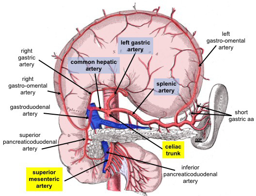
Image Source: Wikimedia Commons (2019). Celiac Trunk and Branches, Licensed under a creative commons licence. Available for free at: https://upload.wikimedia.org/wikipedia/commons/3/36/Celiac_Trunk_and_Branches.jpg (Accessed: 9 January 2024).
The superior mesenteric artery supplies blood from the level of the ampulla of Vater within the duodenum to the proximal 2/3rd of the transverse colon. The inferior mesenteric artery supplies the remainder 1/3rd of the transverse colon up to the anal canal at the level of the pectinate line.
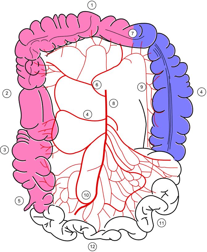
Image Source: Wikimedia Commons (2008). Colonic blood supply, Licensed under a creative commons licence. Available for free at: https://upload.wikimedia.org/wikipedia/commons/9/9f/Colonic_blood_supply.svg (Accessed: 9 January 2024).
Legend: 4 – Right colic artery; 6 – Middle colic artery; 8 – Superior mesenteric artery; 9 – Marginal artery; 10 – Ileocolic artery.

Image Source: Wikimedia Commons (2005). Inferior mesenteric, Licensed under a creative commons licence. Available for free at: https://upload.wikimedia.org/wikipedia/commons/6/64/Inferior_mesenteric_a.gif (Accessed: 9 January 2024).
It is important to note the watershed area created within the splenic flexure, as this is the point of anastomosis between the superior and inferior mesenteric arteries.
The pelvic blood supply mainly arises from the internal iliac artery. The internal iliac artery will provide branches to the bladder, rectum, perineum, and muscles surrounding the gluteal and adductor muscles. It also supplies arterial blood to the vagina and uterus.
Head and neck
The arterial supply to the head and neck arises from the common carotid artery. This arises directly from the arch of the aorta on the left, while the right common carotid artery arises from the brachiocephalic artery. The common carotid artery will bifurcate into the internal and external carotid artery.
The internal carotid artery mainly supplies the intracranial structures, with the ophthalmic artery supplying the structures within the orbit.
The external carotid artery supplies the remainder of the neck and head. The main branches of this include the facial artery, maxillary artery and superficial temporal artery. The external carotid artery branches will provide arterial blood to the structures of the face, tongue, external ear, pharynx, maxilla, mandible, nasal structures and scalp.
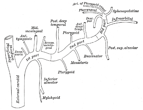
Image Source: Wikimedia Commons (2012). Gray511, Licensed under a creative commons licence. Available for free at: https://upload.wikimedia.org/wikipedia/commons/a/aa/Gray511.png (Accessed: 9 January 2024).
Upper limb
The arterial supply to the upper limb arises from the subclavian artery bilaterally. It travels laterally through the axillae and becomes the axillary artery after it crosses the lateral border of the first rib. The axillary artery provides the anterior and posterior circumflex artery, which wraps around the surgical neck of the humerus, and it can be damaged during fractures of the surgical neck of the humerus. The axillary artery becomes the brachial artery after it crosses the teres major.
The brachial artery travels with the radial nerve within the radial groove of the humerus and provides the bulk of the arterial supply to the structures of the upper arm. As the artery crosses the bicipital aponeurosis, it bifurcates into the radial and ulnar arteries. The radial artery supplies the structures within the posterolateral forearm, while the ulnar artery supplies the anteromedial forearm. Within the hand, the radial and ulnar arteries form two anastomoses, giving rise to the deep and superficial palmar arteries.

Image Source: Wikimedia Commons (2013). Thoracic Upper Limb Arteries Chart, Licensed under a creative commons licence. Available for free at: https://upload.wikimedia.org/wikipedia/commons/c/c0/2128_Thoracic_Upper_Limb_Arteries_Chart.jpg (Accessed: 9 January 2024).
Lower limb
The femoral artery is the lower limb’s main artery, which acts as a direct continuation of the external iliac artery as it crosses the inguinal ligament. Several branches arise from the femoral artery, which supplies the femur and muscles around the thigh. It becomes the popliteal artery as it enters the posterior compartment of the leg close to the knee.
The popliteal artery later divides into the anterior tibial and tibioperoneal trunk. The tibioperoneal artery will further divide into the posterior tibial and fibular arteries. The anterior tibial artery continues along the anterior aspect of the leg and becomes the dorsalis pedis artery. The posterior tibial artery enters the foot via the tarsal tunnel and crosses posterior to the medial malleolus. This contributes to the circulation of the foot via the medial and lateral plantar arteries.
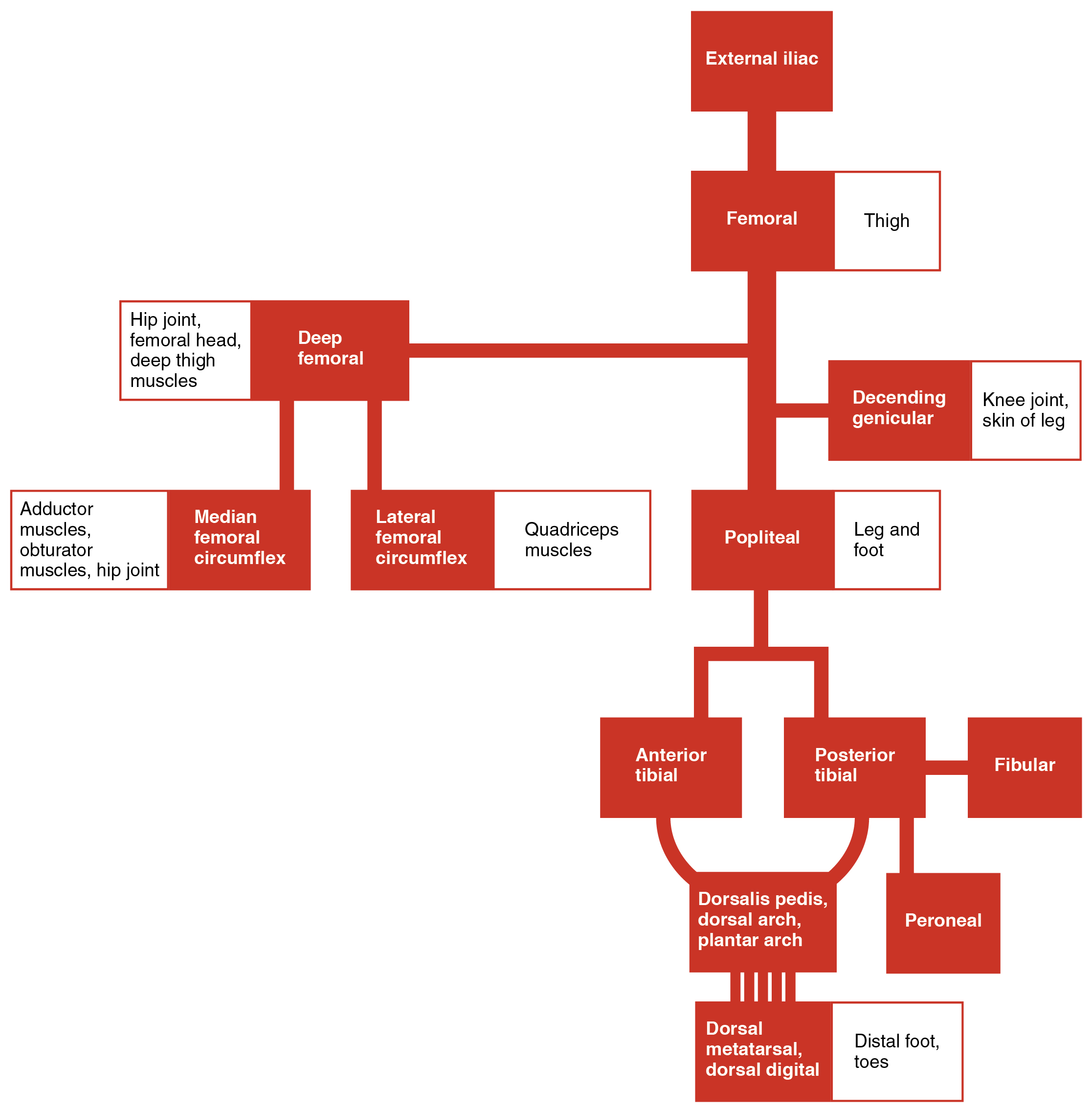 Image Source: Wikimedia Commons (2017). 2130 Lower Limb Arteries Chart, Licensed under a creative commons licence. Available for free at: https://upload.wikimedia.org/wikipedia/commons/0/0d/2130_Lower_Limb_Arteries_Chart.jpg (Accessed: 9 January 2024).
References
- Moore, K. L., Dalley, A. F., & Agur, A. M. R. (2014). Moore’s Clinically Orientated Anatomy (7th ed.). China: Lippincott.
- Kumar & M. Clark (2012). (Eds.), Kumar & Clark’s Clinical Medicine (12 ed.). Spain: Elsevier.
- Wineski, L. E., & Snell, R. S. (2019). Snell’s clinical anatomy by regions.
Complete Guide to Clinical Genetics ST3 Application and Recruitment (2027)
Written by Medibuddy. Last updated: October 2026. Clinical Genetics is

Complete Guide to Clinical Radiology ST3 Application and Recruitment (2027)
Recruitment paused: NHS England is not running Clinical Radiology ST3+ recru

Complete Guide to Paediatric Surgery ST3 Application and Recruitment in United Kingdom (2026)
Written by Medibuddy. Last updated: October 2026. If you’re planning


Take your subscriptions with you
Our mobile app allows you to access your interview and exam question banks wherever you are.

