


 Close
Close
 20th February 2024
Venous anatomy
Upper limb
The venous drainage of the upper limbs can be divided into the superficial and deep systems. The basilic and cephalic veins will drain blood from the dorsal aspect of the hand and pass within the arm’s medial and anterolateral aspects. They are connected within the elbow by the median cubital vein. Both veins drain into the axillary vein.
The deep venous system is situated beside their accompanying arteries and is formed by the forearm’s radial and ulnar veins. These drain into the brachial vein and eventually into the axillary vein. Both axillary veins will drain into the subclavian and, eventually, the brachiocephalic vein.
Lower limb
The venous drainage of the foot starts with the dorsal venous arch dorsally, with the medial and lateral plantar veins located on the plantar aspect. The dorsal venous arch will travel deep into the leg to form the anterior tibial vein. The medial and lateral plantar veins will combine to form the posterior tibial and the fibular veins. All three veins will unite to form the popliteal vein.
As it travels up the leg through the adductor canal, this becomes the femoral vein. The thigh muscles will drain via the deep vein of the thigh, and it will empty into the femoral vein. As the femoral vein travels past the inguinal canal, it becomes the external iliac vein.
Superficial veins are also found in the lower limb and are made of great saphenous and small saphenous veins. The great saphenous vein originates from the dorsal venous arch of the foot and travels superiorly past the medial malleolus and medial condyle of the femur. This empties into the femoral vein inferior to the inguinal ligament. The small saphenous vein travels past the lateral malleolus along the two heads of the gastrocnemius muscle to empty into the popliteal vein via the popliteal fossa.

Image Source: Wikimedia Commons (2013). 2137 Lower Limb Veins Chart, Licensed under a creative commons licence. Available for free at: https://upload.wikimedia.org/wikipedia/commons/8/86/2137_Lower_Limb_Veins_Chart.jpg (Accessed: 9 January 2024).
The superior and inferior gluteal veins will drain gluteal structures and will drain into the internal iliac vein.
Head and neck
The Nervous System section covers the venous return from the cranium, and these vessels will drain into the internal jugular vein. The external face drains into the external jugular vein, and its tributaries are the posterior auricular and retromandibular veins.
The external jugular veins will empty into the subclavian vein. The internal jugular veins combine with the subclavian veins to form the brachiocephalic veins. The brachiocephalic veins will unite at the right side of the superior mediastinum to form the SVC, and this drains directly into the right atrium.
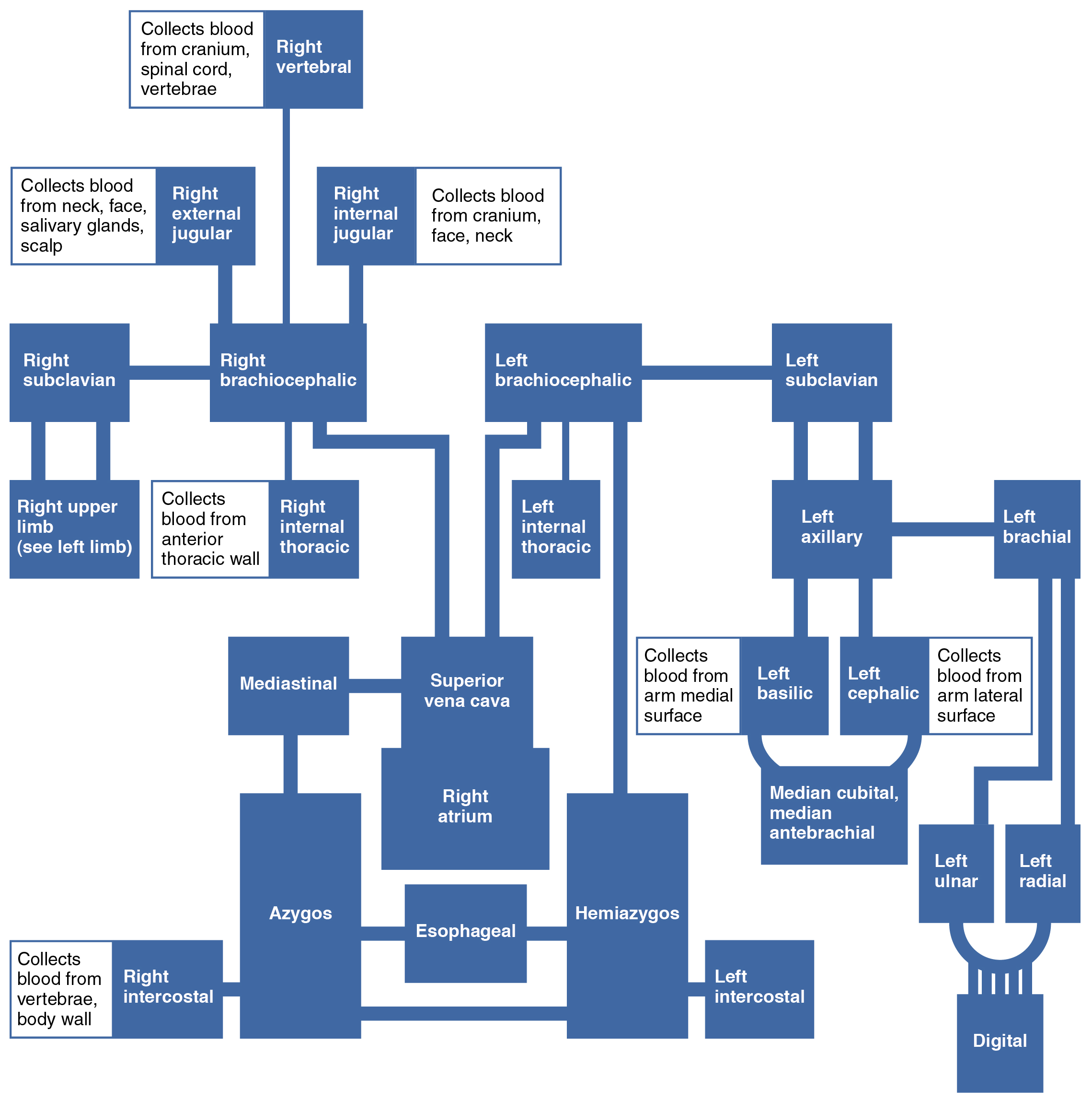 Image Source: Wikimedia Commons (2013). 2135 Veins Draining into Superior Vena Cava Chart, Licensed under a creative commons licence. Available for free at: https://upload.wikimedia.org/wikipedia/commons/d/df/2135_Veins_Draining_into_Superior_Vena_Cava_Chart.jpg (Accessed: 9 January 2024).
Thoracic, abdominal and pelvic cavity
Within the thoracic cavity, the main veins that pass through are the inferior vena cava and the azygos venous system. The azygous venous system consists of the azygous vein, hemiazygos vein and accessory hemiazygos vein. The azygos vein can arise from the lumbar azygos vein or directly from the inferior vena cava at the renal vein level. It courses within the posterior mediastinum and arches over the right pulmonary hilum before draining into the SVC. Its major tributaries are the hemiazygos and accessory hemiazygos vein. The hemiazygos vein drains from the lumbar ascending and subcostal veins from the 9-11 ribs. This will drain back to the azygos vein at the level of T8. The accessory hemiazygos vein will drain blood from the 5th-8th ribs and the left bronchial vein. It empties into the azygos vein at T7.
The pelvic venous drainage occurs through the internal iliac vein. It receives the venous return from the majority of the pelvic organs. It will merge with the external iliac vein at the level of the pubic symphysis to form the common iliac vein.
The systemic venous drainage from the abdomen occurs through the IVC. The IVC is formed from the union of the common iliac veins at the level of L5. This travels superiorly and will drain into the right atrium. It receives blood from the common iliac, lumbar, renal, right gonadal, right suprarenal, and hepatic veins.
 Image Source: Wikimedia Commons (2013). 2132 Thoracic Abdominal Veins, Licensed under a creative commons licence. Available for free at: https://upload.wikimedia.org/wikipedia/commons/2/28/2132_Thoracic_Abdominal_Veins.jpg (Accessed: 9 January 2024).
Venous drainage of the GI tract, spleen, pancreas and gallbladder is carried out via the portal venous system. The union of the splenic vein and superior mesenteric vein forms the portal vein. The portal vein divides into the right and left portal vein as it enters the liver, delivering nutrient-rich venous blood into the liver.
 Image Source: Wikimedia Commons (2013). 2138 Hepatic Portal Vein System, Licensed under a creative commons licence. Available for free at: https://upload.wikimedia.org/wikipedia/commons/c/cf/2138_Hepatic_Portal_Vein_System.jpg (Accessed: 9 January 2024).
References
- Moore, K. L., Dalley, A. F., & Agur, A. M. R. (2014). Moore’s Clinically Orientated Anatomy (7th ed.). China: Lippincott.
- Kumar & M. Clark (2012). (Eds.), Kumar & Clark’s Clinical Medicine (12 ed.). Spain: Elsevier.
- Wineski, L. E., & Snell, R. S. (2019). Snell’s clinical anatomy by regions.
Complete Guide to Clinical Genetics ST3 Application and Recruitment (2027)
Written by Medibuddy. Last updated: October 2026. Clinical Genetics is

Complete Guide to Clinical Radiology ST3 Application and Recruitment (2027)
Recruitment paused: NHS England is not running Clinical Radiology ST3+ recru

Complete Guide to Paediatric Surgery ST3 Application and Recruitment in United Kingdom (2026)
Written by Medibuddy. Last updated: October 2026. If you’re planning


Take your subscriptions with you
Our mobile app allows you to access your interview and exam question banks wherever you are.

