


 Close
Close
 3rd April 2024
Venous Thromboembolism
Venous Thromboembolism
Introduction
Venous thromboembolism (VTE) includes both deep vein thrombosis (DVT) and pulmonary embolism (PE). DVT refers to occluding or non-occluding thrombi in the deep veins which can embolise, causing pulmonary emboli. The annual incidence of VTE is around 2/1000 in the general population. Risk factors include:
- Thrombophilia (factor V Leiden mutation, prothrombin gene mutation, protein C and S deficiency, anti-thrombin deficiency, and antiphospholipid antibody syndrome)
- Previous VTE
- Age over 40 years
- Recent surgery
- Obesity
- Varicose veins
- Prolonged travel
- Acute medical illness
- Cancer
- Immobility
- Pregnancy
- Drugs: 5 fluorouracil, oestrogen-containing contraceptives, hormone replacement therapy, tamoxifen and cisplatin
- Inflammatory bowel disease, nephrotic syndrome, blood transfusions
- Vessel wall damage (venous harvest and central venous catheterisation)
Pathophysiology
Virchow’s triad describes the pathogenesis of DVT and consists of venous stasis, blood hypercoagulability and vessel wall injury. Immobility contributes to venous stasis. Hypercoagulability may be secondary to genetic factors, acute illness or cancer. Vessel wall injury stimulates circulation of pro-coagulants.
A VTE is termed provoked, when caused by an environmental or acquired risk factor and unprovoked, in the absence of these factors. This has implications on treatment duration with longer duration of anticoagulation where VTE has been unprovoked. PE usually results from dislodgement of thrombi from a DVT.
Clinical features
DVT usually occur in the lower limbs but can also present in upper limbs. Presentation may include unilateral localised pain, swelling, tenderness, skin changes and/or distended veins. Signs and symptoms of PE include chest pain, shortness of breath and/or haemoptysis.
Diagnosis
Perform blood tests including FBC, U&Es, LFTs, clotting screen and d-dimer. Where oxygen saturations are low, arterial blood sampling can confirm hypoxaemia and hypocapnia.
In PE, the ECG may show sinus tachycardia, T wave inversion V1-4, atrial fibrillation, nonspecific ST segment changes or may be normal. The classic textbook right heart strain pattern of S1Q3T3 is seen in 20% of cases and is neither sensitive nor specific for PE.
The utility of d-dimer is described below. Where DVT or PE is suspected, a 2-level Wells’ score should be performed (see Tables 1 and 2).
Table 1. Two-level DVT Wells’ score
| Clinical feature | Points |
|---|---|
| Active cancer (treatment ongoing, within 6 months or palliative) | 1 |
| Paralysis, paresis or recent plaster immobilisation of lower extremities | 1 |
| Recently bedridden for 3 days or more, or major surgery within 12 weeks requiring general or regional anaesthesia | 1 |
| Localised tenderness along the distribution of deep venous system | 1 |
| Entire leg swollen | 1 |
| Calf swelling at least 3 cm larger than asymptomatic side | 1 |
| Collateral superficial veins (non-varicose) | 1 |
| Previously documented DVT | 1 |
| An alternative diagnosis is at least as likely as DVT | -2 |
| Clinical probability simplified score | Points |
| DVT likely | >/= 2 points |
| DVT unlikely | </= 1 point |
Management of suspected DVT based on DVT Well’s score (recommendations by NICE)
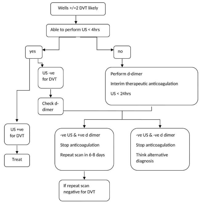
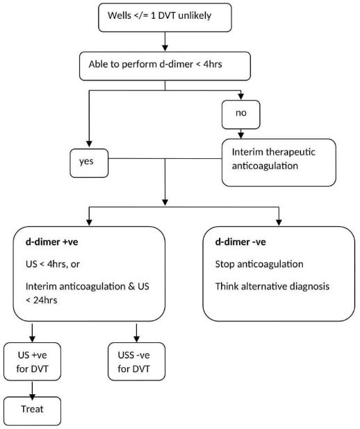
Table 2. 2 level PE Wells’ score
| Clinical features | Points |
|---|---|
| Clinical features and symptoms of DVT (minimum of leg swelling and pain with palpation of the deep veins) | 3 |
| An alternative diagnosis is less likely than PE | 3 |
| Heart rate more than 100/min | 1.5 |
| Immobilisation for more than 3 days or surgery in the previous 4 weeks | 1.5 |
| Previous DVT/ PE | 1.5 |
| Haemoptysis | 1 |
| Active cancer (on treatment, treated in the last 6 months, or palliative) | 1 |
| Clinical probability simplified score | Points |
| PE likely | >4 points |
| PE unlikely | </= 4 points |
Management of suspected PE based on Well’s score (recommendations by NICE)

Management
Offer either a DOAC or LMWH as a choice of anticoagulant based on the clinical scenario. The duration of anticoagulation therapy is for three months in patients with a provoked DVT/PE when the provoking factor is no longer present. Consider anticoagulation beyond three months in unprovoked DVT/PE based on future VTE risk and bleeding risk. If anticoagulation is contraindicated in patients with DVT/PE, an inferior vena cava filter should be considered. For patients with confirmed PE and haemodynamic instability, treatment options include unfractionated heparin infusion and thrombolytic therapy.
Catheter-guided thrombolytic therapy for symptomatic iliofemoral DVT may be considered in those with symptoms < 14 days, with good functional status, life expectancy of more than one year and with low bleeding risk. Do not offer further investigations for cancer to patients unless they have other clinical indications. Do not offer thrombophilia testing for patients continuing on anticoagulation or have had provoked VTE.
Anticoagulation
Various anticoagulation options are available to treat VTE, and choice should be based on clinical scenario and patient preference.
Apixaban and Rivaroxaban can be started at initial suspicion or after diagnosis without bridging LMWH.
- Apixaban 10mg BD for 7 days followed by 5mg BD
- Rivaroxaban 15mg BD for 21 days followed by 20mg OD
Dabigatran and Edoxaban require bridging with LMWH for at least 5 days before initiating.
LMWH should be used concurrently when initiating a Vitamin K antagonist until the INR is at least 2.0 for 2 consecutive readings.
Unfractionated heparin may be used in severe renal impairment or in patients with high bleeding risk.
Prophylaxis
Hospital-acquired VTE refers to a VTE that occurs within 90 days of hospital admission. All patients should have a VTE risk assessment on admission to hospital to ensure adequate prophylaxis, either mechanical or pharmacological, is prescribed. Mechanical prophylaxis includes graduated compression stockings and intermittent pneumatic compression devices (IPC). IPC devices should be used for patients admitted with an acute stroke, initiated within three days and continued for 30 days or until sufficiently mobile. Mechanical prophylaxis is appropriate for patients with major trauma or undergoing cranial, spinal, thoracic, cardiac or ENT surgery. Pharmacological prophylaxis includes heparins (unfractionated or low molecular weight) and DOAC in specific scenarios.
Thrombophlebitis
Background
Superficial thrombophlebitis may also be associated with DVT or PE and is characterised by superficial vein thrombosis with surrounding inflammation. Risk factors for developing thrombophlebitis include:
- Varicose veins
- Thrombophilia
- Intravenous cannulation & drug administration
- Pregnancy
- Malignancy
Management
Pain is managed with NSAIDS, paracetamol and warm compresses. Where varicose veins are implicated, compression stockings may be offered as a treatment and referral to vascular services considered. Arterial insufficiency should be excluded by measuring ankle-brachial pressure index (ABPI) before prescribing compression stockings. In cases of suspected malignancy, patients should be investigated for underlying cancer. Where presentation is recurrent, a thrombophilia screen should be considered. Low molecular weight heparin and fondaparinux have been shown to reduce superficial vein thrombosis extension and recurrence.
References
- National Institute for Health and Care Excellence. (2020). Venous thromboembolic disease: Diagnosis, management and thrombophilia testing [NICE Guideline No. 158]. https://www.nice.org.uk/guidance/ng158/chapter/Recommendations
- VTE pathophysiology – Phillippe HM. Overview of venous thromboembolism. Am J Manag Care. 2017 Dec;23(20 Suppl):S376-S382. PMID: 29297660
- Bennett J, Russell R, Umerah O, Vella C. Pulmonary embolism. BMJ Best Practice. July 2022. https://bestpractice.bmj.com/topics/en-gb/3000115 Last accessed 20 July 2022.
- National Institute for Health and Care Excellence. (2019). Venous thromboembolism in over 16s: reducing the risk of hospital-acquired deep vein thrombosis or pulmonary embolism [NICE Guideline No. 89]. https://www.nice.org.uk/guidance/ng89/chapter/Recommendations
- National Institute for Health and Care Excellence. (2020) Superficial vein thrombosis (Superficial thrombophlebitis) https://cks.nice.org.uk/topics/superficial-vein-thrombosis-superficial-thrombophlebitis/
Complete Guide to Clinical Genetics ST3 Application and Recruitment (2027)
Written by Medibuddy. Last updated: October 2026. Clinical Genetics is

Complete Guide to Clinical Radiology ST3 Application and Recruitment (2027)
Recruitment paused: NHS England is not running Clinical Radiology ST3+ recru

Complete Guide to Paediatric Surgery ST3 Application and Recruitment in United Kingdom (2026)
Written by Medibuddy. Last updated: October 2026. If you’re planning


Take your subscriptions with you
Our mobile app allows you to access your interview and exam question banks wherever you are.

