


 Close
Close
 31st July 2023
Viral Infections
Background to Virology
In order to aid in understanding viral infections in medicine, it is important to be aware of some of the basic sciences which underlie virology.
Taxonomic classification
Taxonomy is one system by which viruses are classified. Viruses are grouped into families, and then each family is broken down into genuses. For example, rabies is caused by the rabies viruses belonging to the Lyssavirus genus in the Rhabdoviridae family.
Some families of viruses have multiple members, which can cause human infection. An example is the Coronaviridae family. Before the advent of COVID-19, little attention was paid to this family in medicine. However, COVID-19 is actually one of 7 distinct coronaviruses of clinical significance. These are SARS-CoV-1, SARS-CoV-2, MERS-CoV, NL63, HKU1, OC43 and 229E – with the latter 4 typically causing no more concern than the common cold.
Molecular classification
Viruses can also be classified based on their genomic structure and the resultant method of viral replication. This is known as the Baltimore Classification System (See image below).
For the MRCP exam, being able to determine where a virus may fit within the 7 categories of the Baltimore system is unlikely to be very important. However, it can help us understand how different viruses replicate and hence cause disease, which is crucial to designing antiviral drugs and vaccines. Hence it is worthwhile being aware of. Basic sciences are on the MRCP curriculum.
Some important viruses, as per their molecular classification, include:
- Group I – herpes simplex virus (HSV)
- Group IV – coronaviruses, dengue virus, and poliovirus
- Group V – Ebola virus
- Group VI – human immunodeficiency virus (HIV)
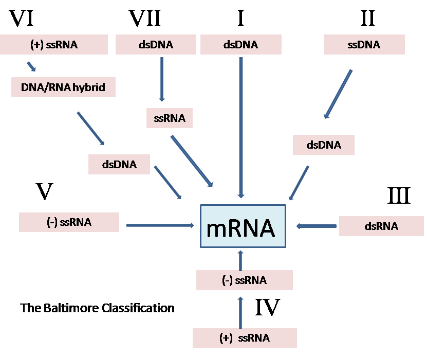
The Baltimore Classification System
Image Source: Wikimedia Commons (2009). Baltimore Classification, Licensed under a creative commons licence. Available for free at: https://commons.wikimedia.org/wiki/File:Baltimore_Classification.png (Accessed: 17 September 2022).
The 7 groups are depicted here, categorised by how they produce the mRNA used to replicate. ds = double stranded, ss = single stranded, (+) = positive sense, (-) = negative sense.
Immunology
The human body has a variety of methods to defend itself against viral infection. These include anatomical and chemical barriers, such as mucus and stomach acid, and chemical mediators of the immune system. The latter can be broken down into intrinsic methods (i.e. interferons, apoptosis, autophagy), the innate immune responses (i.e. complement, cytokines, natural killer cells) and the adaptive immune response (i.e. T cells and B cells). The subject is vast and beyond the remit of this section to fully address.
Interferons (IFNs) are arguably the most important of these defences to know about. IFNs are a crucial part of our intrinsic defence system – they are produced by virus-infected cells and uninfected sentinel cells (e.g. dendritic cells, macrophages and natural killer cells) in response to triggers from viruses.
IFN-α and -β are produced rapidly in the early stages of viral infection, and their binding to receptors triggers the synthesis of multiple cell proteins. Their expression has dramatic physiological consequences, resulting in symptoms such as fever, rigors, malise and nausea, which are well recognised as viral phenomena.
In the reference section, a course providing an overview of viral immunology can be found as further reading.
Clinical Virology
Pathophysiology
The full scope of virus pathophysiology is beyond the remit of this section. Key principles to remember include:
- Viruses rely on a host to replicate and propagate
- Many of the clinical manifestations are the result of the immune response triggered by viral replication
- Host cell lysis will result in cell death locally and also contributes to the clinical illness
Viruses often have a specific action on a particular body system, owing to their individual molecular characteristics – this is known as tropism. Some viruses can cause disease in multiple tissue types, e.g. cytomegalovirus (CMV) in the immunosuppressed individual, which can cause CMV colitis, encephalitis, retinitis and hepatitis.
Assessment and diagnosis
Diagnosis in modern medicine relies predominantly on nucleic acid-based methods and serological assays. However, microscopy-based methods such as immunofluorescence with electron microscopy are also still used.
Samples can be taken from a range of sites specific to the virus in question. For example, if viral encephalitis is suspected, a lumbar puncture is the best method to obtain a sample.
Polymerase chain reaction (PCR)
PCR is a method by which viral DNA can be replicated in order to be detected. Molecular probes known as ‘primers’ are designed that are specific to a certain virus genome. The polymerase enzyme, and heat, are then used to create a reaction in which if any viral DNA is present in the sample tested, it will replicate and be amplified to detectable levels, making the test ‘positive’.
Reverse transcription PCR (RT-PCR) is the same principle but for the detection and amplification of RNA.
Sequencing
Genetic sequencing uses the product amplified from PCR/RT-PCR and is analysed by a sequencing machine. It enables the full sequence of a viral genome to be analysed.
Immunofluorescence
This technique makes use of an antibody specific to the virus suspected in a sample of potentially infected tissue. A chemical, or specific wavelength of light, is applied to the tissue sample. This enables visualisation of the antibody, and hence the virus, if present.
Electron microscopy
Electron microscopy was the traditional mainstay of virology. However, modern techniques require less technical skill and are quicker, so they have overtaken it. The advantage is that through electron microscopy, you can visualise a whole virus, revealing its shape and structure (see image below), and it is useful if viruses do not show up on routine diagnostic tests or if suspicious results need interrogating further.

Electron micrograph of Ebola virus
Image Source: Wikimedia Commons (2011). Ebola virus virion, Licensed under a creative commons licence. Available for free at: https://commons.wikimedia.org/wiki/File:Ebola_virus_virion.jpg (Accessed: 19 September 2022).
When Ebola was first discovered in 1976, it was electron microscopy of the virion that showed it had a different structure to the yellow fever virus, which was initially thought (wrongly) to be the cause of the outbreak in the Democratic Republic of Congo.
Serology
Serological diagnosis is performed through the detection of antibodies and antigens from patient blood samples. IgM antibodies indicate an acute infection. IgG antibodies indicate prior exposure, and hence either vaccination or previous infection.
Antiviral drugs and vaccines
Antivirals
There are relatively few antivirals available in comparison to antibiotics. One of the most common antivirals prescribed in clinical medicine is aciclovir – which is effective against herpes simplex virus and varicella zoster virus (the cause of shingles). Aciclovir and the related agents famciclovir and valaciclovir work through being phosphorylated by viral tyrosine kinase, resulting in early chain termination.
Antiretrovirals
HIV antiretroviral medications are numerous. HIV treatment is based on the principle of utilising multiple methods of attack on the virus simultaneously, known as highly active antiretroviral therapy (HAART). These drugs have revolutionised HIV treatment, making it a manageable, chronic illness. More information can be found in the HIV and Infections in Immunocompromised Hosts topic.
Vaccines
Multiple viruses now have effective vaccines against them. These vaccines can be classified by type. Classical types include live attenuated, toxoid, conjugate, and inactivated vaccines. More recent times have seen the advent of mRNA vaccines, which use the host’s genome to build the proteins that then mount a humoral immune response. Many of the new vaccines against COVID-19 are mRNA vaccines.
Some common vaccines are summarised by type in the table below.
| Type | Examples |
|---|---|
| Live attenuated |
|
| Inactivated |
|
| Toxoid |
|
| Conjugated |
|
Table 1: Examples of viral vaccines by type
Live attenuated vaccines should be avoided in the immunosuppressed. A full UK vaccine schedule can be found on the NHS website and is linked in the References section here.
Influenza antivirals and vaccines
The 2009 swine flu pandemic, caused by the H1N1 influenza virus, led to a rise in the use of oseltamivir (Tamiflu) and, less so, zanamivir (Relenza). These drugs are neuraminidase inhibitors, which block the binding of the neuraminidase antigen of the influenza virus to sialic acid receptors in the lung.
However, there is little evidence that oseltamivir prevents the complications of influenza; although it can help people recover from the illness more quickly. The evidence base for zanamivir is also weak, but it is regularly used to treat influenza A and B infections.
Influenza vaccines are adjusted each year, depending on the seasonal variants that have become dominant worldwide, which is why viral surveillance is so important. Influenza A and influenza B are the most common strains of the virus, and the vaccine is based around these. Most influenza vaccines contain traces of eggs, which is important to know for allergy purposes.
Traveller and Migrant Health
Viral infections are often associated with travelling and migration. This is because these activities can spread disease between different populations, and people from different countries can bring infections not commonly seen in others into a population with a low level of immunity.
Multiple viral outbreaks have had international travel as a contributing factor, with COVID-19, Ebola, Zika and avian flu all recent examples.
HIV is a continued and critical global public health problem, with over 30 million people living with HIV worldwide, and up to half of those estimated to not be aware of their HIV status. Therefore, continual HIV surveillance and expansion of diagnostic and care facilities is critical – not just for HIV but also for diseases that tend to co-exist alongside it. In tropical areas, that often includes tuberculosis and HSV-2.
There will always be exposure to viruses during tropical travel. At the MRCP level, it is especially important to remember viral infections transmitted by mosquitoes – which are highlighted in the table below – as these are frequent question topics.
More information about other important viral infections can be found in the HIV and Infections in Immunocompromised Hosts topic, and other types of infections relevant to traveller and migrant health are also covered in the Parasitic Infections, Fungal Infections and Bacterial Infections topics.
| Virus | Mosquito | Geography | Clinical manifestation |
|---|---|---|---|
| Dengue | Aedes aegypti | Tropics | Fever, arthralgia, retro-orbital headache, maculopapular rash
Possible haemorrhagic transformation |
| Chikungunya | Aedes aegypti | Tropics | Abrupt high fever, debilitating joint aches, maculopapular rash
No haemorrhagic transformation |
| Zika | Aedes aegypti/Aedes albopictus | Tropics – especially Latin America and Polynesia | Mild fever, arthralgia, rash
Microcephaly if congenital |
| Yellow fever | Aedes aegypti | Predominantly Brazil and Subsaharan Africa | Fever, jaundice, progressing into liver failure and haemorrhagic shock |
Table 2: Common mosquito-borne viruses for MRCP. The ‘FAR’ Syndrome of fever, arthralgia and rash is a typical presentation
Complete Guide to Clinical Genetics ST3 Application and Recruitment (2027)
Written by Medibuddy. Last updated: October 2026. Clinical Genetics is

Complete Guide to Clinical Radiology ST3 Application and Recruitment (2027)
Recruitment paused: NHS England is not running Clinical Radiology ST3+ recru

Complete Guide to Paediatric Surgery ST3 Application and Recruitment in United Kingdom (2026)
Written by Medibuddy. Last updated: October 2026. If you’re planning


Take your subscriptions with you
Our mobile app allows you to access your interview and exam question banks wherever you are.

