


 Close
Close
 13th February 2025
Urgent causes of red eye
Orbital cellulitis
Infection of the subcutaneous tissues surrounding the eye may involve the orbit (orbital cellulitis), or tissues anterior to the orbital septum only (periorbital, or preseptal cellulitis). Orbital cellulitis often arises from local spread of paranasal sinus infection or spread of periorbital cellulitis. It is an emergency which can lead to permanent vision loss or even brain abscess and death and, therefore, requires an emergency ophthalmology and/or ENT (Ear, nose and throat) referral.
The eyelid will be inflamed (Figure 1). Ocular signs may include a swollen, red and proptosed eye, with painful and reduced eye movements. Optic nerve dysfunction may be present in severe cases, manifesting as reduced visual acuity, impaired colour vision or relative afferent pupillary defect. The patient may be systemically unwell and pyrexial.
High-resolution CT of the orbit, sinuses and brain is key for diagnosis and to rule out an underlying abscess. Treatment is with IV broad-spectrum antibiotics but sinus or abscess drainage by ENT may also be required.
In contrast to orbital cellulitis, periorbital cellulitis may present with a swollen and red eyelid, with or without pyrexia, but does not usually cause any proptosis, decrease in eye movements or worsening visual acuity. It can usually be treated with oral antibiotics, though due to high risk of orbital extension, especially in children, urgent ophthalmology review is necessary.
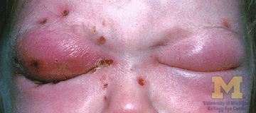
Image Source: Wikimedia Commons (2011). Orbital cellulitis, Licensed under a creative commons licence. Available for free at: https://commons.wikimedia.org/wiki/File:Orbital_cellulitis.jpg#/media/File:Orbital_cellulitis.jpg (Accessed: 18 January 2024).
Infectious keratitis
Infectious keratitis is an infection of the cornea; it is most commonly caused by bacteria but can also have a range of viral, fungal and parasitic aetiologies. All suspected corneal infections or ulcers should be referred to ophthalmology urgently due to risk of potentially sight-threatening complications.
Bacterial
Risk factors include anything that may disrupt the epithelium; the most notable in younger patients is contact lens wear, particularly overnight. Others include trauma, surgery or underlying corneal pathologies such as severe dry eye or bullous keratopathy. Causative organisms are most commonly Staphylococcus aureus, Streptococci species, and Pseudomonas aeruginosa. It should be noted that some organisms are able to penetrate intact corneal epithelium, including Shigella, Neisseria, Listeria and corynebacterium species.
The patient often complains of a foreign body sensation. They may have photophobia and be unable to keep the eye open. On fluorescein staining, an epithelial defect may be visible, with conjunctival injection surrounding the cornea. A corneal opacity may also be seen. Mucopurulent discharge may be present, and in severe cases a hypopyon (pus in the anterior chamber). Treatment is usually with topical antibiotics. Corneal scraping and conjunctival swabs may be carried out for culture and sensitivity to guide further choice of antibiotic.
Herpes zoster ophthalmicus (ophthalmic shingles)
Varicella zoster virus that becomes dormant in the dorsal root ganglia after chickenpox may reactivate in later years to cause herpes zoster (shingles). This can affect the ophthalmic division of the trigeminal nerve (CN V1), causing herpes zoster ophthalmicus (HZO). It is most common in the 6th and 7th decades of life, with more severe disease seen in immunosuppressed patients. There is usually a prodrome lasting around 3-5 days, during which the patient is systemically unwell, with symptoms such as fever, malaise and a headache. This is followed by tingling, burning, or pain which may be mild or severe, and a vesicular rash, both respecting the CN V1 dermatomal distribution (anterior forehead and nose), not crossing the midline (Figure 2).
Ocular manifestations can include conjunctivitis, keratitis, uveitis or episcleritis. Dendritic lesions may be seen on fluorescein staining, which have tapered ends (pseudodendrites) in comparison to the terminal bulbs seen on dendrites in herpes simplex infection. A good predictor of eye disease with shingles is the presence of skin lesions at the side or tip of the nose, indicating involvement of the nasociliary nerve; this is called Hutchinson’s sign.
Oral antivirals are indicated within 72 hours of the rash appearing and reduce the duration of symptoms, severity and risk of chronic pain after rash resolution (post-herpetic neuralgia). Intravenous acyclovir may be required in severe cases or in immunocompromised patients. Antibiotics may also be given to prevent secondary bacterial infection of the eye.
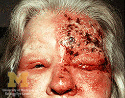
Image Source: Wikimedia Commons (2011). Trigeminal herpes with uveitis and keratitis, Licensed under a creative commons licence. Available for free at: https://commons.wikimedia.org/wiki/File:Trigeminal_herpes_with_uveitis_and_keratitis.jpg#/media/File:Trigeminal_herpes_with_uveitis_and_keratitis.jpg (Accessed: 18 January 2024).
Herpes simplex keratitis
This is most commonly secondary to infection with herpes simplex virus 1 (HSV1). The patient may have a foreign body sensation, photophobia, blurred vision and watering. Examination with fluorescein shows a dendritic ulcer, which is a linear, branching lesion with terminal bulbs, usually in the centre of the cornea (Figure 3). Contrast this to the tapered ends of the pseudodendrites seen in HZO. HSV keratitis is usually treatable with topical antivirals.
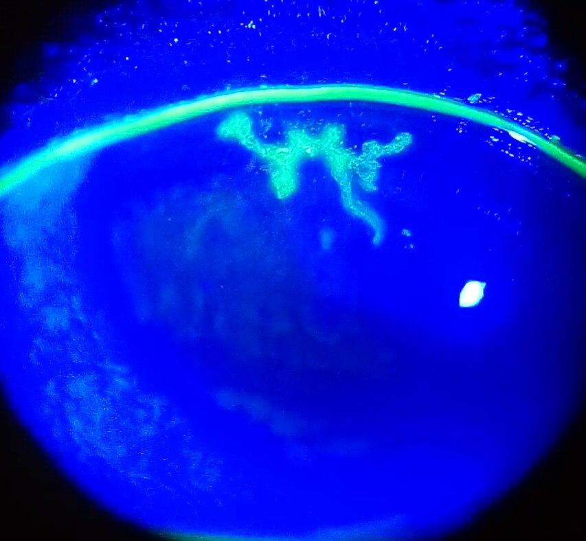
Image Source: Wikimedia Commons (2017). Dendritic corneal ulcer, Licensed under a creative commons licence. Available for free at: https://commons.wikimedia.org/w/index.php?curid=58964337#/media/File:Dendritic_corneal_ulcer.jpg (Accessed: 18 January 2024).
Acanthamoeba keratitis
Vigilance for infection with the rare but potentially sight-threatening Acanthamoeba spp should be maintained, especially in patients who have had limited response to antibiotics. It is a protozoan usually found in soil or fresh water and is associated with poor contact lens hygiene such as swimming or showering in contact lenses. Patients often present with unilateral eye pain that is characteristically out of proportion to clinical findings. Grey-coloured irregular epithelial corneal infiltrates may be seen and in later stages, a grey ring-shaped stromal infiltrate. Infiltrates along corneal nerves (radial keratoneuritis) are pathognomonic.
Scleritis
The sclera is the outer, fibrous layer making up the ‘white’ of the eyes and consists of a thin outermost episclera, containing a rich blood supply, a more dense stromal layer, and a layer continuous with the uveal tract. Scleritis is the inflammation of the stromal layer and can rapidly lead to deterioration and permanent loss of vision due to tissue destruction. Importantly, scleritis is most commonly immune mediated and can represent a host of underlying inflammatory conditions; rheumatoid arthritis is the most common association. Scleritis may also have infective causes, such as herpes zoster, syphilis or tuberculosis. Urgent ophthalmology review is imperative in these cases, in addition to diagnosis and treatment of underlying disease.
The patient may complain of a gradually worsening constant eye pain which becomes severe and keeps them up at night. The pain is often poorly responsive to analgesia and may radiate to the jaw or temple. Photophobia may be present.
In diffuse anterior scleritis, there is a red sclera due to dilatation of the deep vessels, with scleral oedema. Scleritis may also be nodular in pattern (tender single or multiple nodules). Necrotising disease, which can involve an extremely tender sclera and can present with patches of non-perfused overlying episclera, occurs in vaso-occlusive disease. There may also be a blue tinge to the sclera.
Due to disease affecting the deeper scleral vasculature, instillation of 2.5-10% phenylephrine drops will not blanch the redness. This is in contrast to the less concerning diagnosis of episcleritis.
Immune-mediated non-necrotising scleritis is treated with NSAIDs, although often steroids or other immunosuppression are required where this is inadequate (or in necrotising disease).
Complete Guide to Clinical Genetics ST3 Application and Recruitment (2027)
Written by Medibuddy. Last updated: October 2026. Clinical Genetics is

Complete Guide to Clinical Radiology ST3 Application and Recruitment (2027)
Recruitment paused: NHS England is not running Clinical Radiology ST3+ recru

Complete Guide to Paediatric Surgery ST3 Application and Recruitment in United Kingdom (2026)
Written by Medibuddy. Last updated: October 2026. If you’re planning


Take your subscriptions with you
Our mobile app allows you to access your interview and exam question banks wherever you are.

