


 Close
Close
 29th May 2026
Acute Coronary Syndrome (ACS)
Unstable Angina
Background
Unstable angina (UA) is myocardial ischemia occurring at rest or minimal exertion without acute cardiomyocyte injury or necrosis. Key features include:
- Prolonged (>20 minutes) angina at rest.
- New onset of severe angina.
- Increasing frequency, duration, or intensity of angina.
- Angina after a recent myocardial infarction.
Epidemiology
UA is a form of acute coronary syndrome (ACS) with rising prevalence, particularly among individuals with risk factors such as diabetes, hypertension, and smoking. It often indicates a higher risk of progression to myocardial infarction or other severe cardiac events.
Aetiology & Pathophysiology
UA is caused by a non-occlusive thrombus on a ruptured atherosclerotic plaque, leading to partial obstruction of the coronary artery and myocardial ischemia without infarction. The severity of symptoms is determined by the degree of ischemia, collateral circulation, and comorbidities. Key features include plaque rupture at thin-cap regions and thrombus formation that is typically non-occlusive.
Diagnosis
Investigations
- ECG: A 12-lead ECG should be performed within 10 minutes to identify changes such as ST depression, transient ST elevation, or T-wave inversion.
- Troponin: Troponin levels help differentiate UA from NSTEMI. Dynamic or persistently normal troponin levels can rule out myocardial infarction. If there is strong suspicion of unstable angina with negative ECG and troponin, serial measurements are essential to identify progression to NSTEMI and to help with risk stratification.
- Chest X-ray: To rule out alternative diagnoses like pneumothorax or aortic dissection.
- Blood Tests: FBC, U&E, LFTs, glucose, and CRP to assess the patient’s overall health and detect complications.
- Echocardiography: Used to evaluate left ventricular (LV) function and identify regional wall motion abnormalities suggestive of ongoing ischemia.
- Risk Pathways: Troponin levels, clinical features, and ECG findings guide decisions regarding further testing and intervention, including coronary angiography.
Clinical Manifestations
Chest pain in UA is typically retrosternal, lasting for more than 20 minutes, and often occurs at rest or with minimal exertion. Symptoms are frequently accompanied by nausea, sweating, breathlessness, or dizziness.
- Red Flags: Hemodynamic instability, acute LV failure, or arrhythmias.
Management
Initial Treatment
- Aspirin: Administer a loading dose unless contraindicated.
- Anticoagulation: Fondaparinux or unfractionated heparin, especially in patients with renal impairment.
- Pain Relief: Glyceryl trinitrate (GTN) and morphine as needed.
- Monitoring: Continuous monitoring for symptom recurrence, dynamic ECG changes, or rising troponin levels.
- Antiplatelet Therapy: Dual antiplatelet therapy (DAPT) with aspirin and a P2Y12 inhibitor is recommended for high-risk patients. If severe thrombocytopaenia develops, alternative anticoagulation should be considered. In patients at high risk of thrombotic complications, a direct thrombin inhibitor such as bivalirudin should be considered.
- Invasive Strategy: Coronary angiography is indicated within 24-72 hours for high-risk patients based on clinical features and risk stratification.
Long-term Treatment
- Continue aspirin and P2Y12 inhibitors for up to 12 months.
- Consider ACE inhibitors/ARBs, beta-blockers, statins, and aldosterone antagonists for patients with heart failure or diabetes.
- SGLT2 inhibitors (e.g., dapagliflozin) may be used for patients with heart failure, regardless of ejection fraction.
- Stress-testing a few weeks after the event may guide the need for further invasive treatment.
Prevention & Follow-Up
- Primary Prevention: Smoking cessation, a healthy diet, weight management, and regular physical activity are crucial. Statins should be prescribed for high cardiovascular risk patients.
- Secondary Prevention: Cardiac rehabilitation and adherence to long-term medications, including antiplatelet therapy, statins, and ACE inhibitors.
- Follow-Up: Regular outpatient follow-up within 1-2 weeks post-discharge, with lipid monitoring every 6 months. Medication reviews should be conducted at each visit.
Complications
- Short-term: Cardiac arrhythmias, acute heart failure, cardiogenic shock, and post-ischemic pericarditis.
- Variable: Depression and in-stent thrombosis.
- Bleeding: Major bleeding events occur in 3.9% of ACS patients, especially those receiving anticoagulation therapy. Minor bleeding can be managed conservatively without halting anticoagulants or antiplatelet therapy. Major bleeding may require discontinuation of DAPT, as well as measures such as platelet transfusion and resuscitation.
Prognosis
Mortality
- Overall mortality in NSTE-ACS patients is 4.8% over 6 months.
- Mortality rates exceed those of STEMI after 6 months.
- At 12 months, adverse cardiovascular events (recurrent ischemia, MI, and death) occur in up to 10% of patients, likely reflecting widespread atherosclerotic disease.
Prognostic Factors
- Adverse markers include rest pain, comorbidities (e.g., diabetes, hypertension, chronic kidney disease), left ventricular failure, ST depression, raised biomarkers, and multi-vessel or left main disease.
- Short-term risk factors for death or non-fatal MI include:
- Accelerated ischemic symptoms, prolonged rest pain, pulmonary oedema, new mitral regurgitation, age >75, and significant ECG changes (ST-T changes, new bundle branch block, sustained VT).
Risk Assessment
- Risk models (e.g., GRACE Score) predict outcomes for up to 6 months and maintain predictive value for 1-year outcomes such as death or MI.
- The GRACE score takes into account factors such as patient age, cardiac history, ECG findings, signs and symptoms, and gives a score to estimate 6-month mortality.
Non-ST Elevation Myocardial Infarction (NSTEMI)
Background
Non-ST elevation myocardial infarction (NSTEMI) is an acute ischemic event causing myocyte necrosis, detected by troponin elevation. It can be distinguished from unstable angina by a dynamic elevation of troponin above the 99th percentile. The ECG may show ischemic changes (e.g., ST depression, T-wave changes), but it can also be normal or show non-specific changes.
Key Difference from Unstable Angina: NSTEMI involves troponin elevation, while unstable angina does not.
Epidemiology
NSTEMI incidence is rising due to improved diagnostics, early pharmacotherapy, and better STEMI prevention.
Aetiology & Pathophysiology
NSTEMI results from oxygen supply-demand imbalance, often due to non-occlusive thrombus from plaque rupture, vasospasm, or other factors. Severity depends on ischemia duration, collateral flow, and comorbidities. Key features include plaque rupture at thin cap regions, with occlusions often arising from lesions <50% stenotic.
Diagnosis
Investigations
- ECG: 12-lead within 10 minutes; may show ST depression, T-wave inversion, or transient ST elevation.
- Troponin: Dynamic elevation confirms NSTEMI.
- Diagnosis requires rise/fall with ≥99th percentile value.
- Use rapid rule-in/rule-out algorithms (ESC 0/1h or 0/3h) where validated.
- Large elevations (>5× URL) indicate acute Type-1 MI and higher risk.
- Other tests: Chest X-ray, blood tests (FBC, U&E, LFTs, glucose, CRP), and echocardiography if heart failure or instability is suspected.
- Risk Pathways: Use troponin levels and repeat testing to guide management.
Clinical Manifestations
Chest pain >15 minutes, new/worsened, accompanied by nausea, sweating, breathlessness, or hemodynamic instability. Risk factors include diabetes, renal dysfunction, hypertension, smoking, a family history of coronary artery disease, a sedentary lifestyle, and obesity. Red flags include hemodynamic instability, acute LV failure, or arrhythmias.
Management
- Initial Treatment: Aspirin, sublingual GTN, morphine, oxygen (if SpO₂ <90%).
- Anticoagulation: Fondaparinux or heparin.
- Urgent Coronary Angiography (ICA): For unstable patients or those with high-risk features (ongoing pain, instability, arrhythmias, high-risk comorbidities, or high-risk stratification scores).
- Long-term Treatment: Dual antiplatelet therapy (for at least 12 months), beta-blockers, ACE inhibitors/ARBs, statins, aldosterone antagonists (for HF), and SGLT2 inhibitors (for HF).
Prevention & Follow-Up
- Primary Prevention: Smoking cessation, healthy diet, regular physical activity, and statin therapy for high-risk individuals.
- Secondary Prevention: Lifestyle changes, cardiac rehab, and long-term medication adherence.
- Obesity is a significant modifiable risk factor, and weight management and lifestyle modification are key to reducing the risk of future cardiovascular events.
- Smoking is also known to be a well-established risk factor linked to recurrent cardiovascular events, and smoking cessation should be encouraged in all patients.
- Follow-up: Outpatient visit 1-2 weeks post-discharge, lipid monitoring every 6 months, and regular medication reviews.
Complications
- Short-term: Cardiac arrhythmias, acute heart failure, cardiogenic shock, acute mitral regurgitation, post-MI pericarditis, venous thromboembolism (VTE).
- Variable: Depression, in-stent thrombosis.
Prognosis
NSTEMI patients face high morbidity and mortality risks, with a 4-6 times higher risk of sudden death than the general population. Key risk factors include diabetes, smoking, heart failure, and infarct extent. Modern treatments (statins, revascularisation) reduce mortality, but age and other factors temper the benefits. Cardiovascular death risk post-MI is about 10% in the first year, with lifestyle changes and cardiac rehab improving survival.
ST-Elevation Myocardial Infarction (STEMI)
Background
ST-elevation myocardial infarction (STEMI) is a type of acute myocardial infarction caused by complete atherothrombotic occlusion of a coronary artery, leading to myocardial cell death due to oxygen supply-demand mismatch.
Diagnostic Criteria
- Persistent ST-segment elevation in at least two contiguous ECG leads:
- ≥2.5 mm (men <40 years)
- ≥2 mm (men >40 years)
- ≥1.5 mm (women, all ages)
Epidemiology
STEMI incidence has declined over the past two decades, but saw a 16% increase in 2021-22 due to post-COVID-19 effects. In the UK, STEMI accounts for ~3% prevalence and 1.2 million hospitalisations annually.
Risk Factors
- Lifestyle: Smoking, obesity, physical inactivity, poor diet, cocaine use.
- Chronic conditions: Hypertension, diabetes, dyslipidemia, atherosclerosis.
- Demographics: Age >60, family history, prior stent thrombosis, sleep apnea, depression.
Aetiology & Pathophysiology
- Coronary Artery Disease (CAD): Plaque rupture or fissuring leads to thrombus formation and complete occlusion.
- Plaque Formation: LDL accumulation, foam cell formation, smooth muscle migration, and potential rupture.
- Myocardial Ischaemia: Occlusion halts contractile function, leading to necrosis and infarct zone expansion.
Fourth Universal Definition of MI
- Type 1: Atherothrombotic MI (e.g., STEMI, NSTEMI).
- Type 2: Oxygen supply-demand imbalance.
- Type 3: Sudden cardiac death with symptoms before biomarker confirmation.
Diagnosis
Investigations
- Immediate Actions: ECG within 10 minutes, assess hemodynamic status, and consider reperfusion therapy (primary PCI or fibrinolysis).
- Symptoms: Central chest pain, radiating to the arm, neck, or jaw, with nausea or syncope.
- Cardiac Biomarkers: Elevated troponins confirm diagnosis but should not delay reperfusion therapy.
- ECG Findings: ST-elevation criteria based on gender and age, and consider posterior STEMI or left main coronary artery obstruction.
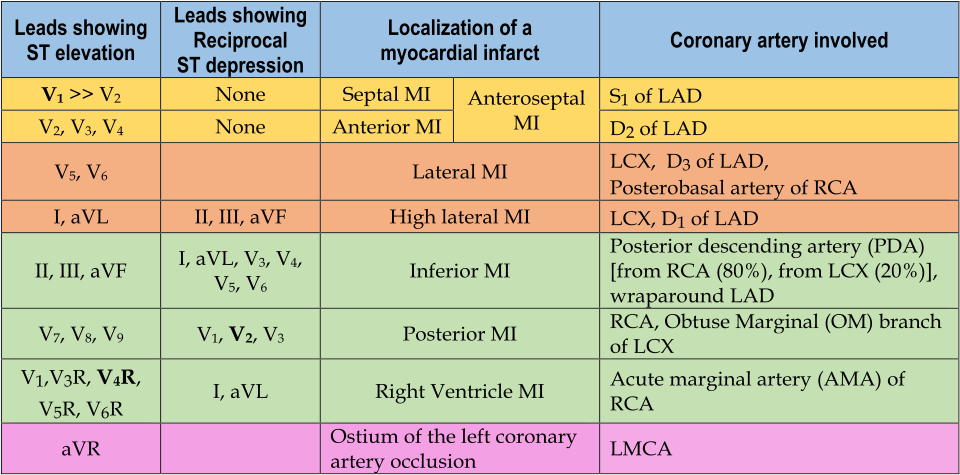
Localisation of ST-elevation MI, from: Image Source: Wikimedia Commons (2020), ECG AVNRT, Licenced under a creative commons licence. Available for free at: https://commons.wikimedia.org/wiki/File:Localisation_of_the_occlusion_in_STEMI.svg (Accessed: 25 April 2026).
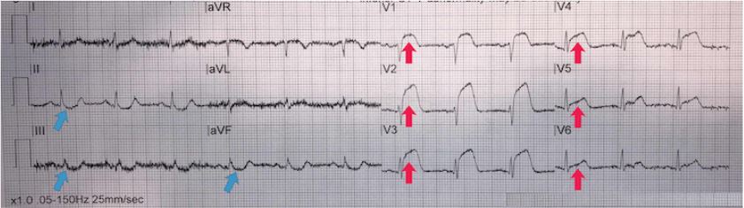
Anterior STEMI. From the personal collection of Dr Aung Myat (used with permission)
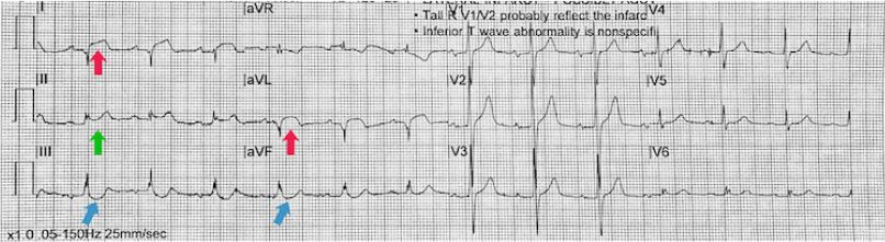
High lateral STEMI. From the personal collection of Dr Aung Myat (used with permission)
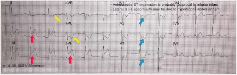
Inferoposterior STEMI example II. From the personal collection of Dr Aung Myat (used with permission)
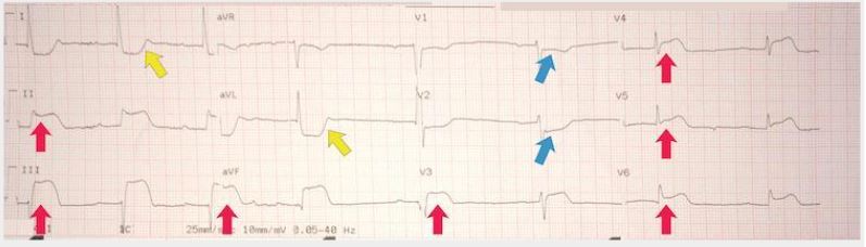
Inferoposterolateral STEMI example III. From the personal collection of Dr Aung Myat (used with permission)
Management
Emerging Treatments:
- Factor Xa Inhibitors: Rivaroxaban reduces cardiovascular events but increases bleeding risk.
- Colchicine: Low-dose colchicine for long-term secondary prevention in ACS patients.
Clinical Diagnosis and Immediate Action
- Diagnose STEMI based on signs of myocardial ischaemia + persistent ST elevation in two or more contiguous leads on ECG.
- Do not wait for cardiac troponin results before initiating treatment.
- Administer a single loading dose of aspirin (unless hypersensitive).
- Begin reperfusion therapy (primary PCI or fibrinolysis) promptly.
- Primary PCI is preferred if feasible within 120 minutes of symptom onset.
- Fibrinolysis if PCI is not timely, using alteplase or tenecteplase.
- Begin dual antiplatelet therapy (aspirin + P2Y12 inhibitor).
- Provide pain relief (opioids are preferred agents) and antiemetics.
- Use oxygen only if oxygen saturation is <90%.
- Consider nitrates for persistent pain or hypertension, but avoid if hypotension is present.
Coronary Reperfusion Therapy
- Primary PCI is the preferred strategy if symptom onset is within 12 hours and PCI can be performed within 120 minutes.
- If PCI cannot be delivered within 120 minutes, fibrinolysis is recommended.
- If fibrinolysis is given, assess success via ECG after 60-90 minutes.
- If fibrinolysis fails, perform immediate coronary angiography and PCI.
- In elderly patients, although bleeding risk is higher, if candidates for fibrinolysis, the benefits of early reperfusion generally outweigh the risks
Contraindications to fibrinolysis
- Absolute:
- Prior intracranial haemorrhage/stroke
- Recent ischemic stroke (<6 months)
- CNS damage/neoplasm/AV malformation
- Recent major trauma/surgery/head injury (<1 month)
- GI bleeding (<1 month)
- Known bleeding disorders
- Aortic dissection
- Non-compressible punctures (<24h)
- Relative
- TIA (<6 months)
- Oral anticoagulant use
- Pregnancy or <1 week postpartum
- Refractory hypertension (SBP >180 mmHg, DBP >110 mmHg)
- Advanced liver disease
- Infective endocarditis
- Active peptic ulcer
- Prolonged or traumatic resuscitation
Anticoagulation and Antithrombotic Therapy
- Avoid anticoagulants if the patient is eligible for primary PCI (PCI team will start anticoagulation).
- Following PCI, DAPT should be commenced in all patients (aspirin and P2Y12 inhibitor combination, e.g. clopidogrel, ticagrelor), and typically continued for at least 12 months.
- If fibrinolysis is used, start anticoagulation (enoxaparin or unfractionated heparin).
Special Considerations
- Out-of-hospital cardiac arrest: If there is ST elevation on post-ROSC ECG, proceed with PCI.
- Cardiogenic shock: Immediate senior support and PCI are recommended.
- Late presentation (>12 hours after symptom onset): Consider coronary angiography or PCI if there is ongoing ischaemia, haemodynamic instability, or arrhythmias.
Timing Targets
- Aim for rapid delivery of reperfusion therapy (PCI or fibrinolysis).
- Optimal outcomes are achieved when reperfusion is done as early as possible.
| Interval | Target |
| First medical contact to ECG and STEMI diagnosis | <10 minutes |
| STEMI diagnosis to primary PCI | <120 minutes
If this time target cannot be met, consider fibrinolysis |
| STEMI diagnosis to primary PCI if the patient’s first medical contact is at a hospital | <60 minutes if the patient presents to or is in a PCI-capable hospital
<90 minutes if the patient presents to or is in a non-PCI-capable hospital and needs transferring |
| STEMI diagnosis to administration of a bolus/infusion of a fibrinolytic drug (if primary PCI cannot be accessed within 120 minutes) | <10 minutes |
| Start of fibrinolysis to ECG assessment of its success or failure | 60-90 minutes |
| If fibrinolysis is successful, time interval from starting fibrinolysis to early coronary angiography | 2-24 hours |
Long-Term Management of STEMI
Medications
- Aspirin: Continue lifelong, with a P2Y12 inhibitor for up to 12 months as part of dual antiplatelet therapy.
- ACE inhibitor: For patients with reduced ejection fraction or heart failure.
- Beta-blocker: To reduce the risk of reinfarction and improve survival.
- Statin: To manage cholesterol and reduce cardiovascular risk.
Cardiac Rehabilitation and Risk Factor Modification
- Enrol in cardiac rehabilitation to improve recovery and reduce the risk of future events.
- Address modifiable risk factors for cardiovascular disease, including:
- Smoking cessation
- Blood pressure control
- Diabetes management
- Dietary changes and exercise
- Weight management
These measures are crucial for improving long-term outcomes, preventing reinfarction, and reducing the risk of complications.
Prevention and Follow-up
- Primary Prevention: Encourage lifestyle changes and medications (statins) for high-risk individuals.
- Secondary Prevention: Lifestyle modifications, cardiac rehabilitation, medications (dual antiplatelet therapy, beta-blockers, statins, ACE inhibitors).
- Follow-up: Survivors should be evaluated within 2-3 weeks post-discharge. Continue optimal medical therapy and assess ejection fraction.
Complications
- Short-term: Sinus bradycardia, heart block, recurrent chest pain, acute mitral regurgitation, ventricular septal defects, and pericarditis.
- Long-term: Congestive heart failure, recurrent ischemia, depression, ventricular arrhythmias.
Prognosis
The prognosis is better with early reperfusion, adherence to therapy, and risk factor modification. In-hospital mortality is around 9%, and elevated troponin levels indicate a worse prognosis. Major bleeding events increase 1-year mortality risk. Evidence-based treatments improve patient outcomes.
References
- Alpert JS, Thygesen K, Antman E, et al. Myocardial infarction redefined – a consensus document of the Joint European Society of Cardiology/American College of Cardiology Committee for the Redefinition of Myocardial Infarction. J Am Coll Cardiol. 2000 Sep;36(3):959-69.Full text Abstract LibKey Get Full Text
- Alpert JS, Thygesen KA, White HD, et al. Diagnostic and therapeutic implications of type 2 myocardial infarction: review and commentary. Am J Med. 2014 Feb;127(2):105-8.Full text Abstract LibKey Get Full Text
- Amsterdam EA, Wenger NK, Brindis RG, et al. 2014 AHA/ACC guideline for the management of patients with non-ST-elevation acute coronary syndromes: a report of the American College of Cardiology/American Heart Association task force on practice guidelines. J Am Coll Cardiol. 2014 Dec 23;64(24):e139-228.Full text Abstract LibKey Get Full Text
- Antman E, Bassand JP, Klein W, et al. Myocardial infarction redefined – a consensus document of The Joint European Society of Cardiology/American College of Cardiology Committee for the redefinition of myocardial infarction. J Am Coll Cardiol. 2000 Sep;36(3):959-69.Full text Abstract LibKey Get Full Text
- Byrne RA, Rossello X, Coughlan JJ, et al. 2023 ESC guidelines for the management of acute coronary syndromes. Eur Heart J. 2023 Oct 12;44(38):3720-826.Full text Abstract LibKey Get Full Text
- Cenko E, Yoon J, Kedev S, et al. Sex differences in outcomes after STEMI: effect modification by treatment strategy and age. JAMA Intern Med. 2018 May 1;178(5):632-9. Abstract LibKey Get Full Text
- Cheema KM, Dicks E, Pearson J, et al. Long-term trends in the epidemiology of cardiovascular diseases in the UK: insights from the British Heart Foundation statistical compendium. Cardiovasc Res. 2022 Jul 27;118(10):2267-80.Full text Abstract LibKey Get Full Text
- Faxon DP, Fuster V, Libby P, et al. Atherosclerotic vascular disease conference: writing group III: pathophysiology. Circulation. 2004 Jun 1;109(21):2617-25.Full text Abstract LibKey Get Full Text
- Hansson GK, Libby P, Tabas I. Inflammation and plaque vulnerability. J Intern Med. 2015 Nov;278(5):483-93.Full text Abstract LibKey Get Full Text
- Libby P. Molecular bases of the acute coronary syndromes. Circulation. 1995 Jun 1;91(11):2844-50.Full text Abstract LibKey Get Full Text
- Moreno PR, Falk E, Palacios IF, et al. Macrophage infiltration in acute coronary syndromes: implications for plaque rupture. Circulation. 1994 Aug;90(2):775-8. Abstract LibKey Get Full Text
- Mozaffarian D, Benjamin EJ, Go AS, et al. Heart disease and stroke statistics – 2016 update: a report from the American Heart Association. Circulation. 2016 Jan 26;133(4):e38-360.Full text Abstract LibKey Get Full Text
- National Institute for Health and Care Excellence. Acute coronary syndromes. November 2020 [internet publication].Full text
- National Institute for Health and Care Excellence. Recent-onset chest pain of suspected cardiac origin: assessment and diagnosis. November 2016 [internet publication].Full text
- National Institute for Health and Care Excellence. Stable angina: management. August 2016 [internet publication].Full text
- Neumann JT, Goßling A, Sörensen NA, et al. Temporal trends in incidence and outcome of acute coronary syndrome. Clin Res Cardiol. 2020 Sep;109(9):1186-92. Abstract LibKey Get Full Text
- Thygesen K, Alpert JS, Jaffe AS, et al. Fourth universal definition of myocardial infarction (2018). Glob Heart. 2018 Dec;13(4):305-38.Full text Abstract LibKey Get Full Text
- Thygesen K, Alpert JS, Jaffe AS, et al; Executive Group on behalf of the Joint European Society of Cardiology (ESC)/American College of Cardiology (ACC)/American Heart Association (AHA)/World Heart Federation (WHF) Task Force for the Universal Definition of Myocardial Infarction. Fourth universal definition of myocardial infarction (2018). J Am Coll Cardiol. 2018 Oct 30;72(18):2231-64.Full text Abstract LibKey Get Full Text
- Timmis A, Vardas P, Townsend N, et al. European Society of Cardiology: cardiovascular disease statistics 2021. Eur Heart J. 2022 Feb 22;43(8):716-99.Full text Abstract LibKey Get Full Text
- Tsao CW, Aday AW, Almarzooq ZI, et al. Heart disease and stroke statistics – 2023 update: a report from the American Heart Association. Circulation. 2023 Feb 21;147(8):e93-621.Full text Abstract LibKey Get Full Text
- World Health Organization. Cardiovascular diseases (CVDs). June 2021 [internet publication].Full text
- World Health Organization. Cardiovascular diseases (CVDs). June 2021 [internet publication].
- Full textWorld Health Organization. Global status report on noncommunicable diseases, 2014. 2014 [internet publication].Full text
Complete Guide to Clinical Genetics ST3 Application and Recruitment (2027)
Written by Medibuddy. Last updated: October 2026. Clinical Genetics is

Complete Guide to Clinical Radiology ST3 Application and Recruitment (2027)
Recruitment paused: NHS England is not running Clinical Radiology ST3+ recru

Complete Guide to Paediatric Surgery ST3 Application and Recruitment in United Kingdom (2026)
Written by Medibuddy. Last updated: October 2026. If you’re planning


Take your subscriptions with you
Our mobile app allows you to access your interview and exam question banks wherever you are.

