


 Close
Close
 27th May 2025
Adrenal Disorders
Primary adrenal insufficiency (Addison’s disease)
Background
In primary adrenal insufficiency (PAI) there is ablation of the adrenal cortex resulting in a deficiency of all steroid hormones. The hypothalamic-pituitary axis (HPA) aims to correct low cortisol levels through increased adrenocorticotropic hormone (ACTH) production, where α-melanocyte-stimulating-hormone is produced as a by-product. Therefore insidious PAI commonly presents with mucocutaneous hyperpigmentation, as well as fatigue, weakness, hypotension, anorexia, nausea and vomiting. It can also present acutely with hypovolaemic shock, acute abdominal pain, fever and vomiting.
Epidemiology
The prevalence of primary adrenal insufficiency is low at 40 cases per million in the UK. In developed countries, autoimmunity is the most common cause of primary adrenal insufficiency (up to 96% of cases), affecting women to a greater degree than men. There is also an association with other autoimmune endocrine disorders including Graves’ disease, type 1 diabetes mellitus and Hashimoto’s thyroiditis.
Aetiology
Anatomic destruction of the adrenal glands can result in acute or chronic PAI:
- Autoimmunity: antibodies formed against 21-hydroxylase.
- Infection: tuberculosis, P. aeruginosa and meningococcal septicaemia, histoplasmosis, HIV, cryptococcosis.
- Surgery: bilateral adrenalectomy e.g. for phaeochromocytoma.
- Haemorrhagic infarction: antiphospholipid syndrome, anticoagulation, Waterhouse-Friderichson syndrome.
- Trauma.
- Infiltrative disease: e.g. amyloidosis, haemochromatosis, sarcoidosis.
- Metastatic: e.g. lung, breast, kidney cancers.
Metabolic failure tends to be more insidious in onset:
- Genetic conditions: e.g. congenital adrenal hyperplasia (described below), adrenoleukodystrophy
- Drug-induced: inhibitors of cortisol production e.g. ketoconazole or etomidate, or inducers of cortisol metabolism e.g. phenytoin, rifampicin.
- ACTH or glucocorticoid resistance.
- Cytotoxic agents.
Diagnosis
Symptoms of PAI include:
- Fatigue
- Weakness
- Myalgia and arthralgia
- Weight loss
- Nausea, vomiting, abdominal pain
Signs of PAI include:
- Mucocutaneous hyperpigmentation – commonly on areas of friction e.g. palms, knuckles.
- Hypotension and postural hypotension.
- Less commonly vitiligo, premature greying, goitre (with Hashimoto’s thyroiditis).
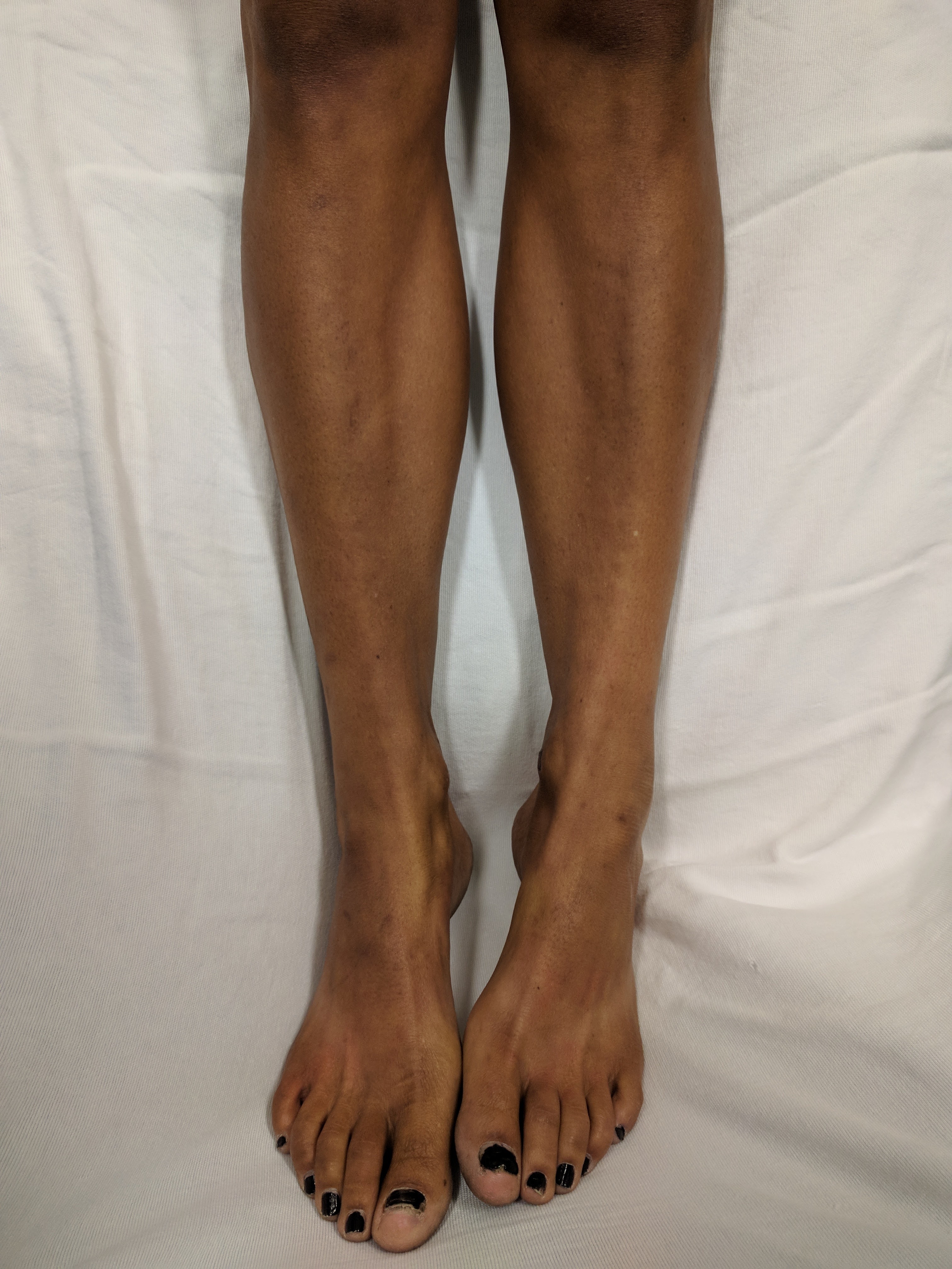
Image Source: Wikimedia Commons (2018) Addison’s Leg’s, Licensed under a creative commons licence. Available for free at: https://upload.wikimedia.org.wikipedia/commons/6/69/Addison%Legs.jpg (Accessed 13 May 2025).
The following laboratory findings are typical in PAI:
| Biochemical test | Findings in PAI |
|---|---|
| Sodium | ↓ in 90% of cases |
| Potassium | ↑ in 50% of cases |
| Calcium | ↑ in 10-20% of cases |
| FBC | Anaemia, mild eosinophilia and lymphocytosis |
| Glucose | ↓ often in children |
| LFTs | ↑ transaminases |
| Morning cortisol | ↓ levels <100 nmol/L warrant urgent admission, 100-500 nmol/L warrants endocrinology referral |
| ACTH | ↑ in primary adrenal insufficiency but ↓ in secondary |
| RAAS | ↑ renin and ↓ aldosterone in PAI |
Table 1: laboratory findings in primary adrenal insufficiency.
Ancillary investigations include an ACTH stimulation (synacthen) test where synthetic ACTH is administered and should result in a rise in cortisol for a physiologic response. However, in primary adrenal insufficiency this does not occur i.e. the cortisol will remain low when re-checked following administration of synacthen, as the adrenal glands do not respond to stimulation.
To investigate the underlying aetiology, the following investigations should be considered:
- Adrenal autoantibody testing.
- Chest x-ray to investigate lung neoplasm or tuberculosis.
- Tuberculosis skin prick or ELISA testing.
- Abdominal x-ray which may show adrenal calcification.
- CT or MRI of adrenal glands.
- MRI of hypothalamus and pituitary if suspicion of a central cause.
Management
In acute adrenal crisis, the mainstay of treatment includes:
- IV hydrocortisone, typically given as 100 mg STAT followed by an infusion of 200 mg/24 hours. N.B. if co-existing hypothyroidism, thyroid hormones must not be replaced prior to glucocorticoids, as this can precipitate adrenal crisis.
- Fluid resuscitation with monitoring of blood pressure, fluid status and electrolytes.
- Correction of hypoglycaemia.
- Treatment of the underlying cause once stable.
- Education regarding symptoms and risk factors for adrenal crisis, issue of a steroid card or medical alert bracelet.
In intercurrent illness, those with PAI will require corticosteroid supplementation. In circumstances of severe stress or high risk of crisis (e.g. acute gastroenteritis, surgery, critical illness), IV hydrocortisone as described above is recommended. However, for minor stressors (e.g. febrile illness, minor procedures requiring fasting), doubling of the patient’s maintenance dose of corticosteroid is recommended.
For the management of PAI in stable patients, oral glucocorticoid (e.g. hydrocortisone or prednisolone) and mineralocorticoid (e.g. fludrocortisone) replacement therapy is given. This is titrated according to clinical symptoms until a maintenance dose is found. Mineralocorticoid dosing will be adjusted based on symptoms such as postural hypotension and salt-craving.
Complications
Long-term PAI can result in acute adrenal crisis with hypovolaemic shock, hypoglycaemia, and electrolyte abnormalities as described above. However it is preventable with good education and dose adjustment during intercurrent illness.
Other complications are related to excessive glucocorticoid replacement, resulting in secondary Cushing’s syndrome, osteoporosis and hypertension. Cushing’s syndrome manifests as weight gain, moon facies and peripheral muscle wasting.
Secondary adrenal failure
Background
In secondary adrenal insufficiency (SAI), the lack of steroid hormone production by the adrenal glands is secondary to inadequate pituitary or hypothalamic stimulation, resulting in reduced ACTH and corticotropin-releasing hormone (CRH) production respectively and thus reduced endogenous cortisol production.
Epidemiology
SAI is more common than primary adrenal insufficiency, with a prevalence of 150-280 per million, with a preponderance towards the female sex and commonly presenting between the 5th to 6th decades of life.
Aetiology
The most common cause is suppression of the HPA by exogenous steroid use, which may present clinically if steroids are rapidly weaned. Other causes of adrenal suppression include antipsychotics e.g. chlorpromazine and paraneoplastic steroid secretion.
Causes of SAI include:
- Congenital e.g. aplasia.
- Tumours e.g. adenomas, meningiomas, gliomas, craniopharyngiomas.
- Panhypopituitarism e.g. postpartum pituitary necrosis (aka Sheehan’s syndrome where hypovolaemic shock occurring in post-partum haemorrhage can cause pituitary infarction), pituitary apoplexy.
- Infection e.g. TB, meningitis, encephalitis, syphilis.
- Infiltration e.g. sarcoidosis, haemochromatosis, hypophysitis.
- Radiotherapy e.g. high-dose pituitary or whole-brain radiation.
- Surgery e.g. hypophysectomy.
- Trauma e.g. skull base fractures.
- Isolated ACTH deficiency.
- Corticotropin-releasing hormone (CRH) deficiency.
Diagnosis
The history is key to diagnosis of SAI, particularly regarding exogenous steroid exposure. Risk factors include systemic glucocorticoids, high doses and prolonged treatment (>3 weeks). Systemic glucocorticoid exposure can result in suppression of the HPA after 4-5 days, whereas intra-articular glucocorticoids can result in suppression in 1-4 weeks depending on dose and frequency. Adrenal crisis can occur when glucocorticoids are rapidly weaned, abruptly ceased or not increased in intercurrent illness.
Symptoms may be similar to PAI, but signs on examination can suggest SAI. For example, in SAI, there may be Cushingoid features from long-term exogenous glucocorticoids such as moon facies, abdominal striae, dorsocervical fat pads and proximal muscle wasting. Another differentiating factor is the absence of hyperpigmentation, which is seen in PAI due to α-melanocyte-stimulating-hormone produced as a byproduct of ACTH.
First-line investigations are as above for PAI, including FBC, U&Es, bone profile, LFTs, glucose, morning cortisol, ACTH and ACTH stimulation (synacthen) testing. The insulin tolerance test (ITT) can be impractical but can assess for partial adrenal suppression, where a normal ACTH stimulation test may be obtained (as the adrenal glands respond to ACTH stimulation) but ITT will reveal an inability of the adrenals to respond to the stress of hypoglycaemia induced in the ITT. Here 0.1 to 0.15 units/kg short-acting insulin IV is given, and plasma glucose and cortisol levels are measured at 0, 30 and 60 minutes. In partial adrenal suppression, despite symptomatic hypoglycaemia (<2.2 mmol/L) an insufficient cortisol response (<497 nmol/L) is mounted. An alternative to this test is the overnight metyrapone test.
Management
Management of SAI is the same as PAI with respect to acute crisis and in the context of intercurrent illness. There are also recommendations for tapering of glucocorticoids based on the length of treatment. The consensus is that if the duration is <1 week, glucocorticoids can be stopped without need to taper. For a duration of 1 to 3 weeks, tapering should be considered if the dose exceeds 7.5 mg prednisolone per day (or equivalent). For long-term regimens exceeding 3 weeks, it is recommended to taper by 2.5 mg prednisolone every 3 to 4 days until a physiological dose is reached. Once a dose of 2.5 mg prednisolone is reached, morning cortisol levels can be checked, and if normal, glucocorticoids can be stopped.
Complications
Complications are related to exogenous glucocorticoids as described above for PAI.
Phaeochromocytoma
Background
Phaeochromocytoma is a catecholamine secreting tumour derived from chromaffin cells of the adrenal medulla (85%) or extra-adrenal paragangliomas. The classic triad of symptoms is palpitations, diaphoresis, and headaches, which can occur in episodes lasting seconds to hours, and worsening with tumour progression. It also can result in life-threatening, intractable hypertension and arrhythmia. Most cases are benign (at least 75%), and paraganglioma are more likely to metastasise compared to phaeochromocytoma.
Epidemiology
The annual incidence of phaeochromocytoma is low at 2 to 9.1 per 1 million adults. It occurs with equal prevalence in men and women, and across different ethnicities. The peak age of onset is in the 3rd to 5th decades of life. Approximately 20% of cases are found in children, owing to underlying genetic conditions. It is estimated that 50% of phaeochromocytomas are only diagnosed at autopsy, as many cases remain asymptomatic.
Aetiology
Whilst the majority occur sporadically, hereditary syndromes such as the following account for a 40% of cases in adults and 80% of cases in children:
- Multiple endocrine neoplasia type 2A and 2B due to the RET gene – occurring bilaterally in 70% of MEN syndromes.
- Von Hippel-Lindau disease due to the VHL gene – also associated with cerebellar haemangioblastomas and renal cell carcinomas.
- Neurofibromatosis type 1 due to the NF1 gene – with an incidence of 1% of phaechromocytoma.
- Paraganglioma syndromes types 1, 3 and 4.
Germline or somatic mutations can affect over 20 different genes, affecting metabolism e.g. succinate dehydrogenase or signalling pathways e.g. kinase and Wnt.
Diagnosis
A history suggestive of paraganglioma will typically include symptoms such as diaphoresis, palpitations, headache and tremor as the predominant symptoms. Other less common symptoms include nausea, weakness, epigastric pain, anxiety and weight loss. Risk factors, such as a family history of the aforementioned hereditary syndromes, should be elicited. Signs upon examination include hypertension, postural hypotension, pallor, tremor, arrhythmia, fever and abdominal masses in later stages of disease.
First-line investigations are as follows:
- FBC, U&Es and bone profile which may reveal erythrocytosis, hypokalaemia and hypercalcaemia (in MEN2) respectively.
- 24-hour urine collection for catecholamines, creatinine, metanephrines, normetanephrines and vanillylmandelic acid (VMA). The latter three are breakdown products of catecholamines. False positive results can be reduced by informing the patient to avoid caffeine, tobacco or strenuous exercise.
- Serum free metanephrines and normetanephrines.
- Plasma catecholamines.
In phaeochromocytoma, catecholamines and their breakdown products will be elevated. In such cases, genetic testing is warranted to investigate potential hereditary syndromes and allow for genetic counselling. Imaging will also be necessary to localise the tumour, with CT being the initial imaging modality of choice and MRI preferred to localise metastases. If CT and MRI are unable to localise a tumour, I-123 metaiodobenzylguanidine (MIBG) scintigraphy may be performed, as I-123 MIBG uptake will be high in catecholamine-secreting tumours. I-123 MIBG scintigraphy is beginning to be superseded by FDG PET imaging.
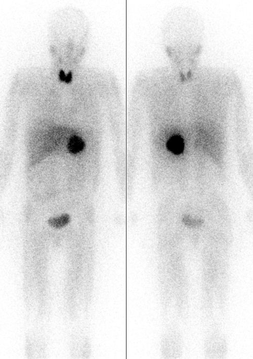
Image Source: Wikimedia Commons (2010). Pheochromocytoma Scan, Licensed under a creative commons licence. Available for free at: https://commons.wikimedia.org/wiki/File:Pheochromocytoma_Scan.jpg (Accessed 13 May 2025).
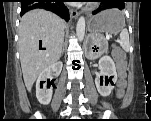
Image Source: Wikimedia Commons (2010). Phaeochromozytoma CT coronal labelled, Licensed under a creative commons licence. Available for free at https://commons.wikimedia.org/wiki/File:Phaeochromozytoma_CT_coronal_labelled.jpg (Accessed 13 May 2025).
Management
In benign disease, surgical resection is the first-line treatment. However hypertension must be managed pre-operatively, using the following:
- Alpha-blockers e.g. phenoxybenzamine but this can cause reflex tachycardia due to blockage of alpha-2 receptors. Prazosin and doxazosin are selective alpha-1 antagonists which do not result in reflex tachycardia but do not provide complete alpha blockade.
- Beta-blockers e.g. atenolol, propranolol or metoprolol. These can only be started after sufficient alpha blockade, otherwise it can precipitate hypertensive crisis through unopposed alpha receptor action causing vasoconstriction.
- Calcium channel blocker e.g. nifedipine or amlodipine. These can supplement the above to achieve adequate control of hypertension.
Alpha-blockade can result in volume contraction and orthostatic hypotension, and this can be counteracted by fluid resuscitation and a high salt diet.
In benign disease, surgical methods of resection include:
- Open or laparoscopic total adrenalectomy.
- Adrenal-sparing or partial adrenalectomy. Cortical-sparing adrenalectomy is done in those with hereditary phaeochromocytomas and can avoid need for lifelong corticosteroid replacement, however it requires monitoring for disease recurrence.
- Robot-assisted adrenalectomy.
Malignant tumours can be surgically debulked to provide symptomatic relief, however the mainstay of treatment is CVD (cyclophosphamide, vincristine and dacarbazine) chemotherapy with a complete and partial response rate of up to 57%. Radioactive iodine (iobenguane I-121), external beam radiotherapy and radiofrequency ablation are adjunctive therapies that can be used.
Complications
Acute hypertensive crisis is the most common complication of phaeochromocytoma. It presents with severe hypertension, shock and circulatory failure. End organ damage can occur including myocardial infarction, cerebral haemorrhage and heart failure. Precipitating factors include anaesthesia, opiates, cocaine, tricyclic antidepressants, contrast media, dopamine antagonists, pregnancy and childbirth. The first-line treatment is phentolamine 5-20mg IV, or nitroprusside 0.3 to 0.5 mcg/kg/min IV infusion.
Other complications include hypertensive encephalopathy due to long-term severe hypertension, presenting with altered cognition and mental status, focal neurology and seizures. The risk of cerebral haemorrhage and cerebrovascular accidents is also increased in hypertensive crises.
Primary hyperaldosteronism
Background
In primary hyperaldosteronism (PH), aldosterone production is increased, usually in an autonomous fashion independent of the renin aldosterone angiotensin (RAAS) system in primary aldosteronism, whereas secondary hyperaldosteronism occurs due to high renin levels.
High aldosterone levels lead to excessive sodium retention in the distal renal tubule, and concomitant urinary excretion of potassium and hydrogen ions. This leads to water retention, causing hypertension and suppression of the RAAS system, alongside hypokalaemia and metabolic alkalosis in severe cases.
Epidemiology
The most common cause of secondary hypertension in middle-aged adults is PH, accounting for 5 – 10% of cases. Approximately 20% of patients will have hypokalaemia, and therefore screening in hypertension is warranted regardless of potassium levels.
Aetiology
Excessive aldosterone production occurs due to adrenal adenoma (also known as Conn’s syndrome) in 80% of cases, idiopathic bilateral adrenal hyperplasia in 15% of cases, familial hyperaldosteronism type I and II, or adrenal carcinoma. Adrenal adenomas are more common in females, whilst bilateral adrenal hyperplasia more commonly occurs in males past the age of 40.
Diagnosis
Risk factors in the history that could indicate primary hyperaldosteronism include:
- Early age of onset of hypertension (<35 years).
- Positive family history of PH, or early onset hypertension and/or stroke.
- Accelerated and/or resistant hypertension (e.g. resistant to >3 drugs).
- Unusual symptoms e.g. weakness, sweating attacks.
- Hypokalaemia prior to diuresis.
The first-line investigations often centre around testing of the RAAS system, and therefore drugs that may influence this system must be stopped in advance e.g. ACE inhibitors, spironolactone and angiotensin II receptor blockers. Typical findings are:
- Raised plasma aldosterone : renin ratio (ARR).
- Raised plasma aldosterone levels.
- Suppressed plasma renin.
- Hypokalaemia which can cause weakness.
- Urinary potassium loss.
- Metabolic alkalosis and elevated bicarbonate.
To differentiate whether an adrenal adenoma or bilateral adrenal hyperplasia is responsible for excess aldosterone secretion, a CT or MRI is warranted. Selective adrenal venous sampling can be done to localise the cause of primary aldosteronism.
Confirmatory tests include a fludrocortisone suppression test, where fludrocortisone (0.1 mg / 6 hours), sodium and potassium supplementation are administered for 4 days. On the 4th day, a cortisol level is measured at 7 am, and renin, cortisol and aldosterone levels are measured at 10 am. A serum aldosterone >6 ng/dL, renin < 1 ng/mL/hr and potassium in normal range is diagnostic. If cortisol is lower at 10 am than at 7 am, this also excludes a confounding ACTH effect. A saline infusion test is an alternative confirmatory test, whereby renin, aldosterone, cortisol and potassium are measured prior to and after administration of 2 L of normal saline. A serum aldosterone > 10 ng/dL is diagnostic, whilst < 5 ng/dL rules out PH and values in between are indeterminate. Other confirmatory tests include oral sodium loading and the captopril challenge test.
Management
Management will depend on whether PH is due to a unilateral issue e.g. adenoma or bilateral, and whether the patient is fit for surgery. In the instance of unilateral causes, laparoscopic adrenalectomy may be performed and can resolve hypertension in 70% of patients. Even for bilateral causes, unilateral laparoscopic adrenalectomy can be of benefit by reducing the amount of adrenal tissue capable of secreting aldosterone, and therefore reduce hypertension. If unfit for surgery, lifelong aldosterone antagonists may be prescribed (such as spironolactone, amiloride or eplerenone) and these can take weeks to months to improve hypertension. Treatment must be titrated to avoid excessive volume depletion that can result in pre-renal acute kidney injury and hyperkalaemia. Dietary sodium restriction is also of benefit.
Complications
The main complications of PH are due to hypertension and include increased risk of cerebrovascular accident, myocardial infarction, heart failure, atrial fibrillation and chronic kidney failure. Excessive aldosterone can result in myocardial fibrosis, but this is ameliorated with treatment with aldosterone antagonists.
Congenital adrenal hyperplasia
Background
Congenital adrenal hyperplasia (CAH) is a family of autosomal recessive disorders where inherited enzyme deficiencies impair cortisol production. The primary cause is 21-hydroxylase deficiency (21-HD) in 95% of cases, characterised by cortisol deficiency, androgen excess and there may or may not be aldosterone deficiency. hydroxysteroid dehydrogenase deficiency.
Epidemiology
Previous studies have found that CAH affects 1 in 18,000 newborns. Although it affects similar numbers of females and males, males are more likely to present with severe complications such as a salt-wasting crisis. There is an increased prevalence seen in the Ashkenazi Jewish population at 1 in 27.
Aetiology
As an autosomal recessive disorder, CAH most commonly affects the gene encoding 21-hydroxylase (CYP21A2) which is on chromosome 6. The second most common gene affected is that encoding 11-beta-hydroxylase (CYP11B1). As a result of insufficient cortisol production, increased secretion of CRH and ACTH occur, where the latter leads to adrenal hyperplasia and excessive androgen secretion.
The presentation will vary depending on the severity of mutations present:
- Classical salt-wasting CAH: severe mutations cause females to have virilised atypical genitalia, and severe mineralocorticoid and glucocorticoid deficiency. These can result in salt-wasting crises and adrenal crises respectively.
- Classical simple virilising CAH: moderate mutations result in virilisation, but the ability to retain salt is preserved.
- Non-classical CAH: mild to moderate mutations result in signs of hyperandrogenism but no virilisation of genitalia.
Diagnosis
In classic CAH, diagnosis in female infants can often be found before salt-wasting crisis occurs due to identification of virilised genitalia e.g. clitoral enlargement. However, in male infants, signs will be subtle and therefore diagnosis can be missed until a salt-wasting crisis occurs, presenting with vomiting, dehydration, lethargy, weight loss, hyponatremia and hyperkalaemia. Non-classical CAH presents with precocious puberty (e.g. early pubic hair and advanced bone age) and abnormalities of the gonadal hormones (e.g. oligo/amenorrhea, PCOS, hirsutism and impaired fertility). A positive family history will be common.
First-line investigations include:
- FBC, U&Es, blood glucose: hyponatremia, hyperkalaemia and hypoglycaemia may be seen.
- Serum 17-hydroxyprogesterone: this is a substrate for 21-hydroxylase, and will be high level in 21-HD.
- ACTH stimulation test: gold standard for diagnosis of the non-classic form, where stimulated levels of 17-hydroxyprogesterone are plotted on a nomogram.
- Genetic analysis and karyotyping.
- Ultrasound of pelvis and adrenals.
- Bone age to assess for advanced skeletal maturation.
Management
For classical CAH, the recommended treatment is as follows:
- Glucocorticoid: lifelong glucocorticoids e.g. hydrocortisone prevent further virilisation, and the target dose should maintain physiologic androgen levels.
- Mineralocorticoid: to correct electrolytes and plasma renin activity.
- Sodium chloride: to supplement salt-wasting CAH.
- Growth hormone + gonadotropin releasing hormone analogue: this can be given to infants and children to improve growth rate and final height.
- Genital surgery: this can be offered to remove redundant erectile tissue in virilised females.
For non-classical CAH, the recommended treatment is as follows:
- Glucocorticoid: only recommended for symptomatic non-classical CAH, and when there is risk of premature bone maturation affecting height.
- Growth hormone + gonadotropin releasing hormone analogue: as above.
- Oral contraceptive: can reduce hirsutism.
Complications
Short-term complications include:
- Salt-wasting crisis: hypotension, hyponatremia and hyperkalemia.
- Adrenal crisis: as described in previous sections.
Longer-term complications include:
- Precocious puberty: premature epiphyseal closure and short stature.
- Hyperandrogenism also carries a risk of PCOS, insulin resistance and hypertension.
- Infertility: particularly in forms of classical CAH.
- Side effects related to long-term glucocorticoids: e.g. osteoporosis.
- Testicular adrenal rest tumours: found in males who have undertreated CAH.
Cushing’s syndrome
Background
Cushing’s syndrome arises from long-term exposure to excessive glucocorticoids of either endogenous or exogenous source. Exogenous steroids are the most common cause, and this should be excluded first. The presentation can be subtle, but presents with central obesity, proximal muscle weakness, dorsal and supraclavicular fat pads, abdominal violaceous striae, bruising and moon facies. Metabolic syndrome can arise from Cushing’s disease, as well as metabolic bone disease and hypertension.
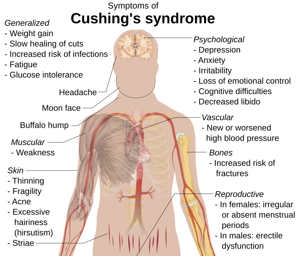
Image Source: Wikimedia Commons (2015). Cuching’s Syndrome (Vector image), Licensed under a creative commons licence. Available for free at: https//commons.wikimedia.org/wiki/File:Cushing%27s_syndrome_(vector_image).svg (Accessed 13 May 2025).
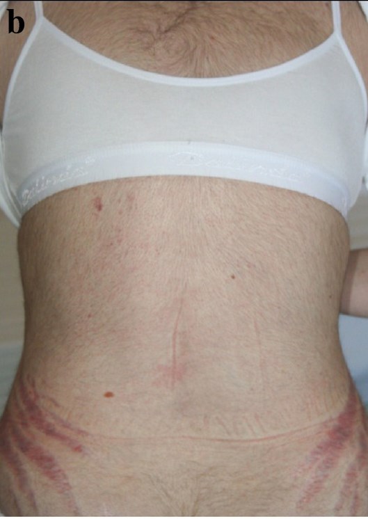
Image Source: Wikimedia Commons (2012). Cushing’s Body, Licensed under a creative commons licence. Available for free at: https://en.wikipedia.org/wiki/Cushing%27s_syndrome#/media/File:CushingsBody.jpg (Accessed 13 May 2025).
Epidemiology
Cushing’s syndrome is uncommon with an incidence of 40 per million. It more commonly affects females than males (3:1). Cushing’s disease (from pituitary secretion of ACTH) affects females to a greater degree (3:1 to 5:1), with a peak age of onset between 25 to 40 years of age. A greater prevalence of 2% to 5% is found in obese patients with type 2 diabetes mellitus.
Aetiology
There are two groups of causes:
- ACTH-dependent disease: excessive ACTH secretion by the pituitary (known as Cushing’s disease) is the most common endogenous cause responsible for 70 – 80% of cases. Other causes include ectopic ACTH-secreting tumours (e.g. in small cell lung cancer) or excess ACTH administration.
- ACTH-independent disease: the most common cause overall is excess exogenous glucocorticoids. Other causes include adrenal adenomas, adrenal carcinomas and macronodular adrenal hyperplasia.
Diagnosis
The signs and symptoms of Cushing’s syndrome are summarised in Figure 4.
The first-line investigations are:
- FBC, U&Es and glucose: may show raised WCC, hypokalaemia, and hyperglycaemia respectively.
- 24-hour urinary cortisol: levels will be elevated to >50 mcg/24 hours. This test should not be used in those with renal failure.
- Low-dose dexamethasone suppression test: 1mg dexamethasone is given at 11pm, and plasma cortisol is measured at 8am the following morning, where levels elevated to >50 nmol/L indicate Cushing’s syndrome.
- Midnight cortisol levels: collected between 11pm and 1am, and will be elevated due to loss of normal diurnal variation in cortisol levels.
- Plasma ACTH: suppressed ACTH and high cortisol indicates an ACTH-independent cause such as adrenal adenoma. Elevated ACTH can suggest ACTH-dependent Cushing’s disease.
If plasma ACTH is detectable, there is benefit in the following tests:
- High-dose dexamethasone suppression test: 8mg dexamethasone is given at 11pm. Initial plasma cortisol is measured, and again at 8am the following morning. Suppression of cortisol <50% of baseline, indicates ACTH-dependent Cushing’s disease. High or normal cortisol, with a low ACTH suggests ACTH-independent causes such as adrenal adenoma. Whereas high-normal cortisol with a high ACTH suggests ectopic ACTH secretion.
- Inferior petrosal sinus sampling: blood is sampled peripherally and from the inferior petrosal sinus. If there is an elevated central : peripheral ACTH ratio, this indicates a pituitary cause.
- MRI pituitary gland: should be requested for suspected ACTH-dependent disease to investigate for pituitary adenoma.
- CT chest, abdomen and pelvis: to assess adrenals to identify adrenal adenoma, carcinoma or hyperplasia. It is also warranted to look for tumours that may be a source of ectopic ACTH secretion e.g. small cell lung cancer.
Management
The management of Cushing’s syndrome depends on the aetiology. Management in each case is summarised below:
| Aetiology | Management | Comments |
|---|---|---|
| Cushing’s disease (ACTH-secreting pituitary adenoma) | Transsphenoidal pituitary adenomectomy | Performed endoscopically or microscopically. |
| Somatostatin analogues e.g. pasireotide, steroidogenesis inhibitors e.g. osilodrostat, glucocorticoid receptor antagonists e.g. mifepristone | These are used to address short-term severe hypercortisolism whilst awaiting definitive treatment, or if the patient is not fit for surgery. | |
| Hormonal replacement | Due to pituitary resection, replacement of thyroxine, gonadal hormones, somatropin and desmopressin may be needed. | |
| Pituitary radiotherapy | Indicated if high cortisol persists post-tumour resection. | |
| Ectopic ACTH | Surgical resection of tumour/metastases | Resection of ectopic tumours responsible for ACTH (or CRH) production. |
| Steroidogenesis inhibitors, glucocorticoid receptor antagonists | As above. | |
| Chemotherapy and radiotherapy | As above. | |
| Adrenal carcinoma or adenoma | Laparoscopic adrenalectomy or tumour resection | Unilateral resection is often curative without subsequent adrenal insufficiency. |
| Steroidogenesis inhibitors, glucocorticoid receptor antagonists | As above. | |
| Chemotherapy and radiotherapy | Adrenal carcinoma will often require this, as it often will have progressed at time of diagnosis. | |
| Bilateral adrenal hyperplasia or adenoma | Bilateral adrenalectomy | Resection is often curative without subsequent adrenal insufficiency. In some cases mineralocorticoid and corticosteroid replacement may be indicated. |
| Steroidogenesis inhibitors, glucocorticoid receptor antagonists | As above. | |
| Exogenous glucocorticoids | Gradual withdrawal of glucocorticoids | Aiming to discontinue or reduce maintenance dose. |
Table 2: Management of Cushing’s syndrome dependent on aetiology.
Complications
Complications of Cushing’s syndrome include adrenal insufficiency following surgery, due to adrenal suppression as described in the Secondary Adrenal Insufficiency chapter. This can necessitate replacement glucocorticoid therapy.
Hypercortisolism also carries an increased risk of metabolic syndrome (diabetes mellitus, hypertension and obesity), cardiovascular disease, thrombophilia, osteoporosis and impaired immunity.
Treatment-related complications include Nelson’s syndrome following bilateral adrenalectomy, where loss of adrenal feedback causes growth of pre-existing ACTH-secreting pituitary adenomas. Transsphenoidal pituitary resection can also cause deficiency of other pituitary hormones e.g. thyrotropin, somatotropin, desmopressin and androgens.
References
- https://bestpractice.bmj.com/topics/en-gb/56
- https://patient.info/doctor/adrenal-insufficiency-and-addisons-disease
- https://cks.nice.org.uk/topics/addisons-disease
- https://bestpractice.bmj.com/topics/en-gb/863
- https://patient.info/doctor/phaeochromocytoma-pro
- https://bestpractice.bmj.com/topics/en-gb/163
- https://bestpractice.bmj.com/topics/en-gb/253
- https://patient.info/doctor/hyperaldosteronism
- https://bestpractice.bmj.com/topics/en-gb/699
- https://patient.info/doctor/congenital-adrenal-hyperplasia-pro
- https://bestpractice.bmj.com/topics/en-gb/205
- https://patient.info/doctor/cushings-syndrome-pro
Complete Guide to Clinical Genetics ST3 Application and Recruitment (2027)
Written by Medibuddy. Last updated: October 2026. Clinical Genetics is

Complete Guide to Clinical Radiology ST3 Application and Recruitment (2027)
Recruitment paused: NHS England is not running Clinical Radiology ST3+ recru

Complete Guide to Paediatric Surgery ST3 Application and Recruitment in United Kingdom (2026)
Written by Medibuddy. Last updated: October 2026. If you’re planning


Take your subscriptions with you
Our mobile app allows you to access your interview and exam question banks wherever you are.

