


 Close
Close
 27th May 2025
Cushing’s syndrome
Background
Cushing’s syndrome arises from long-term exposure to excessive glucocorticoids of either endogenous or exogenous source. Exogenous steroids are the most common cause, and this should be excluded first. The presentation can be subtle, but presents with central obesity, proximal muscle weakness, dorsal and supraclavicular fat pads, abdominal violaceous striae, bruising and moon facies. Metabolic syndrome can arise from Cushing’s disease, as well as metabolic bone disease and hypertension.
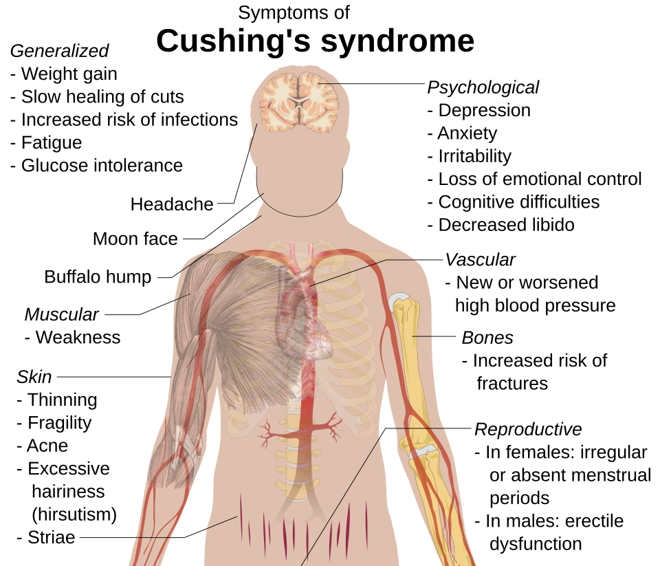
Image Source: Wikimedia Commons (2015). Cuching’s Syndrome (Vector image), Licensed under a creative commons licence. Available for free at: https//commons.wikimedia.org/wiki/File:Cushing%27s_syndrome_(vector_image).svg (Accessed 13 May 2025).
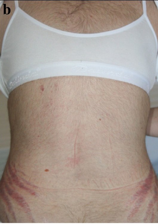
Image Source: Wikimedia Commons (2012). Cushing’s Body, Licensed under a creative commons licence. Available for free at: https://en.wikipedia.org/wiki/Cushing%27s_syndrome#/media/File:CushingsBody.jpg (Accessed 13 May 2025).
Epidemiology
Cushing’s syndrome is uncommon with an incidence of 40 per million. It more commonly affects females than males (3:1). Cushing’s disease (from pituitary secretion of ACTH) affects females to a greater degree (3:1 to 5:1), with a peak age of onset between 25 to 40 years of age. A greater prevalence of 2% to 5% is found in obese patients with type 2 diabetes mellitus.
Aetiology
There are two groups of causes:
- ACTH-dependent disease: excessive ACTH secretion by the pituitary (known as Cushing’s disease) is the most common endogenous cause responsible for 70 – 80% of cases. Other causes include ectopic ACTH-secreting tumours (e.g. in small cell lung cancer) or excess ACTH administration.
- ACTH-independent disease: the most common cause overall is excess exogenous glucocorticoids. Other causes include adrenal adenomas, adrenal carcinomas and macronodular adrenal hyperplasia.
Diagnosis
The signs and symptoms of Cushing’s syndrome are summarised in Figure 4.
The first-line investigations are:
- FBC, U&Es and glucose: may show raised WCC, hypokalaemia, and hyperglycaemia respectively.
- 24-hour urinary cortisol: levels will be elevated to >50 mcg/24 hours. This test should not be used in those with renal failure.
- Low-dose dexamethasone suppression test: 1mg dexamethasone is given at 11pm, and plasma cortisol is measured at 8am the following morning, where levels elevated to >50 nmol/L indicate Cushing’s syndrome.
- Midnight cortisol levels: collected between 11pm and 1am, and will be elevated due to loss of normal diurnal variation in cortisol levels.
- Plasma ACTH: suppressed ACTH and high cortisol indicates an ACTH-independent cause such as adrenal adenoma. Elevated ACTH can suggest ACTH-dependent Cushing’s disease.
If plasma ACTH is detectable, there is benefit in the following tests:
- High-dose dexamethasone suppression test: 8mg dexamethasone is given at 11pm. Initial plasma cortisol is measured, and again at 8am the following morning. Suppression of cortisol <50% of baseline, indicates ACTH-dependent Cushing’s disease. High or normal cortisol, with a low ACTH suggests ACTH-independent causes such as adrenal adenoma. Whereas high-normal cortisol with a high ACTH suggests ectopic ACTH secretion.
- Inferior petrosal sinus sampling: blood is sampled peripherally and from the inferior petrosal sinus. If there is an elevated central : peripheral ACTH ratio, this indicates a pituitary cause.
- MRI pituitary gland: should be requested for suspected ACTH-dependent disease to investigate for pituitary adenoma.
- CT chest, abdomen and pelvis: to assess adrenals to identify adrenal adenoma, carcinoma or hyperplasia. It is also warranted to look for tumours that may be a source of ectopic ACTH secretion e.g. small cell lung cancer.
Management
The management of Cushing’s syndrome depends on the aetiology. Management in each case is summarised below:
| Aetiology | Management | Comments |
|---|---|---|
| Cushing’s disease (ACTH-secreting pituitary adenoma) | Transsphenoidal pituitary adenomectomy | Performed endoscopically or microscopically. |
| Somatostatin analogues e.g. pasireotide, steroidogenesis inhibitors e.g. osilodrostat, glucocorticoid receptor antagonists e.g. mifepristone | These are used to address short-term severe hypercortisolism whilst awaiting definitive treatment, or if the patient is not fit for surgery. | |
| Hormonal replacement | Due to pituitary resection, replacement of thyroxine, gonadal hormones, somatropin and desmopressin may be needed. | |
| Pituitary radiotherapy | Indicated if high cortisol persists post-tumour resection. | |
| Ectopic ACTH | Surgical resection of tumour/metastases | Resection of ectopic tumours responsible for ACTH (or CRH) production. |
| Steroidogenesis inhibitors, glucocorticoid receptor antagonists | As above. | |
| Chemotherapy and radiotherapy | As above. | |
| Adrenal carcinoma or adenoma | Laparoscopic adrenalectomy or tumour resection | Unilateral resection is often curative without subsequent adrenal insufficiency. |
| Steroidogenesis inhibitors, glucocorticoid receptor antagonists | As above. | |
| Chemotherapy and radiotherapy | Adrenal carcinoma will often require this, as it often will have progressed at time of diagnosis. | |
| Bilateral adrenal hyperplasia or adenoma | Bilateral adrenalectomy | Resection is often curative without subsequent adrenal insufficiency. In some cases mineralocorticoid and corticosteroid replacement may be indicated. |
| Steroidogenesis inhibitors, glucocorticoid receptor antagonists | As above. | |
| Exogenous glucocorticoids | Gradual withdrawal of glucocorticoids | Aiming to discontinue or reduce maintenance dose. |
Table 2: Management of Cushing’s syndrome dependent on aetiology.
Complications
Complications of Cushing’s syndrome include adrenal insufficiency following surgery, due to adrenal suppression as described in the Secondary Adrenal Insufficiency chapter. This can necessitate replacement glucocorticoid therapy.
Hypercortisolism also carries an increased risk of metabolic syndrome (diabetes mellitus, hypertension and obesity), cardiovascular disease, thrombophilia, osteoporosis and impaired immunity.
Treatment-related complications include Nelson’s syndrome following bilateral adrenalectomy, where loss of adrenal feedback causes growth of pre-existing ACTH-secreting pituitary adenomas. Transsphenoidal pituitary resection can also cause deficiency of other pituitary hormones e.g. thyrotropin, somatotropin, desmopressin and androgens.
Complete Guide to Clinical Genetics ST3 Application and Recruitment (2027)
Written by Medibuddy. Last updated: October 2026. Clinical Genetics is

Complete Guide to Clinical Radiology ST3 Application and Recruitment (2027)
Recruitment paused: NHS England is not running Clinical Radiology ST3+ recru

Complete Guide to Paediatric Surgery ST3 Application and Recruitment in United Kingdom (2026)
Written by Medibuddy. Last updated: October 2026. If you’re planning


Take your subscriptions with you
Our mobile app allows you to access your interview and exam question banks wherever you are.

