


 Close
Close
 27th May 2025
Phaeochromocytoma
Background
Phaeochromocytoma is a catecholamine secreting tumour derived from chromaffin cells of the adrenal medulla (85%) or extra-adrenal paragangliomas. The classic triad of symptoms is palpitations, diaphoresis, and headaches, which can occur in episodes lasting seconds to hours, and worsening with tumour progression. It also can result in life-threatening, intractable hypertension and arrhythmia. Most cases are benign (at least 75%), and paraganglioma are more likely to metastasise compared to phaeochromocytoma.
Epidemiology
The annual incidence of phaeochromocytoma is low at 2 to 9.1 per 1 million adults. It occurs with equal prevalence in men and women, and across different ethnicities. The peak age of onset is in the 3rd to 5th decades of life. Approximately 20% of cases are found in children, owing to underlying genetic conditions. It is estimated that 50% of phaeochromocytomas are only diagnosed at autopsy, as many cases remain asymptomatic.
Aetiology
Whilst the majority occur sporadically, hereditary syndromes such as the following account for a 40% of cases in adults and 80% of cases in children:
- Multiple endocrine neoplasia type 2A and 2B due to the RET gene – occurring bilaterally in 70% of MEN syndromes.
- Von Hippel-Lindau disease due to the VHL gene – also associated with cerebellar haemangioblastomas and renal cell carcinomas.
- Neurofibromatosis type 1 due to the NF1 gene – with an incidence of 1% of phaechromocytoma.
- Paraganglioma syndromes types 1, 3 and 4.
Germline or somatic mutations can affect over 20 different genes, affecting metabolism e.g. succinate dehydrogenase or signalling pathways e.g. kinase and Wnt.
Diagnosis
A history suggestive of paraganglioma will typically include symptoms such as diaphoresis, palpitations, headache and tremor as the predominant symptoms. Other less common symptoms include nausea, weakness, epigastric pain, anxiety and weight loss. Risk factors, such as a family history of the aforementioned hereditary syndromes, should be elicited. Signs upon examination include hypertension, postural hypotension, pallor, tremor, arrhythmia, fever and abdominal masses in later stages of disease.
First-line investigations are as follows:
- FBC, U&Es and bone profile which may reveal erythrocytosis, hypokalaemia and hypercalcaemia (in MEN2) respectively.
- 24-hour urine collection for catecholamines, creatinine, metanephrines, normetanephrines and vanillylmandelic acid (VMA). The latter three are breakdown products of catecholamines. False positive results can be reduced by informing the patient to avoid caffeine, tobacco or strenuous exercise.
- Serum free metanephrines and normetanephrines.
- Plasma catecholamines.
In phaeochromocytoma, catecholamines and their breakdown products will be elevated. In such cases, genetic testing is warranted to investigate potential hereditary syndromes and allow for genetic counselling. Imaging will also be necessary to localise the tumour, with CT being the initial imaging modality of choice and MRI preferred to localise metastases. If CT and MRI are unable to localise a tumour, I-123 metaiodobenzylguanidine (MIBG) scintigraphy may be performed, as I-123 MIBG uptake will be high in catecholamine-secreting tumours. I-123 MIBG scintigraphy is beginning to be superseded by FDG PET imaging.
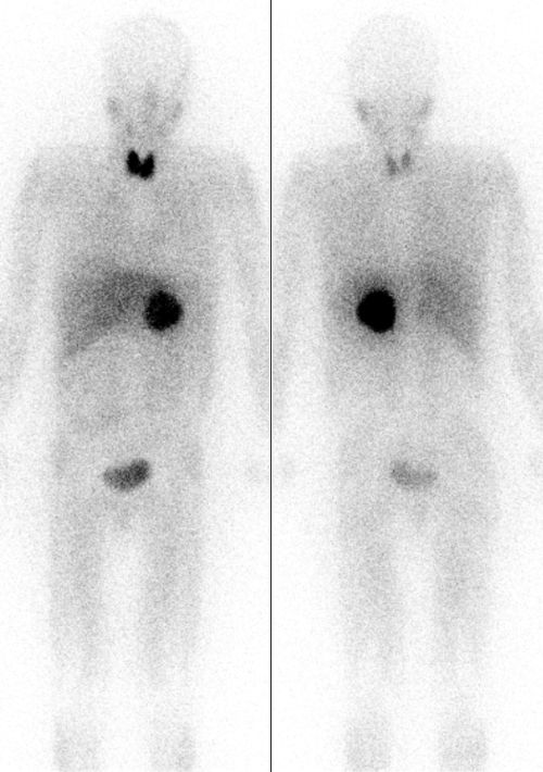
Image Source: Wikimedia Commons (2010). Pheochromocytoma Scan, Licensed under a creative commons licence. Available for free at: https://commons.wikimedia.org/wiki/File:Pheochromocytoma_Scan.jpg (Accessed 13 May 2025)
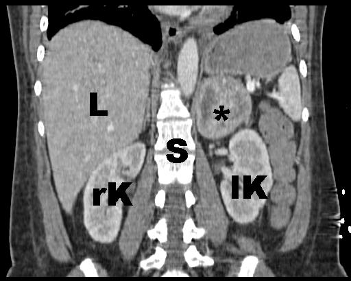
Image Source: Wikimedia Commons (2010). Phaeochromozytoma CT coronal labelled, Licensed under a creative commons licence. Available for free at https://commons.wikimedia.org/wiki/File:Phaeochromozytoma_CT_coronal_labelled.jpg (Accessed 13 May 2025)
Management
In benign disease, surgical resection is the first-line treatment. However hypertension must be managed pre-operatively, using the following:
- Alpha-blockers e.g. phenoxybenzamine but this can cause reflex tachycardia due to blockage of alpha-2 receptors. Prazosin and doxazosin are selective alpha-1 antagonists which do not result in reflex tachycardia but do not provide complete alpha blockade.
- Beta-blockers e.g. atenolol, propranolol or metoprolol. These can only be started after sufficient alpha blockade, otherwise it can precipitate hypertensive crisis through unopposed alpha receptor action causing vasoconstriction.
- Calcium channel blocker e.g. nifedipine or amlodipine. These can supplement the above to achieve adequate control of hypertension.
Alpha-blockade can result in volume contraction and orthostatic hypotension, and this can be counteracted by fluid resuscitation and a high salt diet.
In benign disease, surgical methods of resection include:
- Open or laparoscopic total adrenalectomy.
- Adrenal-sparing or partial adrenalectomy. Cortical-sparing adrenalectomy is done in those with hereditary phaeochromocytomas and can avoid need for lifelong corticosteroid replacement, however it requires monitoring for disease recurrence.
- Robot-assisted adrenalectomy.
Malignant tumours can be surgically debulked to provide symptomatic relief, however the mainstay of treatment is CVD (cyclophosphamide, vincristine and dacarbazine) chemotherapy with a complete and partial response rate of up to 57%. Radioactive iodine (iobenguane I-121), external beam radiotherapy and radiofrequency ablation are adjunctive therapies that can be used.
Complications
Acute hypertensive crisis is the most common complication of phaeochromocytoma. It presents with severe hypertension, shock and circulatory failure. End organ damage can occur including myocardial infarction, cerebral haemorrhage and heart failure. Precipitating factors include anaesthesia, opiates, cocaine, tricyclic antidepressants, contrast media, dopamine antagonists, pregnancy and childbirth. The first-line treatment is phentolamine 5-20mg IV, or nitroprusside 0.3 to 0.5 mcg/kg/min IV infusion.
Other complications include hypertensive encephalopathy due to long-term severe hypertension, presenting with altered cognition and mental status, focal neurology and seizures. The risk of cerebral haemorrhage and cerebrovascular accidents is also increased in hypertensive crises.
Complete Guide to Clinical Genetics ST3 Application and Recruitment (2027)
Written by Medibuddy. Last updated: October 2026. Clinical Genetics is

Complete Guide to Clinical Radiology ST3 Application and Recruitment (2027)
Recruitment paused: NHS England is not running Clinical Radiology ST3+ recru

Complete Guide to Paediatric Surgery ST3 Application and Recruitment in United Kingdom (2026)
Written by Medibuddy. Last updated: October 2026. If you’re planning


Take your subscriptions with you
Our mobile app allows you to access your interview and exam question banks wherever you are.

