


 Close
Close
 19th May 2025
Mechanisms of drug-induced kidney injury
There are various methods of drug toxicity which are influenced by the pharmacological agent’s mechanism of action, metabolism, and excretion. Drug-induced nephrotoxicity can be classified into 3 broad categories as follows:
- Pre-renal
- Intrinsic
- Intrinsic acute renal failure can be further classified as:
- acute tubular necrosis
- acute interstitial nephritis
- thrombotic microangiopathy
- glomerular disease.
- Post-renal
| Pre-renal | Afferent arteriolar vasoconstriction | Non-steroidal anti-inflammatory drugs (NSAIDs), selective COX-2 inhibitors e.g. celecoxib, calcineurin inhibitors, vasopressors |
| Efferent arteriolar vasodilation | Angiotensin-converting enzyme inhibitors (ACEis), angiotensin receptor blockers (ARBs) | |
| Intrinsic | Acute tubular necrosis | Aminoglycosides, contrast agents, amphotericin B, cisplatin, cephalosporins, nucleotide analogues e.g. tenofovir, zoledronate, pentamidine, cisplatin, calcineurin inhibitors, intravenous immunoglobulins, statins |
| Acute interstitial nephritis | Beta-lactam antibiotics, quinolones, NSAIDs, proton pump inhibitors e.g. omeprazole, lithium, H2-receptor antagonists e.g. cimetidine, cephalosporins, sulphonamides, phenytoin, amphotericin B, allopurinol, rifampicin, 5-aminosalicylates e.g. mesalamine | |
| Thrombotic microangiopathy | Calcineurin inhibitors, gemcitabine, mitomycin C, bleomycin, cisplatin, clopidogrel, oestrogen-containing oral contraceptive pill, quinine | |
| Glomerular disease | NSAIDs, selective COX-2 inhibitors, lithium, penicillins, quinolone, penicillamine, gold, infliximab, pamidronate | |
| Post-renal | Crystal formation in tubules / ureters | Aciclovir, sulphonamides, methotrexate, triamterene |
| Renal calculus formation | Guaifenesin, acetazolamide | |
| Retroperitoneal fibrosis | Methysergide |
Table 1: Mechanisms of drug-induced kidney injury
Pre-renal nephrotoxicity
Pre-renal drug-induced acute kidney injury is a result of reduction in renal perfusion and thus glomerular filtration rate (GFR). Factors putting patients at particular risk of drug-induced pre-renal AKI are:
- Reduced intravascular volume (e.g. inadequate fluid intake, vomiting, diarrhoea)
- Reduced effective intravascular volume (e.g. congestive cardiac failure, liver cirrhosis)
- Older age
- Bilateral renal artery stenosis
Offending drugs may either reduce renal perfusion through inducing afferent arteriolar vasoconstriction, or efferent arteriolar vasodilation. Drugs which reduce prostaglandin synthesis via inhibition of cyclo-oxygenase (COX) enzymes, such as NSAIDs, reduce the vasodilatation effect of prostaglandins on the afferent arteriole hence causing vasoconstriction and reducing renal perfusion. Calcineurin inhibitors such as ciclosporin and tacrolimus promote afferent arteriolar constriction by stimulating the production of angiotensin II, thromboxane A2, leukotrienes and endothelin, and inhibiting the release of endothelial-derived nitric oxide.
In contrast, angiotensin-converting enzyme inhibitors (ramipril, lisinopril, perindopril) and angiotensin receptor blockers (candesartan, losartan) promote vasodilatation of the efferent arteriole by limiting the action of angiotensin II, which ordinarily acts to promote vasoconstriction and ultimately increases efferent arteriolar resistance. In patients with underlying volume depletion this causes a reduction in trans-glomerular capillary pressure and a resultant drop in glomerular filtration rate.
In pre-renal AKI, urine volume is low with low sodium concentration and high osmolality. Urinalysis is expected to be negative for red blood cells, leukocytes or protein. The approach to management should include stopping or lowering the dose of the offending drug and any other nephrotoxic medications, administration of intravenous fluids to restore circulating volume, and treating contributing comorbidities.
Intrinsic nephrotoxicity
The aetiology of intrinsic drug-induced AKI may be further split into acute tubular necrosis (ATN), acute interstitial nephritis (AIN), thrombotic microangiopathy (TMA), and glomerular disease. The table below discusses the clinical findings and approach to management of each of these four mechanisms. Further detail on the underlying pathophysiology of each is provided in the text below.
| Disease process | Clinical findings | Management |
| Acute tubular necrosis (ATN) |
|
|
| Acute interstitial nephritis (AIN) |
|
|
| Thrombotic microangiopathy |
|
|
| Glomerular disease |
|
|
Table 2: Mechanisms, clinical findings and management of intrinsic drug-induced kidney injury
Acute tubular necrosis
In acute tubular necrosis (ATN), the culprit drug or its metabolite has a directly toxic effect on the renal tubules; the proximal tubule is particularly vulnerable due to its role in increasing the concentration of the glomerular filtrate. This is typically a dose-dependent effect, and arises due to apical contact of the tubular epithelium with drugs and / or their metabolites. There are various pathophysiological mechanisms implicated in causing ATN, dependent on the causative drug; among these are deranged metabolism of phospholipids, alterations in cell membrane permeability and transmembrane ionic gradients (particularly calcium ions), mitochondrial disruption, and generation of oxygen free radicals. Of note, aminoglycosides may cause ATN as a small proportion of the drug is retained in the epithelial cells of the proximal tubules, which can result in disruption of the structure and permeability of the cell membranes, inhibition of lysosomal phospholipase function, and reduction in the release of liposomal hydrolases. Ultimately, aminoglycoside-induced ATN leads to inhibition of mitochondrial function, generation of oxygen free radicals and consequently cell death. Once-daily dosing, dose adjustment according to serum levels, and limiting the duration of use, can reduce the likelihood of aminoglycoside-induced ATN.
Statins may cause ATN by triggering rhabdomyolysis and consequent myoglobinuria; the myoglobin released from myocytes forms casts in the distal convoluted tubule, resulting in occlusion of the tubule, increase in intratubular pressure and ultimately reduction in glomerular filtration rate.
Renal histology in ATN will show degenerative changes in the tubules such as loss of the brush border, nuclear enlargement and pleomorphism, and apoptotic changes. Patients with infection, malignancy, or cardiovascular disease are at higher risk of developing ATN when causative drugs are administered.
Acute interstitial nephritis
Acute interstitial nephritis (AIN) results from an inflammatory infiltrate in the renal tubule interstitium. The most common causative drugs are beta-lactam antibiotics, fluoroquinolones, proton pump inhibitors and NSAIDs. Patients over the age of 65 years and those with underlying inflammatory disease (e.g. inflammatory bowel disease) are at higher risk. Drug-induced AIN is the result of a hypersensitivity reaction, and the resulting inflammatory infiltrate consists of eosinophils, lymphocytes, mast cells and plasma cells, which can be demonstrated by renal biopsy. Patients present clinically with evidence of a hypersensitivity reaction with fevers, rash, joint pain, and occasionally uveitis. However, they may also be asymptomatic. Blood tests will show AKI and an eosinophilia and there may be elevated circulating IgE levels.
Thrombotic microangiopathy
Thrombotic microangiopathy (TMA) refers to abnormalities within arteriolar and capillary walls which predispose to the formation of microvascular hyaline thrombi, and represents a spectrum of disease that encompasses thrombotic thrombocytopenic purpura (TTP) and haemolytic uraemic syndrome (HUS). In the non-immune-mediated form of the disease, the triggering drug causes endothelial damage within the renal microvasculature leading to platelet activation and aggregation. In the immune form, the offending drug (e.g. clopidogrel) leads to the production of autoantibodies against a metalloproteinase responsible for processing von Willebrand Factor, causing the accumulation of multimers that activate platelets and trigger their aggregation. Both pathogenetic mechanisms of small-vessel platelet thrombi formation result in a microangiopathic haemolytic anaemia and thrombocytopenia. AKI is often severe and mortality from thrombotic microangiopathy is high. In addition to withdrawing the triggering drug, approach to management may warrant renal dialysis, which may be required lifelong. Chemotherapeutic agents such as gemcitabine are classically implicated.
Glomerular disease
Drug-induced glomerular disease is a relatively rare form of drug-induced renal disease. It has been broadly split into two categories in the literature: direct cellular injury and immune-mediated injury. The three targets of drug-induced cellular injury within the glomerulus are the podocytes, endothelial cells and mesangial cells (Figure 1). Podocyte injury can lead to minimal change disease or focal segmental glomerulosclerosis. Endothelial cell damage manifests as thrombotic microangiopathy (see above). Mesangial cell damage causes nodular glomerulosclerosis. Immune-mediated drug injury to the glomerulus can take the form of immune complex disease (which includes membranous nephropathy and drug-induced lupus glomerulonephritis) or small vessel glomerulonephritis (segmental necrotising and crescenteric glomerulonephritis). Examples of drugs causing each of these forms of glomerular damage are provided in the table below.
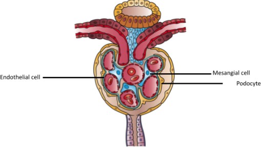
Figure 1: Diagram illustrating cells within the glomerulus which may be affected by glomerular drug injury. Image reproduced with thanks to original artwork creator Holly Fischer, available at: http://open.umich.edu/educational/med/resources/second-look-series/materials – Urinary Tract Slide 10. License for image use available at: https://creativecommons.org/licenses/by/3.0/legalcode. Figure annotations added after acquiring image for use in the current publication. – Can’t find this reference Drug-induced glomerular disease type |
Examples of causative agents | ||
| Direct cellular toxicity: | |||
| Podocytes: | Minimal change disease | NSAIDs, selective COX inhibitors, lithium, penicillins, quinolone | |
| Focal segmental glomerulosclerosis | Pamidronate, morphine, lithium, interferon therapy | ||
| Endothelial cells: | Thrombotic microangiopathy | Gemcitabine, clopidogrel, quinine | |
| Mesangial cells: | Nodular glomerulosclerosis | Smoking | |
| Immune-mediated toxicity: | |||
| Immune-complex disease: | Membranous nephropathy | Gold, penicillamine, NSAIDs | |
| Drug-induced lupus glomerulonephritis | Hydralazine, sulfasalazine, procainamide, infliximab, propylthiouracil | ||
| Small vessel glomerulonephritis: | Segmental necrotising glomerulonephritis & crescenteric glomerulonephritis | Propylthiouracil, minocycline, ciprofloxacin, hydrazlazine | |
Table 3: Toxicity and Causative Agents
Post-renal nephrotoxicity
Three major mechanisms of drug-induced post-renal nephrotoxicity are outlined below:
- Tubule / ureter crystal formation – drugs and / or their metabolites which are insoluble in urine may directly result in crystal formation, most commonly within the distal renal tubules, e.g. methotrexate, ciprofloxacin, aciclovir. This causes damage through local inflammation and necrosis via oxygen free radical formation.
- Renal calculus formation – drugs and / or their metabolites may themselves form components of renal tract calculi (ciprofloxacin, sulfonamides, triamterene), or they may influence purine or calcium oxalate metabolism, the accumulation of which leads to calculus formation (carbonic anhydrase inhibitors e.g. acetazolamide, topiramate, loop diuretics).
- Retroperitoneal fibrosis – this is a rare inflammatory process resulting in the deposition of fibrotic material around the infrarenal abdominal aorta and iliac vessels, resulting in an obstructive nephropathy. Drugs which are believed to possibly demonstrate this effect include bromocriptine, L-dopa, hydralazine, and methysergide (a specific serotonin 5HT2A receptor antagonist historically used to treat migraine and cluster headaches).
Complete Guide to Clinical Genetics ST3 Application and Recruitment (2027)
Written by Medibuddy. Last updated: October 2026. Clinical Genetics is

Complete Guide to Clinical Radiology ST3 Application and Recruitment (2027)
Recruitment paused: NHS England is not running Clinical Radiology ST3+ recru

Complete Guide to Paediatric Surgery ST3 Application and Recruitment in United Kingdom (2026)
Written by Medibuddy. Last updated: October 2026. If you’re planning


Take your subscriptions with you
Our mobile app allows you to access your interview and exam question banks wherever you are.

