


 Close
Close
 5th May 2025
Atrial Arrhythmias
Background
Atrial arrhythmias are arrythmias that are generated above or at the level of the atrioventricular node (AVN) and follow the normal His-Purkinje bundle system to rapidly activate the ventricles. These encompass a wide variety of abnormal heart rhythms caused by a multitude of underlying aetiologies. When assessing and managing atrial arrhythmias, follow the standard resus council guidelines for managing arrhythmias:
- Is there evidence of haemodynamic instability? If so, needs synchronised DC shock
- Is the rhythm fast or slow?
- Are the QRS complexes broad or narrow (i.e. <120ms)? Atrial arrhythmias are generally narrow complex (although beware of atrial tachycardias with variable block/aberrant conduction which may appear broad complex!)
- Is the rhythm regular or irregular?
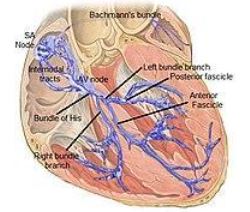
Image Source: Wikimedia Commons (2017). Cardiac Conduction System, Licensed under a creative commons licence. Available for free at: https://commons.wikimedia.org/wiki/File:Cardiac_conduction_system.jpg (Accessed 1 May 2025).
Epidemiology
Narrow complex tachycardias can be divided into:
| Type of narrow complex tachycardia | ECG morphology |
|---|---|
| Sinus tachycardia | Normal p wave followed by normal QRS |
| Atrial fibrillation (AF) | Absent p wave, irregular QRS complexes – irregularly irregular |
| Atrial flutter | Atrial rate usually 300 bpm – “flutter” or “sawtooth” baseline, ventricular rate often 150bpm (2:1 block) but can be 100bpm (3:1 block), etc |
| Atrial tachycardia | Abnormally shaped p waves which may outnumber QRS complexes in a less regular fashion than atrial flutter |
| Multifocal atrial tachycardia | 3 or more p wave morphologies with irregular QRS complexes |
| Supraventricular tachycardia (SVT) – encompasses diagnoses below | P waves absent or inverted after QRS |
| Junctional tachycardia (AV nodal reentrant tachycardias) | P wave either buried in QRS or after QRS complex, rate often 150-200bpm |
| AV reentrant tachycardias (e.g. Wolff-Parkinson-White or accessory pathway) | P wave inverted after QRS, delta wave in WPW, rate often 150-250 bpm. QRS can be broad |
Table 1: Stratification of narrow complex tachycardias.
Atrial fibrillation is the most common cardiac arrhythmia with a prevalence in England of 2.5%. Approximately 1.4 million people were living with AF in England in 2016 with approximately 425,000 undiagnosed and untreated. Prevalence is higher in men (2.9%) than women (2.0%).
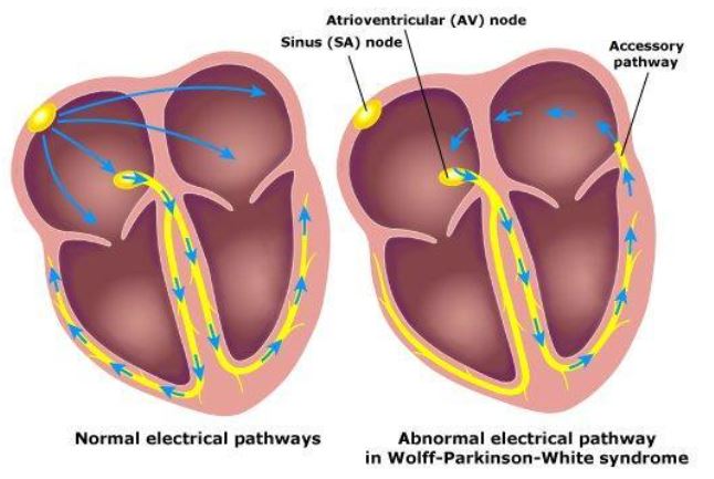
Image Source: Wikimedia Commons (2011). WPW, Licensed under a creative commons licence. Available for free at: https://commons.wikimedia.org/wiki/File:WPW.jpeg (Accessed 1 May 2025)
Aetiology
Atrial arrhythmias are caused by a wide variety of diseases and it is useful to stratify this using a surgical sieve as in the table below. Due to this, a thorough history and examination is of paramount importance in ascertaining the cause of arrhythmia.
| Surgical sieve | Cause |
|---|---|
| Intracardiac
(Encompassing vascular, congenital, traumatic, degenerative and inflammatory causes) |
Myocardial infarction and coronary artery disease
LV aneurysm Valvular disease: particularly mitral regurgitation (MR) Most common valvular lesion responsible for AF. Pericarditis/myocarditis Aberrant conduction pathways and pre-excitation syndromes (e.g. Wolff-Parkinson-White) Sick sinus syndrome Congenital heart disease Amyloidosis Pericardial effusion |
| Infection | Systemic infection
Infective endocarditis |
| Metabolic/autonomic | Electrolyte derangement (Mg, K, Ca, Na)
Hypoxia Hypercapnia Metabolic acidosis Thyroid disease Vagal nerve dysfunction Diabetes mellitus |
| Iatrogenic | β2 agonists, digoxin, levodopa, tricyclic antidepressants, doxorubicin, catecholamines, thyroxine (list not exhaustive) |
| Dietary/lifestyle | Caffeine, alcohol, smoking, obesity
Drugs e.g. cocaine, ecstasy, amphetamines |
| Neoplastic | Thyroid adenoma – hyperthyroidism
Phaeochromocytoma – excess catecholamines Solid organ cancers metastasising to pericardium (breast, lung, malignant melanoma) Atrial myxoma Neuroendocrine tumours |
| Vascular | Pulmonary embolism |
Table 2: Causes of atrial arrhythmias.
Diagnosis
Definitive diagnosis of an atrial arrhythmia will be based on ECG evidence, however as with all conditions a detailed history and examination should precede investigations (in a haemodynamically stable, alert patient).
Symptoms (history)
- Palpitations
- Chest pain
- Shortness of breath
- Orthopnoea/paroxysmal nocturnal dyspnoea (evidence of cardiac decompensation)
- Loss of consciousness/blackouts (no warning)
- Dizziness/light-headedness
- Stroke symptoms
Signs (examination)
It is important to look for signs pertaining to possible underlying causes for the arrhythmia (based on history and presentation). Signs may include:
- Fever
- Hypotension
- Loss of consciousness
- Pallor
- Cachexia
- Tachypnoea
- Irregular, fast pulse
- Elevated JVP if in heart failure
- Cardiac murmurs if associated structural disease
- Evidence of thyroid disease – tremor, weight loss, exophthalmos, goitre, anxiety
Investigations
Investigations should be tailored to the suspected underlying cause. Below is a non-exhaustive table of immediate investigations to help ascertain the type of arrhythmia and possible underlying cause.
| Investigation | Positive findings |
|---|---|
| Urine dip +/- culture | Evidence of infection
Proteinuria/haematuria indicating renal/cardiac failure |
| Blood tests | ABG/VBG – acidosis, hypoxia, electrolyte abnormalities, lactate, hypercapnia
FBC – infection, anaemia U&E – electrolyte derangement, renal failure CRP – infection LFTs – cholestasis, hepatitis, poor nutrition Mg – hypomagnesaemia TFTs – hyper/hypothyroidism Bone profile – hypo/hypercalcaemia, phosphate INR/coag – may determine treatment Troponin – evidence of strain/ischaemia |
| CXR | Evidence of infection/fluid overload |
| ECG | Characterise arrhythmia (see below) |
| Echocardiogram | View structural abnormalities, systolic function and assess for right heart strain (PE) |
Table 3: Investigations in patients presenting with cardiac arrhythmias
ECGs
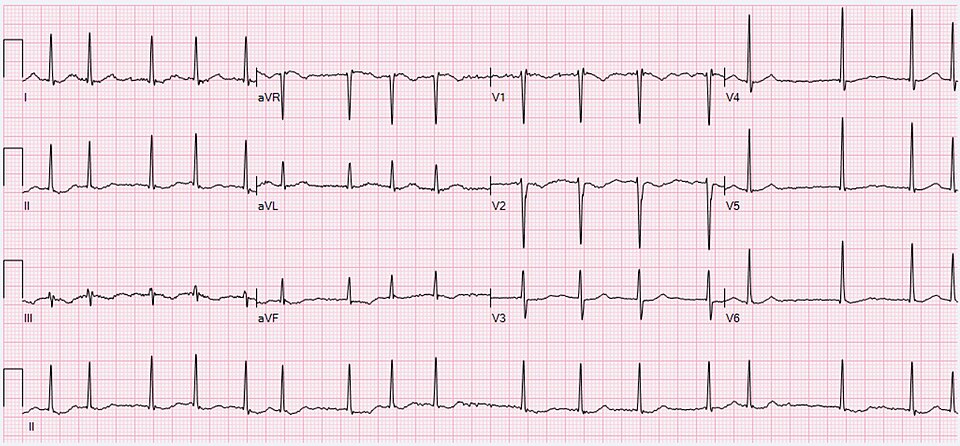 Image Source: Wikimedia Commons (2020). Atrial Fibrillation, Licensed under a creative commons licence. Available for free at: https://commons.wikimedia.org/wiki/File:ECG_Atrial_Fibrillation_98_bpm.jpg (Accessed 1 May 2025).
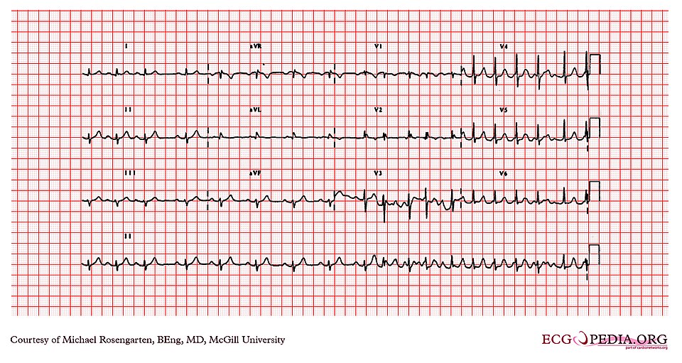 Image Source: Wikimedia Commons (2012). Cardio Networks ECGpedia, Licensed under a creative commons licence. Available for free at: https://commons.wikimedia.org/wiki/File:E234_(CardioNetworks_ECGpedia).jpg (Accessed 1 May 2025).
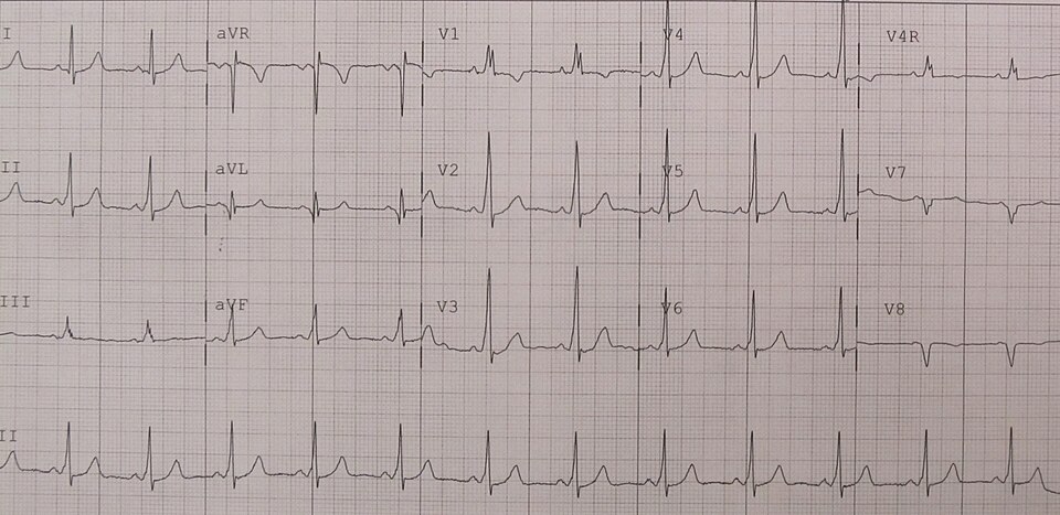 Image Source: Wikimedia Commons (2012). WPW 2012, Licensed under a creative commons licence. Available for free at: https://commons.wikimedia.org/wiki/File:WPW2012.jpg (Accessed 1 May 2025).
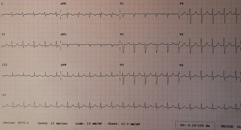 Image Source: Wikimedia Commons (2014). Junctional Tachycardia, Licensed under a creative commons licence. Available for free at: https://commons.wikimedia.org/wiki/File:Junctional_Tachycardia.jpg (Accessed 1 May 2025)
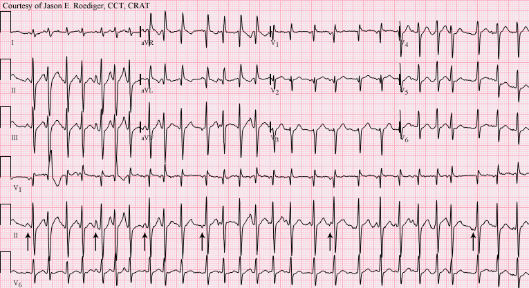 Image Source: Wikimedia Commons (2012). Multifocal Atrial Tachycardia, Licensed under a creative commons licence. Available for free at:
https://commons.wikimedia.org/wiki/File:Multifocal_atrial_tachycardia_-_MAT.png (Accessed 1 May 2025).
Management
Management of patients presenting with any tachyarrhythmia should be preceded by an immediate ABCDE assessment and then follow the resus council guidelines for adult tachycardia. The primary treatment for any tachycardia is to identify and treat reversible causes e.g. electrolyte abnormalities, hypovolaemia etc.
Life-threatening features
If a patient presenting with an atrial tachycardia has been adequately resuscitated medical and has ongoing evidence of haemodynamic compromise, syncope, myocardial ischaemia or severe heart failure then proceed as below:
- Deliver up to 3 synchronised DC shocks (sedate patient if conscious)
- If unsuccessful – give amiodarone 300mg over 10-20 minutes
- Reattempt synchronised DC cardioversion
Regular narrow-complex tachycardias
For patients who are haemodynamically stable and have a regular narrow-complex tachycardia, follow the steps below:
- Attempt vagal manoeuvres. This can be sufficient to cardiovert the patient back to sinus rhythm.
- If unsuccessful and there is no evidence of a pre-excitation syndrome (e.g. WPW), give adenosine under direct ECG monitoring. The doses are 6mg rapid IV bolus, then 12mg if unsuccessful, then 18mg if unsuccessful.
- If unsuccessful, give verapamil or beta-blocker (metoprolol is the drug of choice in acute situations due to its fast onset of action)
- If ineffective, attempt DC cardioversion as above (note atrial flutter is unlikely to chemically cardiovert and will often need DC cardioversion if treatment of underlying causes is unsuccessful).
Patients with a pre-excitation syndrome such as Wolff-Parkinson-White should not be given digoxin or verapamil, as these can provoke fatal arrhythmias through blockade of the SA node, encouraging conduction down the aberrant pathway.. If cardioversion is unsuccessful, patients should be referred for consideration of radio-frequency ablation of their aberrant conduction pathway.
| Class | Mechanism |
|---|---|
| Class 1 | Inhibits fast Na+ channels during depolarisation, reducing depolarisation and conduction velocity. Membrane stabilisers |
| Class 1a
e.g. quinidine, procainamide |
Prolonged action potential duration by reducing the excitability of non-nodal regions of the heart during phase 0 depolarisation |
| Class 1b
e.g. lignocaine, phenytoin |
Reduce action potential duration by decreasing the rate of spontaneous phase 4 in myocardial cells (reduces automaticity) |
| Class 1c
e.g. flecainide, encainide |
Potent Na+ channel blockers – little effect on duration of action potential in ventricular myocardium, shortens action potential in Purkinje fibres. Decreases speed of conduction of cardiac impulses. |
| Class 2 (beta-blockers)
e.g. metoprolol, esmolol |
Decrease rate of depolarisation and increase effective refractory period of AV node & decreased automaticity, decreased QT duration. Decreases heart rate and oxygen consumption. |
| Class 3
e.g. amiodarone, sotalol |
Inhibit K+ channels to prolong the refractory period, cardiac depolarisation and action potential duration. Effectively decreases the amount of time cardiac cells are excitable.
Note amiodarone has some class 1, 2, 3 and 4 effects. |
| Class 4 (calcium-channel block)
e.g. verapamil, diltiazem, nifedipine |
Inhibit slow Ca2+ channels – slow calcium ion currents may contribute towards development of VT. Block L-type Ca2+ channels to impair SA node pacemaker activity. Decrease duration of action potential but no effect on automaticity. |
Table 4: Vaughan-Williams classification of anti-arrhythmic drugs.
Irregular narrow-complex tachycardias (i.e. atrial fibrillation)
Atrial fibrillation is the most common tachyarrhythmia in the UK. In the acute setting the priority should be on rapid pharmacological rate control of the stable patient, and then subsequent decision regarding rate vs rhythm control. There is no evidence to suggest mortality is any worse following a rate control rather than a rhythm control strategy for patients, if high-risk patients are anticoagulated. However, patients in sinus rhythm may report a better quality of life than those in AF. The mainstay of treatment in AF therefore is rate control and prevention of thromboembolic events.
Atrial fibrillation can be classified as paroxysmal (less than 7 days, then terminating), persistent (greater than 7 days, would last indefinitely unless cardioverted) and permanent (greater than 7 days, sinus rhythm not possible).
- Rate control
- Usually with a standard beta-blocker (e.g. bisoprolol) or rate-limiting calcium-channel blocker (diltiazem or verapamil)
- Consider digoxin if above contra-indicated
- Amiodarone should not be used for long-term rate control
- Anticoagulation
- Use CHA2DS2VASc and ORBIT scores to assess patient for suitability of anticoagulation
- First-line anticoagulation for AF is a direct oral anticoagulant (DOAC) such as apixaban, dabigatran, edoxaban or rivaroxaban
- Vitamin K antagonists such as warfarin can be used if DOACs are contraindicated
| CHA₂DS₂VASc stroke risk score | Points |
|---|---|
| Congestive cardiac failure
Reduced ejection fraction or requiring hospitalisation regardless of EF |
1 |
| Hypertension
Systolic BP >140 mmHg and/or Diastolic BP >90 mmHg On at least 2 occasions, or on antihypertensive medication |
1 |
| Age greater than 75 years | 2 |
| Diabetes mellitus
Fasting plasma glucose >7.0 mmol/l or treatment with oral hypoglycaemic drugs +/- insulin |
1 |
| Stroke/TIA | 2 |
| Vascular disease
Previous MI, peripheral arterial disease, or aortic plaque |
1 |
| Age 65-74 years | 1 |
| Sex – female | 1 |
Table 6: ORBIT scoring system to assess bleeding risk when considering a patient for anticoagulation. A score of 1-2 is deemed low risk, 3 is medium risk and 4-7 high risk for a significant bleed if started on anticoagulation.
- Cardioversion
- DCCV may be appropriate for patients presenting with fast AF if onset is within 48 hours of presentation.
- If the history is suggestive of symptoms beginning earlier than this, or the patient is asymptomatic with no clear time of onset, cardioversion is contraindicated due to the risk of stroke from atrial thrombus formation.
- It is important to note that chemical cardioversion with flecainide or amiodarone carries the same risk, and therefore in patients presenting longer than 48 hours after development of AF, the management strategy should focus on rate or rhythm control, as detailed above.
References
- https://www.uptodate.com/contents/narrow-qrs-complex-tachycardias-clinical-manifestations-diagnosis-and-evaluation?search=atrial%20arryhthmia&topicRef=2118&source=see_link
- https://litfl.com/arrhythmias/
- https://cks.nice.org.uk/topics/atrial-fibrillation/
- Longmore et al. Oxford Handbook of Clinical Medicine. 8th Edition. Oxford University Press 2010.
- Ramrakha et al. Oxford Handbook of Acute Medicine. 3rd Edition. Oxford University Press 2010.
- https://www.resus.org.uk/sites/default/files/2021-04/Tachycardia%20Algorithm%202021.pdf
Complete Guide to Clinical Genetics ST3 Application and Recruitment (2027)
Written by Medibuddy. Last updated: October 2026. Clinical Genetics is

Complete Guide to Clinical Radiology ST3 Application and Recruitment (2027)
Recruitment paused: NHS England is not running Clinical Radiology ST3+ recru

Complete Guide to Paediatric Surgery ST3 Application and Recruitment in United Kingdom (2026)
Written by Medibuddy. Last updated: October 2026. If you’re planning


Take your subscriptions with you
Our mobile app allows you to access your interview and exam question banks wherever you are.

