


 Close
Close
 5th May 2025
Diagnosis
Definitive diagnosis of an atrial arrhythmia will be based on ECG evidence, however as with all conditions a detailed history and examination should precede investigations (in a haemodynamically stable, alert patient).
Symptoms (history)
- Palpitations
- Chest pain
- Shortness of breath
- Orthopnoea/paroxysmal nocturnal dyspnoea (evidence of cardiac decompensation)
- Loss of consciousness/blackouts (no warning)
- Dizziness/light-headedness
- Stroke symptoms
Signs (examination)
It is important to look for signs pertaining to possible underlying causes for the arrhythmia (based on history and presentation). Signs may include:
- Fever
- Hypotension
- Loss of consciousness
- Pallor
- Cachexia
- Tachypnoea
- Irregular, fast pulse
- Elevated JVP if in heart failure
- Cardiac murmurs if associated structural disease
- Evidence of thyroid disease – tremor, weight loss, exophthalmos, goitre, anxiety
Investigations
Investigations should be tailored to the suspected underlying cause. Below is a non-exhaustive table of immediate investigations to help ascertain the type of arrhythmia and possible underlying cause.
| Investigation | Positive findings |
|---|---|
| Urine dip +/- culture | Evidence of infection
Proteinuria/haematuria indicating renal/cardiac failure |
| Blood tests | ABG/VBG – acidosis, hypoxia, electrolyte abnormalities, lactate, hypercapnia
FBC – infection, anaemia U&E – electrolyte derangement, renal failure CRP – infection LFTs – cholestasis, hepatitis, poor nutrition Mg – hypomagnesaemia TFTs – hyper/hypothyroidism Bone profile – hypo/hypercalcaemia, phosphate INR/coag – may determine treatment Troponin – evidence of strain/ischaemia |
| CXR | Evidence of infection/fluid overload |
| ECG | Characterise arrhythmia (see below) |
| Echocardiogram | View structural abnormalities, systolic function and assess for right heart strain (PE) |
Table 3: Investigations in patients presenting with cardiac arrhythmias
ECGs
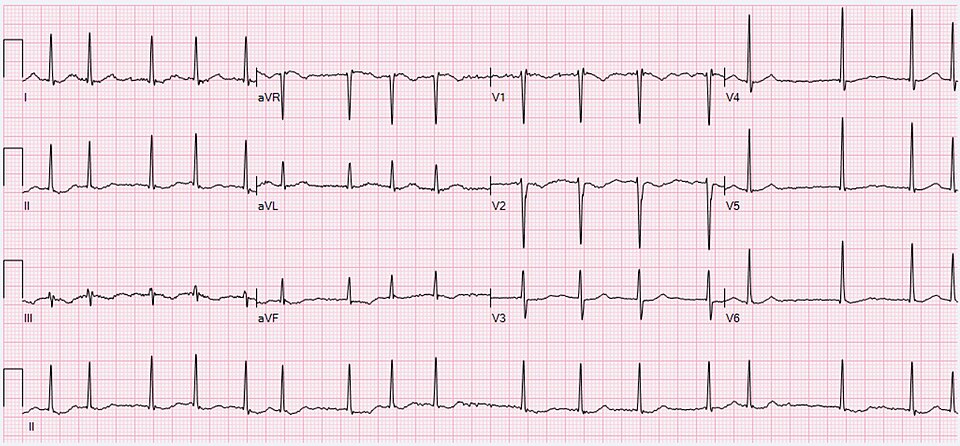
Image Source: Wikimedia Commons (2020). Atrial Fibrillation, Licensed under a creative commons licence. Available for free at: https://commons.wikimedia.org/wiki/File:ECG_Atrial_Fibrillation_98_bpm.jpg (Accessed 1 May 2025).
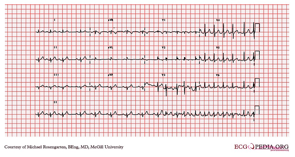 Image Source: Wikimedia Commons (2012). Cardio Networks ECGpedia, Licensed under a creative commons licence. Available for free at: https://commons.wikimedia.org/wiki/File:E234_(CardioNetworks_ECGpedia).jpg (Accessed 1 May 2025).
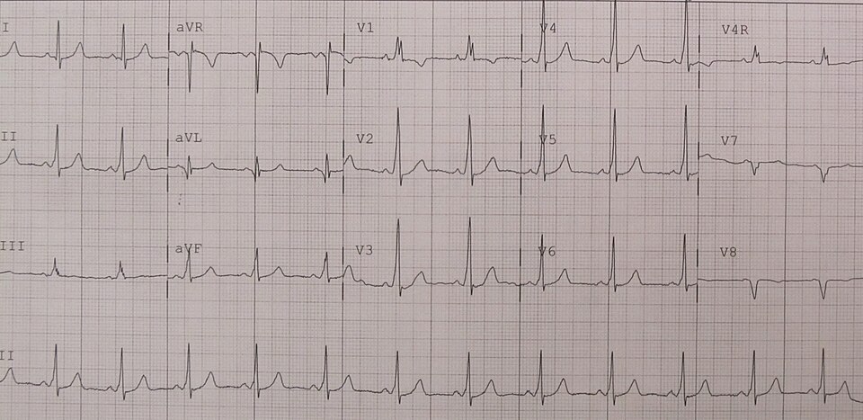
Image Source: Wikimedia Commons (2012). WPW 2012, Licensed under a creative commons licence. Available for free at: https://commons.wikimedia.org/wiki/File:WPW2012.jpg (Accessed 1 May 2025).
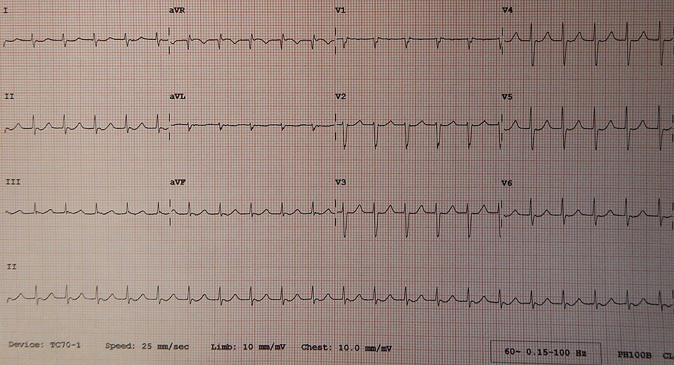
Image Source: Wikimedia Commons (2014). Junctional Tachycardia, Licensed under a creative commons licence. Available for free at: https://commons.wikimedia.org/wiki/File:Junctional_Tachycardia.jpg (Accessed 1 May 2025)
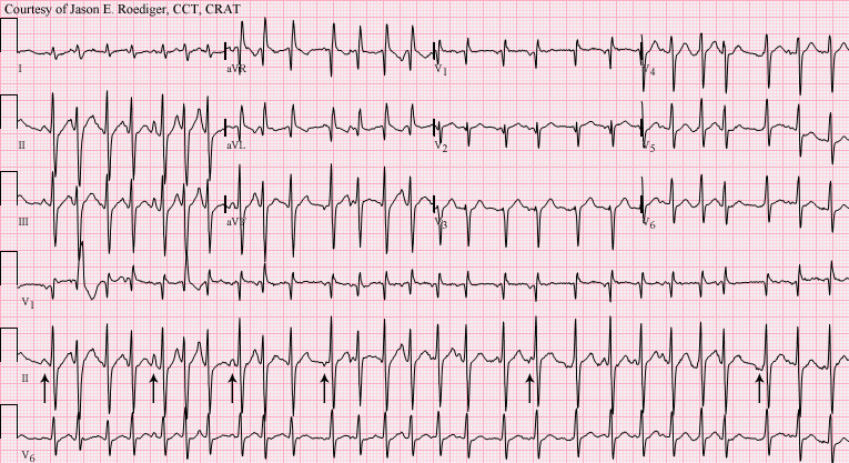
Image Source: Wikimedia Commons (2012). Multifocal Atrial Tachycardia, Licensed under a creative commons licence. Available for free at:
https://commons.wikimedia.org/wiki/File:Multifocal_atrial_tachycardia_-_MAT.png (Accessed 1 May 2025).
Complete Guide to Clinical Genetics ST3 Application and Recruitment (2027)
Written by Medibuddy. Last updated: October 2026. Clinical Genetics is

Complete Guide to Clinical Radiology ST3 Application and Recruitment (2027)
Recruitment paused: NHS England is not running Clinical Radiology ST3+ recru

Complete Guide to Paediatric Surgery ST3 Application and Recruitment in United Kingdom (2026)
Written by Medibuddy. Last updated: October 2026. If you’re planning


Take your subscriptions with you
Our mobile app allows you to access your interview and exam question banks wherever you are.

