


 Close
Close
 5th May 2025
Epidemiology
Narrow complex tachycardias can be divided into:
| Type of narrow complex tachycardia | ECG morphology |
|---|---|
| Sinus tachycardia | Normal p wave followed by normal QRS |
| Atrial fibrillation (AF) | Absent p wave, irregular QRS complexes – irregularly irregular |
| Atrial flutter | Atrial rate usually 300 bpm – “flutter” or “sawtooth” baseline, ventricular rate often 150bpm (2:1 block) but can be 100bpm (3:1 block), etc |
| Atrial tachycardia | Abnormally shaped p waves which may outnumber QRS complexes in a less regular fashion than atrial flutter |
| Multifocal atrial tachycardia | 3 or more p wave morphologies with irregular QRS complexes |
| Supraventricular tachycardia (SVT) – encompasses diagnoses below | P waves absent or inverted after QRS |
| Junctional tachycardia (AV nodal reentrant tachycardias) | P wave either buried in QRS or after QRS complex, rate often 150-200bpm |
| AV reentrant tachycardias (e.g. Wolff-Parkinson-White or accessory pathway) | P wave inverted after QRS, delta wave in WPW, rate often 150-250 bpm. QRS can be broad |
Table 1: Stratification of narrow complex tachycardias.
Atrial fibrillation is the most common cardiac arrhythmia with a prevalence in England of 2.5%. Approximately 1.4 million people were living with AF in England in 2016 with approximately 425,000 undiagnosed and untreated. Prevalence is higher in men (2.9%) than women (2.0%).
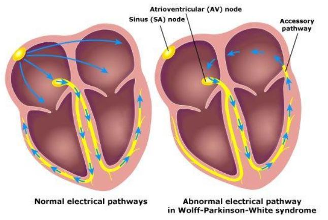
Image Source: Wikimedia Commons (2011). WPW, Licensed under a creative commons licence. Available for free at: https://commons.wikimedia.org/wiki/File:WPW.jpeg (Accessed 1 May 2025)
Complete Guide to Clinical Genetics ST3 Application and Recruitment (2027)
Written by Medibuddy. Last updated: October 2026. Clinical Genetics is

Complete Guide to Clinical Radiology ST3 Application and Recruitment (2027)
Recruitment paused: NHS England is not running Clinical Radiology ST3+ recru

Complete Guide to Paediatric Surgery ST3 Application and Recruitment in United Kingdom (2026)
Written by Medibuddy. Last updated: October 2026. If you’re planning


Take your subscriptions with you
Our mobile app allows you to access your interview and exam question banks wherever you are.

