


 Close
Close
 4th June 2025
Skin manifestations of anaemia
Aetiology and epidemiology
Anaemia is defined as a low concentration of haemoglobin (Hb); it can be further categorised by mean cell volume (MCV) of the red blood cell (i.e. microcytic, normocytic or macrocytic) and each of these subtypes has their own aetiology. The skin manifestations of anaemia vary depending on the underlying cause. Approximately 33% of the population globally are thought to be anaemic.
Microcytic anaemia
- The most common cause of anaemia globally is iron-deficiency anaemia (IDA) – which is also by far the most common form of microcytic anaemia.
- IDA is more common in low-income countries (reduced dietary iron intake, parasitic worm infections causing blood loss), young children (who require greater amounts of iron for growth) and pre-menopausal women (due to blood loss during menstruation).
- Other uncommon causes of microcytic anaemia include the thalassaemias (genetic diseases of abnormal Hb production, these are more common in people of Mediterranean, Middle-Eastern or Southeast Asian descent), and sideroblastic anaemia. When discussing microcytic anaemia in this topic, this will largely be referring to IDA.
- Aetiology
- Reduced dietary intake of iron (poverty leading to malnutrition, meat-free diet)
- Reduced absorption of iron (inflammatory bowel disease, coeliac disease, helicobacter pylori infection)
- Bleeding (menstruation, gastrointestinal blood loss (malignancy, peptic ulcer disease, haemorrhoids), parasitic intestinal infections)
Normocytic anaemia
- The prevalence of normocytic anaemia increases with age; the most common form of normocytic anaemia is anaemia of chronic disease. Normocytic anaemia has been reported in up to 44% of men older than 85 years (in the USA).
- The causes of normocytic anaemia are numerous, including:
- Anaemia of chronic disease
- Acute blood loss
- Bone marrow failure
- Renal failure
- Pregnancy
Macrocytic anaemia
- The most common cause of macrocytic anaemia is B12 and/or folate deficiency; this will produce a megaloblastic anaemia, where abnormal red blood cell precursors are found in the bone marrow.
- Macrocytic anaemia may also be non-megaloblastic.
- Megaloblastic macrocytic anaemia: causes of B12 and folate deficiency include:
- Vegan diet (the main dietary sources of B12 are animal products such as meat, eggs, fish and dairy)
- Pernicious anaemia (most frequently diagnosed aged 60, associated with other autoimmune conditions, F>M at a ratio of 1.6:1)
- Disease of the terminal ileum (e.g. Crohn’s disease, tapeworm infection, small bowel bacterial overgrowth)
- Causes of a non-megaloblastic macrocytic anaemia:
- Alcohol excess
- Liver disease
- Myelodysplastic syndromes
- Hypothyroidism
Clinical findings in skin manifestations of anaemia
The skin manifestations of anaemia can be subtle and may not be evident until there is a significant drop in haemoglobin.
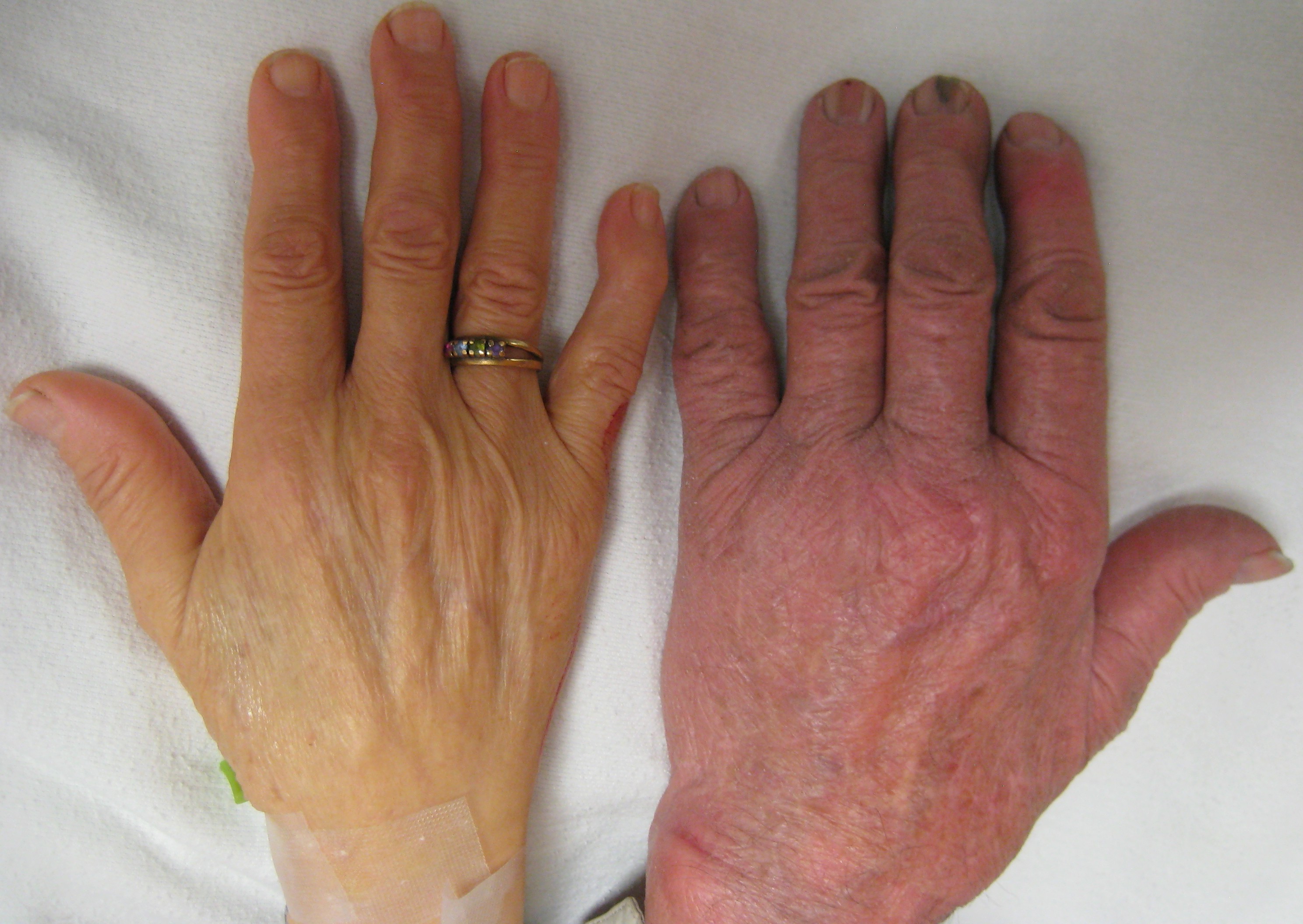
Skin pallor – including palm creases and mucous membranes (conjunctiva)
- The absence of pallor is not thought to be a reliable clinical sign in anaemia as it may be absent even in the severely anaemic; however the presence of pallor is more useful and can indicate severe anaemia.
- Occurs as blood is directed away from the skin and peripheries, and towards internal organs (to enhance oxygenation when the oxygen-carrying capacity of the blood is reduced).
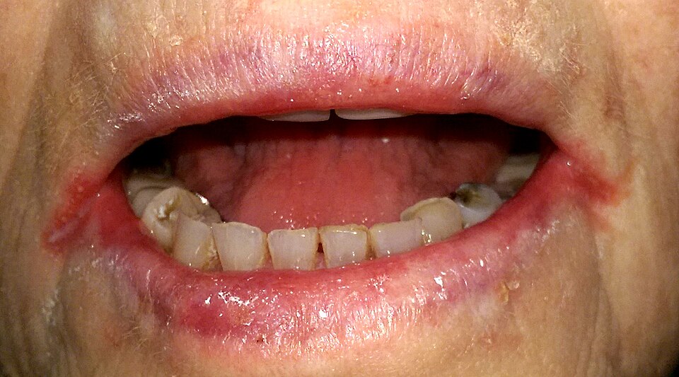
Angular stomatitis
- It is thought that up to 1/4 of cases of angular stomatitis are secondary to nutritional deficiencies (including iron and B12).
- Overexposure of the delicate skin at the mouth corners (commissures) to saliva leads to skin breakdown, saliva is an irritant and contains many digestive enzymes. It is not completely understood how anaemia leads to angular stomatitis, however it is suggested that nutritional deficiencies reduce the body’s ability to regenerate cells and repair tissues – increasing the risk of conditions such as angular stomatitis.
- Manifests clinically as erythema and cracking and crusting of the mouth corners
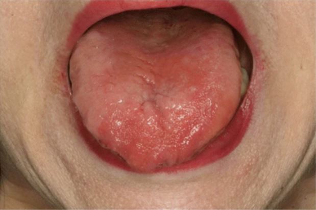
Atrophic glossitis – (loss of papillae, leading to a smooth tongue, may be painful)
- Epidemiology: atrophic glossitis is strongly associated with nutritional deficiencies such as IDA and B12 deficiency (causing a macrocytic anaemia). Some studies suggest atrophic glossitis is present in up to 1% of adults and up to 7% of women (due to the increased prevalence of iron deficiency anaemia).
- Aetiology: iron, B12 and folate (along with other nutrients) are required for the production and repair of healthy oral epithelial cells. Therefore anaemia (from such nutritional deficiencies) can cause atrophy of the epithelial surface of the tongue, leading to loss of papillae and atrophic glossitis.
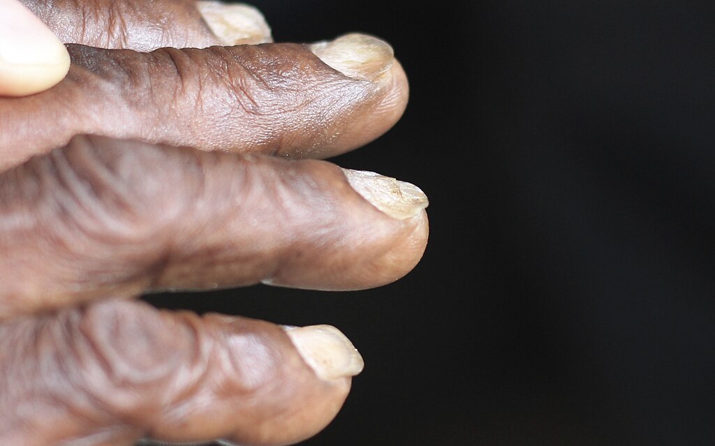
Koilonychia
- Up to 28% of people with IDA have nail changes.
- Kolionychia refers to spoon-shaped nails, and is usually preceded by thinning and flattening of the nails
- Epidemiology: koilonychia can be acquired, inherited or idiopathic, however the most common cause is IDA. Studies have found that up to 5.4% of patients with IDA have koilonychia.
- Aetiology: the pathophysiology of koilonychia is not fully understood, however it is hypothesised that reduced peripheral blood flow (secondary to anaemia) leads to disruption in the normal growth of the connective tissue of the nail bed.
Telogen effluvium
- A form of non-scarring alopecia, associated with increased hair shedding
- Epidemiology: anaemia has long been associated with increased hair loss, however studies have been conflicting. One recent meta-analysis (with over 10,000 total participants) concluded that women with non-scarring alopecia were significantly more likely to be iron deficient.
- Aetiology: as with other manifestations of anaemia, the pathophysiology of hair loss is not fully understood. Hair follicles require significant blood supply and nutrients for hair growth – these may both be compromised in anaemia.
https://en.wikipedia.org/wiki/File:Anemia.JPG
https://en.wikipedia.org/wiki/File:Angular_cheilitis1.jpg
https://en.wikipedia.org/wiki/Koilonychia#/media/File:Koilonychia_iron_deficiency_anemia.jpg
https://upload.wikimedia.org/wikipedia/commons/thumb/3/37/Image_of_the_tongue_in_a_B12-
Medical management
If a patient is found to be anaemic, the underlying cause should be identified and treated (to prevent recurrence). For example in women with IDA secondary to menorrhagia, contraceptive devices and/or tranexamic acid may be used to reduce blood loss.
Patients with IDA will likely require iron replacement to restore iron stores and correct their anaemia.
Treatment options include:
- Increased dietary iron. Haem iron is found in red meat – this is easily absorbed; the absorption of non-haem iron (in vegetables and grains) is increased with vitamin C.
- Oral iron supplementation
- Intravenous iron infusions
- Blood transfusion (in exceptional circumstances)
Complications
Once anaemia is treated, the skin, hair and nail manifestations should slowly recover with time.
Complete Guide to Clinical Genetics ST3 Application and Recruitment (2027)
Written by Medibuddy. Last updated: October 2026. Clinical Genetics is

Complete Guide to Clinical Radiology ST3 Application and Recruitment (2027)
Recruitment paused: NHS England is not running Clinical Radiology ST3+ recru

Complete Guide to Paediatric Surgery ST3 Application and Recruitment in United Kingdom (2026)
Written by Medibuddy. Last updated: October 2026. If you’re planning


Take your subscriptions with you
Our mobile app allows you to access your interview and exam question banks wherever you are.

