


 Close
Close
 5th May 2025
Bradyarrhythmias
Overview
Bradyarrhythmias are heart rhythms with a ventricular rate below 60 beats per minute. Bradyarrhythmias can be physiological (e.g. in athletes or during sleep) or pathological – commonly due to conduction pathway abnormalities or sinoatrial / atrioventricular nodal disease. Clinical manifestations of bradyarrhythmias can range from mild symptoms such as lethargy and exertional dyspnoea, to frank syncope. Up to 40% of patients report a syncopal episode with bradycardia, which is recurring in just over 10% of patients. In the elderly, bradyarrhythmias are a common cause of falls and are a common reason for pacemaker insertion. There is an increased risk of sudden cardiac death due to loss of cardiac output or asystole. Early detection of bradyarrhythmias in patients (ideally, when asymptomatic) is crucial to effective intervention – both in the acute and chronic settings. In this chapter we will look at some common cardiac causes of bradyarrhythmias and their specific management.
Aetiology
The common causes of bradyarrhythmias are summarised in Table 1. In elderly patients, ischaemic heart disease and age-related fibrosis of the cardiac conducting system are common causes of bradyarrhythmias. In younger patients, congenital and autoinflammatory causes such as cardiomyopathies, sarcoidosis and infections are more commonly found. In approximately one quarter of patients who present to the emergency department (ED) with bradycardia, adverse drug reaction or intoxication is identified. For purposes of simplicity, we will break down the causes into ‘cardiac’ and ‘non-cardiac’ causes and explore the common cardiac causes of bradycardia.
| Table 1 – Causes of bradyarrhythmia. Adapted from Honarbakhsh et al; 2018. |
|---|
| Cardiac |
| Age related degeneration of conduction fibres / Sinus node dysfunction
Ischaemic heart disease (particularly affecting right coronary territory) Hypertensive heart disease Genetic (e.g. ryanodine receptor mutations) / Cardiomyopathies Iatrogenic – catheter ablations or coronary intervention |
| Metabolic |
| Thyrotoxicosis / Myxoedema
Electrolyte imbalance Alcohol |
| Inflammatory |
| Sarcoidosis
Amyloidosis |
| Infection |
| Rheumatic fever
Lyme disease |
| Drugs & Toxins |
| Calcium channel blockers
Tricyclic antidepressants Psychoactive drugs (e.g. SSRIs) Chemotherapy drugs |
Clinical Assessment
A detailed clinical history and physical examination is key to formulating an accurate differential diagnosis. A physiological bradycardia may be normal for athletic individuals. Bradycardia should be investigated if associated with clinical symptoms or haemodynamic compromise. Furthermore, bradycardia may be associated with escape rhythms and may be a sign of an underlying pathological cause. In these circumstances, bradycardia should be investigated further with a clinical assessment of the patient.
Patients with symptomatic bradycardia may present with:
- shortness of breath (at rest or exertional)
- chest pain
- fatigue
- lethargy
- confusion
- nausea
- dizziness or syncope
- clinical signs suggestive of an underlying cause (e.g. pretibial myxoedema in hypothyroidism, peripheral pitting oedema or added heart sounds in heart failure)
In cases of severe and prolonged cerebral hypoperfusion, patients may even experience grand mal seizures or transient loss of consciousness – known as Stokes-Adams attacks. Asking about onset, duration and frequency of symptoms may help differentiate cardiac from non-cardiac aetiologies. It is important to ask about symptoms of acute coronary syndrome to exclude an ischaemic cause. Moreover, a known previous personal or family history of cardiac disease, sudden cardiac death (especially if age <50) increases the chances of a cardiac cause underlying the bradyarrhythmia.
Diagnostic workup of the patient starts with bedside investigations such as a 12-lead electrocardiogram (ECG) and basic blood tests including thyroid function, troponin, NT-proBNP and serum electrolytes. Transthoracic echocardiography is of value in evaluating suspected structural heart disease and ambulatory-ECG (Holter) monitoring may be implemented in cases of exercise-induced or intermittent symptoms. An implantable cardiac monitor (ICM) device may be considered in cases of intermittent symptoms not picked up on Holter monitoring. An electrophysiology study may be carried out in presence of ongoing diagnostic uncertainty and cardiac magnetic resonance imaging with late gadolinium enhancement may be used to exclude cardiomyopathy and infiltrative disease.
Cardiac Causes
Nodal disease
Sinus node dysfunction (SND), also known as ‘Sick Sinus Syndrome’ or ‘Tachy-Brady Syndrome’, encompasses a spectrum of conditions that affect the specialised cardiac conduction tissue in the sinoatrial node, the atrial conduction tissue and the atrioventricular node. It commonly tends to be a disease of the older patient, with multiple postulated mechanisms whereby malfunction of the cardiac conduction system (mostly from fibrosis) occurs leading to slower sinoatrial conduction times. Electrocardiographic (ECG) features of this can be varied – from sinus bradycardia, sinus pause (>3 seconds), sinus arrest with intermittent atrial tachycardia. There is often an alternating tachycardia-bradycardia phenomenon seen in this, with intermittent sinus rhythm or sinus bradycardia. SND is usually detected on Holter monitoring but may also be picked up on serial 12-lead ECG. Definitive treatment is with pacemaker insertion, mostly for symptom management.
Heart block
Atrioventricular (AV) block, or heart block, occurs when the electrical impulse travelling from the atria to the ventricles is delayed or completely halted. There are three types of AV-block classically described, based on their ECG features – these are illustrated in Figure 1. Certain drugs (e.g. beta blockers, calcium channel blockers, tricyclic antidepressants) can cause all three types of heart block. In 1st degree AV-block, there is prolongation of the PR-interval beyond 200milliseconds with preservation of subsequent QRS complexes. No specific intervention is required for this isolated 1st degree AV-block and it is often found in asymptomatic and healthy individuals; however, in cases of symptomatic 1ˢᵗ degree AV-block with PR>300ms, a pacemaker may be advocated. In 2nd degree AV-block, there is loss of subsequent QRS complexes. This may be following progressive prolongation of the PR-interval, in the case of Mobitz Type 1 AV-block (also known as Wenckebach phenomenon); or, with a constant PR-interval with occasional loss of QRS complex, in the case of Mobitz Type 2 AV-block. In 3ʳᵈ degree AV-block, also known as complete heart block, there is complete disassociation of the P waves with QRS complexes. In this bradyarrhythmia, there is often a ventricular escape (junctional) rhythm characterised by a slower rate (depending on how distal to the AV-node the escape rhythm originates from) and broad QRS complexes. Due to a higher risk of asystole, pacemaker intervention is indicated in Mobitz Type 2 and 3ʳᵈ degree AV-block.
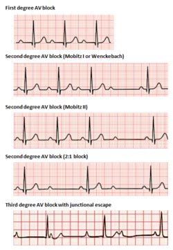
Image Source: Wikimedia Commons (2017). Atrioventricular Block, Licensed under a creative commons licence. Available for free at: https://en.wikipedia.org/wiki/Atrioventricular_block (Accessed 1 May 2025).
Ventricular disease
Abnormal conduction can also occur within the ventricles themselves, in the setting of the bundle branch block. However, this is typically distinct from any abnormal AV-conduction and is often associated with sinus rhythm.
Non-Cardiac Causes
There is an extensive list of non-cardiac causes of bradyarrhythmia, some of which are listed in Table 1. It is crucial to identify any reversible causes in the clinical history and examination as this will help determine need for further investigations and guide the most appropriate intervention. Always take the same systematic ‘A to E’ approach when assessing any patient with a suspected non-cardiac bradyarrhythmia. Consider immediate tests which you can do at the bedside (lying-standing blood pressure to assess for postural hypotension, venous blood gas to check electrolytes before replacing, urine dipstick for infection and checking the medication chart for culprit drugs) before moving onto non-bedside investigations (e.g. chest radiograph, formal blood tests).
Management
All patients with symptomatic bradyarrhythmia should be referred to Cardiology. Clinically stable patients are normally managed in an ambulatory outpatient setting. Those with red flag symptoms should be referred to secondary care for urgent specialist assessment.
Red flag features include:
- Chest pain
- Hypotension
- Impaired consciousness
- Ongoing pre-syncope or syncope with a bradyarrhythmia
- Syncope with evidence of: abnormal ECG features, heart failure, exertional dyspnoea, unexplained prodrome, exertional syncope, family history of arrhythmias or sudden cardiac death
- Ongoing Mobitz type 2 or 3ʳᵈ degree AV-block (1st degree and Mobitz type 1 are not acute indications for intervention but pacing may be considered in patients who are symptomatic)
Following correction of any reversible causes (drugs, electrolyte imbalance, metabolic acidosis, thyroid dysfunction), the definitive treatment for symptomatic bradyarrhythmia is insertion of a pacemaker. In acute settings, isoprenaline or adrenaline may be used as a bridge to pacing in patients who are clinically unwell and require intervention in the interim period – these are usually given in a high-dependency or coronary care unit environment. Patients who have an ischaemic event as the underlying cause of bradyarrhythmias should be monitored in a coronary care unit (CCU) for at least 24 hours following revascularization to ensure no further bradyarrhythmias occur. If the bradyarrhythmia persists, these patients may also be counselled for pacemaker intervention. Any patients who present in extremis or with signs of haemodynamic compromise should also be monitored in a CCU following pacemaker insertion, to ensure no further bradyarrhythmias occur.
Pacing
A pacemaker provides permanent treatment of a bradyarrhythmia. Overall, the commonest indication for pacemaker intervention is high degree AV-block, followed by SND. Patients with AV-block who are conservatively managed have been shown to have poorer survival, when compared with pacemaker-treated patients. In contrast, patients with SND often have an unpredictable prognostic trajectory without intervention; pacing is used for symptom control and to optimise quality of life . The 2021 ESC guidelines recommend that pacing in asymptomatic SND has not been shown to affect prognosis.
Types of pacing
Pacing is classically done via an endocardial approach, whereby a pulse generator device is placed in the pectoral region with transvenous lead(s) implanted into the myocardium. Epicardial pacing, whereby lead(s) are placed onto the myocardial surface via minimally invasive techniques, may be used in certain circumstances – i.e. patients with congenital disease with difficult venous access. Permanent pacemakers (PPM) can be single chamber, dual chamber or leadless. Single chamber PPMs have one lead placed in the right ventricle (single lead right atrial pacing is no longer recommended). A single lead PPM may be used in AV-block or AF with bradycardia. Dual chamber pacemakers pace two cardiac chambers (usually the right atrium and the right ventricle) and are the preferred form of pacing for AV-block and SND. In cases where LV-impairment or a high RV-pacing burden may be seen, a biventricular pacemaker may be used which paces the LV and the RV. An extra atrial lead can be added in conditions with atrial asynchrony (AV-block/SND). Leadless pacing is a relatively new revolutionary form of pacing whereby a miniaturized intracardiac device is inserted via the femoral vein and implanted directly into the RV wall. Although in theory, this would reduce the risk of lead displacement, infection and generator defects, randomised controlled trials directly comparing leadless pacing with conventional pacing are still pending.
Complications
The commonest complications of pacemakers include device infection and lead failure. In the acute phase following PPM insertion, lead displacement, lead failure or perforation can occur and should be monitored for. Although most cardiac centres use a post-insertion antibiotic prophylaxis regime (based on local Trust guidelines), mostly covering for Gram positive bacteria (i.e. Staphylococci spp.), some patients can still experience ongoing pacemaker insertion site pain, discharge or otherwise unexplained fevers. In these patients, a pacemaker site infection should be considered and a decision made based on the clinical status of the patient if they can have ambulatory antimicrobial treatment or whether they require inpatient intravenous treatment and telemetry monitoring for any arrhythmias. Depending on the approach taken, subclavian vein or superior vena cava thrombosis or occlusion can occur. One of the risks of single chamber (i.e. right ventricular) pacing is a delay in activation of the left ventricle, as the RV lead bypasses the His-Purkinje system, causing an asynchrony similar to that of left bundle branch block. In some cases, this asynchrony between atrium-ventricle or ventricle-ventricle can cause a congestive cardiac failure-like clinical syndrome – known as pacemaker syndrome. This can be managed either medically (with diuretics) or by the addition of an atrial lead, or using biventricular pacing. Follow-up every 6–12 months in a pacing clinic is recommended to ensure PPM device integrity and function.
Acute Bradyarrhythmias
In the peri-arrest setting, bradyarrhythmias are managed as per Advanced Life Support guidelines. Initial assessment should always begin with a systematic ‘A to E’ approach followed by ensuring an oxygen supply (if appropriate) and IV access are established. Initial investigations include oxygen saturations, blood pressure and 12-lead ECG monitoring. Evaluation and treatment of any reversible causes (electrolyte derangement, drugs & toxins) is crucial at this stage. Atropine may be used initially to block parasympathetic tone, followed by a positive chronotropic and inotropic agent such as isoprenaline or adrenaline. Non-invasive temporary pacing may be implemented for bradycardias that cause haemodynamic compromise and are unresponsive to atropine – illustrated in Figure 2 below. Pulseless electrical activity is defined as ‘organised cardiac electrical activity in the absence of any palpable pulses. In PEA, there is myocardial contraction sufficient to cause some detectable electrical potential but not sufficient to produce any cardiac output. Management of PEA arrest is as per ALS guidelines (CPR and alternate cycles of adrenaline every 2-minutes). Unless PEA arrest is due to a reversible cause, survival is often unlikely. Asystole is defined as ‘absence of electrical activity on the ECG trace’. When diagnosing asystole, care must be taken to not miss ventricular standstill, which may be reversible and is discussed below.
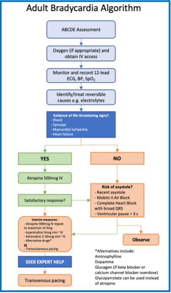
Image Source: Wikimedia Commons (2021). Bradycardia Algorithm, Licensed under a creative commons licence. Available for free at: https://www.resus.org.uk/sites/default/files/2021-04/Bradycardia%20Algorithm%202021.pdf (Accessed 1 May 2025).
Ventricular standstill (VS) is a rare arrhythmia wherein there is no ventricular activity. ECG classically shows p waves with no QRS – it is also hence referred to as ‘p-wave asystole’. Due to there being no flow of blood through the ventricles, there is very high risk of sudden cardiac death. In the acute setting, VS requires temporary pacing wire insertion to prevent further ischaemic damage to the myocardium. Definitive treatment of VS is with permanent pacemaker insertion.
References
- Honarbakhsh S, Hunter L, Chow A, Hunter RJ. Bradyarrhythmias and pacemakers. BMJ. 2018 Mar 15;360:k642. doi: 10.1136/bmj.k642. PMID: 29545242.
- Glikson M, Nielsen JC, Kronborg MB, Michowitz Y, Auricchio A, Barbash IM, Barrabés JA, Boriani G, Braunschweig F, Brignole M, Burri H, Coats AJS, Deharo JC, Delgado V, Diller GP, Israel CW, Keren A, Knops RE, Kotecha D, Leclercq C, Merkely B, Starck C, Thylén I, Tolosana JM; ESC Scientific Document Group. 2021 ESC Guidelines on cardiac pacing and cardiac resynchronization therapy. Eur Heart J. 2021 Sep 14;42(35):3427-3520. doi: 10.1093/eurheartj/ehab364. Erratum in: Eur Heart J. 2022 May 1;43(17):1651. PMID: 34455430.
- Kusumoto FM, Schoenfeld MH, Barrett C, Edgerton JR, Ellenbogen KA, Gold MR, Goldschlager NF, Hamilton RM, Joglar JA, Kim RJ, Lee R, Marine JE, McLeod CJ, Oken KR, Patton KK, Pellegrini CN, Selzman KA, Thompson A, Varosy PD. 2018 ACC/AHA/HRS Guideline on the Evaluation and Management of Patients With Bradycardia and Cardiac Conduction Delay: Executive Summary: A Report of the American College of Cardiology/American Heart Association Task Force on Clinical Practice Guidelines, and the Heart Rhythm Society. J Am Coll Cardiol. 2019 Aug 20;74(7):932-987. doi: 10.1016/j.jacc.2018.10.043. Epub 2018 Nov 6. Erratum in: J Am Coll Cardiol. 2019 Aug 20;74(7):1014-1016. PMID: 30412710.
- Mond HG, Proclemer A. The 11th world survey of cardiac pacing and implantable cardioverter-defibrillators: calendar year 2009–a World Society of Arrhythmia’s project. Pacing Clin Electrophysiol. 2011 Aug;34(8):1013-27. doi: 10.1111/j.1540-8159.2011.03150.x. Epub 2011 Jun 27. PMID: 21707667.
- Soar J, Böttiger BW, Carli P, Couper K, Deakin CD, Djärv T, Lott C, Olasveengen T, Paal P, Pellis T, Perkins GD, Sandroni C, Nolan JP. European Resuscitation Council Guidelines 2021: Adult advanced life support. Resuscitation. 2021 Apr;161:115-151. doi: 10.1016/j.resuscitation.2021.02.010. Epub 2021 Mar 24. Erratum in: Resuscitation. 2021 Oct;167:105-106. PMID: 33773825.
Complete Guide to Clinical Genetics ST3 Application and Recruitment (2027)
Written by Medibuddy. Last updated: October 2026. Clinical Genetics is

Complete Guide to Clinical Radiology ST3 Application and Recruitment (2027)
Recruitment paused: NHS England is not running Clinical Radiology ST3+ recru

Complete Guide to Paediatric Surgery ST3 Application and Recruitment in United Kingdom (2026)
Written by Medibuddy. Last updated: October 2026. If you’re planning


Take your subscriptions with you
Our mobile app allows you to access your interview and exam question banks wherever you are.

