


 Close
Close
 5th May 2025
Clinical Assessment
A detailed clinical history and physical examination is key to formulating an accurate differential diagnosis. A physiological bradycardia may be normal for athletic individuals. Bradycardia should be investigated if associated with clinical symptoms or haemodynamic compromise. Furthermore, bradycardia may be associated with escape rhythms and may be a sign of an underlying pathological cause. In these circumstances, bradycardia should be investigated further with a clinical assessment of the patient.
Patients with symptomatic bradycardia may present with:
- shortness of breath (at rest or exertional)
- chest pain
- fatigue
- lethargy
- confusion
- nausea
- dizziness or syncope
- clinical signs suggestive of an underlying cause (e.g. pretibial myxoedema in hypothyroidism, peripheral pitting oedema or added heart sounds in heart failure)
In cases of severe and prolonged cerebral hypoperfusion, patients may even experience grand mal seizures or transient loss of consciousness – known as Stokes-Adams attacks. Asking about onset, duration and frequency of symptoms may help differentiate cardiac from non-cardiac aetiologies. It is important to ask about symptoms of acute coronary syndrome to exclude an ischaemic cause. Moreover, a known previous personal or family history of cardiac disease, sudden cardiac death (especially if age <50) increases the chances of a cardiac cause underlying the bradyarrhythmia.
Diagnostic workup of the patient starts with bedside investigations such as a 12-lead electrocardiogram (ECG) and basic blood tests including thyroid function, troponin, NT-proBNP and serum electrolytes. Transthoracic echocardiography is of value in evaluating suspected structural heart disease and ambulatory-ECG (Holter) monitoring may be implemented in cases of exercise-induced or intermittent symptoms. An implantable cardiac monitor (ICM) device may be considered in cases of intermittent symptoms not picked up on Holter monitoring. An electrophysiology study may be carried out in presence of ongoing diagnostic uncertainty and cardiac magnetic resonance imaging with late gadolinium enhancement may be used to exclude cardiomyopathy and infiltrative disease.
Cardiac Causes
Nodal disease
Sinus node dysfunction (SND), also known as ‘Sick Sinus Syndrome’ or ‘Tachy-Brady Syndrome’, encompasses a spectrum of conditions that affect the specialised cardiac conduction tissue in the sinoatrial node, the atrial conduction tissue and the atrioventricular node. It commonly tends to be a disease of the older patient, with multiple postulated mechanisms whereby malfunction of the cardiac conduction system (mostly from fibrosis) occurs leading to slower sinoatrial conduction times. Electrocardiographic (ECG) features of this can be varied – from sinus bradycardia, sinus pause (>3 seconds), sinus arrest with intermittent atrial tachycardia. There is often an alternating tachycardia-bradycardia phenomenon seen in this, with intermittent sinus rhythm or sinus bradycardia. SND is usually detected on Holter monitoring but may also be picked up on serial 12-lead ECG. Definitive treatment is with pacemaker insertion, mostly for symptom management.
Heart block
Atrioventricular (AV) block, or heart block, occurs when the electrical impulse travelling from the atria to the ventricles is delayed or completely halted. There are three types of AV-block classically described, based on their ECG features – these are illustrated in Figure 1. Certain drugs (e.g. beta blockers, calcium channel blockers, tricyclic antidepressants) can cause all three types of heart block. In 1st degree AV-block, there is prolongation of the PR-interval beyond 200milliseconds with preservation of subsequent QRS complexes. No specific intervention is required for this isolated 1st degree AV-block and it is often found in asymptomatic and healthy individuals; however, in cases of symptomatic 1st degree AV-block with PR>300ms, a pacemaker may be advocated. In 2nd degree AV-block, there is loss of subsequent QRS complexes. This may be following progressive prolongation of the PR-interval, in the case of Mobitz Type 1 AV-block (also known as Wenckebach phenomenon); or, with a constant PR-interval with occasional loss of QRS complex, in the case of Mobitz Type 2 AV-block. In 3rd degree AV-block, also known as complete heart block, there is complete disassociation of the P waves with QRS complexes. In this bradyarrhythmia, there is often a ventricular escape (junctional) rhythm characterised by a slower rate (depending on how distal to the AV-node the escape rhythm originates from) and broad QRS complexes. Due to a higher risk of asystole, pacemaker intervention is indicated in Mobitz Type 2 and 3ʳᵈ degree AV-block.
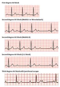
Image Source: Wikimedia Commons (2017). Atrioventricular Block, Licensed under a creative commons licence. Available for free at: https://en.wikipedia.org/wiki/Atrioventricular_block (Accessed 1 May 2025).
Ventricular disease
Abnormal conduction can also occur within the ventricles themselves, in the setting of the bundle branch block. However, this is typically distinct from any abnormal AV-conduction and is often associated with sinus rhythm.
Non-Cardiac Causes
There is an extensive list of non-cardiac causes of bradyarrhythmia, some of which are listed in Table 1. It is crucial to identify any reversible causes in the clinical history and examination as this will help determine need for further investigations and guide the most appropriate intervention. Always take the same systematic ‘A to E’ approach when assessing any patient with a suspected non-cardiac bradyarrhythmia. Consider immediate tests which you can do at the bedside (lying-standing blood pressure to assess for postural hypotension, venous blood gas to check electrolytes before replacing, urine dipstick for infection and checking the medication chart for culprit drugs) before moving onto non-bedside investigations (e.g. chest radiograph, formal blood tests).
Complete Guide to Clinical Genetics ST3 Application and Recruitment (2027)
Written by Medibuddy. Last updated: October 2026. Clinical Genetics is

Complete Guide to Clinical Radiology ST3 Application and Recruitment (2027)
Recruitment paused: NHS England is not running Clinical Radiology ST3+ recru

Complete Guide to Paediatric Surgery ST3 Application and Recruitment in United Kingdom (2026)
Written by Medibuddy. Last updated: October 2026. If you’re planning


Take your subscriptions with you
Our mobile app allows you to access your interview and exam question banks wherever you are.

